The passing of the years inevitably leaves an imprint on the face. In order for the mirror to continue to please even in adulthood, you can and should use all the resources of your own body, in particular, fat cells. Specialists at the Medial aesthetic medicine clinic invite you to a lipofilling procedure that will help get rid of unwanted age-related changes. By resorting to injections of your own fat, you can correct congenital asymmetry of the face, eliminate cosmetic defects in the temporal zone, cheekbones and cheeks, lower eyelids and chin.
Benefits of the procedure:
- minimum risk:
During the procedure, it is not a foreign graft that is used, but the patient’s adipose tissue, which is not rejected by the body and does not cause allergic reactions. Even if some of the fat cells do not take root, they will be eliminated from the body without any health consequences; - natural result:
After the procedure, regardless of which area of the face the procedure is performed on (eyelids, cheekbones and cheeks, chin or all problem areas), the “tweaked” area will look as natural as possible; - long-lasting effect:
Unlike a number of other cosmetic procedures that require periodic correction, the results of volumetric modeling last for many years; - quick recovery:
The procedure does not require general anesthesia. All manipulations in the temporal region, cheekbones and cheeks, eyelids and chin are performed under local anesthesia. The procedure is low-traumatic, well tolerated and does not require long-term rehabilitation; - Deep skin rejuvenation:
Adipose tissue is rich in stem cells. After the procedure, metabolic processes are activated, as a result of which local blood flow improves, the number of facial wrinkles decreases, skin turgor increases, and the face looks healthy and fresh.
Correction zones
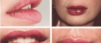
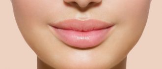
- Lips. Procedures to enlarge and reshape lips are now at the peak of popularity. However, correction with gels has to be repeated constantly, but lipofilling will only need to be done once. This is especially true if you do not want to increase the volume of your lips, but would not mind returning them to their former juiciness.
- Dark circles under the eyes. “Dips” and dark circles under the eyes are usually difficult to correct, but lipofilling easily copes with this task: in addition to replenishing volume, fat transfer leads to a noticeable improvement in the quality of the skin.
- Cheeks and cheekbones. The drooping of the tissues of the middle zone of the face leads to the appearance of jowls and deep nasolabial folds, blurring the contours of the lower jaw. To return a clear contour to the oval of the face, you will need to uniformly inject fat into the recessed areas.
Point injections smooth out nasolabial folds, nasolacrimal depressions, “dips” in the cheekbones and temples, create a gentle oval on thin, pointed faces, and improve the relief of the neck and décolleté. Lipofilling is absolutely indispensable for rejuvenating the hands when you need to smooth out protruding veins and tendons.
Contraindications to the procedure:
- chronic diseases in the acute stage;
- acute diseases of viral and infectious etiology;
- oncological diseases;
- diabetes mellitus type I and II, other endocrine disorders;
- connective tissue pathologies,
- disorders of the blood clotting system,
- lack of sufficient adipose tissue in the patient.
Please make an appointment for a medical consultation in advance and provide the doctor with truthful and as detailed information as possible about your medical condition. This will not only avoid possible complications, but will also increase the effectiveness of the procedure.
How long does it take for the fat to take root and when can you see the final result?
Engrafted fat is already present a month after lipofilling. But there are some fat cells that have not yet taken root as well. The entire metabolic process of engraftment and utilization of destroyed fat ends by 6 months and then we can see the final result. As for facial lipofilling, the result can be seen even earlier, because there is a good blood supply and small volumes of injection, and buttock lipofilling - no earlier than 6 months. From my experience, on average, 50% fat survives, so during lipofilling, you need to deliberately introduce excess fat, more than necessary, given that the volume of fat will decrease.
To prove the effectiveness of lipofilling, I can say that in Brazil and Colombia this is operation No. 1. Patients (often at a young age, so there is no sagging skin) have fat taken from the waist in a circle and injected into the buttocks on average 1-1, 5 liters each. And after 2-3-4 years, no one says that the buttocks have become much smaller. If fat had not taken root so massively, this technique would have been abandoned long ago. But there are also dissatisfied patients, to whom the fat was injected either poorly, or, on the contrary, it was too much (when a lot of it was injected), these are the 2 main problems of lipofilling.
List of necessary tests
Procedures on certain areas of the face are performed under local anesthesia. So that the doctor can predict the result as accurately as possible and assess the condition of your body, you will need to undergo the following tests:
- general clinical blood test (complete + platelets);
- coagulogram (blood clotting time, bleeding time, INR, prothrombin index, fibrinogen, APTT);
- blood sugar (glucose) test;
- examinations for hepatitis B, hepatitis C, HIV, syphilis
*To assess the condition of the body and outline the optimal amount of correction, the doctor can individually prescribe an extended list of studies for you.
Plastic surgeons at the clinic
Yakimov Dmitry Konstantinovich
Experience - 38 years
Doctor of the highest category. Candidate of Medical Sciences. Member of the Society of Plastic Surgeons. Author of more than 30 works in the field of surgery, teacher at the Military Medical Academy.
Gvaramiya Eka Yurievna
Experience - 17 years
Participant of congresses, conferences and seminars on plastic surgery. Priority areas: mammoplasty (breast plastic), blepharoplasty, tummy tuck (abdominoplasty), liposuction, lipofilling, face lift, lip plastic, ear plastic, intimate plastic, body plastic, reconstructive plastic, solving aesthetic problems.
Abzaleva Guzel Rinatovna
Experience - 15 years
Participant of congresses, conferences and seminars on plastic surgery. Priority areas: blepharoplasty, lipofilling, intimate plastic surgery, abdominoplasty, breast plastic surgery.
Kalita Valeria Denisovna
Experience - 4 years
Participant of congresses, conferences and seminars on plastic surgery. Priority areas: mammoplasty, blepharoplasty, liposuction, abdominoplasty.
Makarov Andrey Vitalievich
Experience - 18 years
Certified specialist in plastic surgery, maxillofacial surgery, otolaryngology, general surgery. Participant in master classes on facial plastic surgery and rhinoplasty in Russia and abroad.
Garifulin Marat Sagitovich
Experience - 19 years
Priority areas: mammoplasty and gynecomastia (including in men), waist shaping, blepharoplasty, otoplasty, abdominoplasty.
Karpuschenko Maxim Alekseevich
Experience - 18 years
Specializes in nasal plastic surgery: rhinoplasty, septum correction, surgery after a nasal fracture.
Stages of the procedure
Volume modeling of the face using your own fat is carried out according to an approved medical protocol, which includes 3 stages: sampling of fat cells, cleaning the donor tissue and introducing it into the problem area.
- The doctor selects a donor area and manually removes fat tissue through a thin cannula with a blunt tip. The puncture size is about 3 mm; no hematomas are formed during fat sampling; the subcutaneous vessels maintain their integrity.
- Only intact, well-cleaned cells with high survival rates are suitable for the procedure. Therefore, adipose tissue must be cleaned and washed with special solutions.
- The introduction of purified donor tissue into the problem area is carried out by injection. 2-3 mm punctures are made in natural skin folds or hair growth areas. To perform the procedure, a special thin cannula is used, which allows the dosage of the injected filler to be calculated as accurately as possible. By evenly distributing fat cells, you can achieve impressive long-lasting results.
Facial lipofilling: before and after photos
The facial lipofilling procedure is very responsible, because in the facial area there are nerves and blood vessels that can be damaged during the operation. Therefore, we recommend having lipofilling done in Minsk in a center where the technique of performing this operation performed by highly qualified surgeons will be safe, controlled, and will give a long-term, stable result.
Our doctors will restore youth and freshness to your face, because in professional hands, facial lipofilling is a godsend for the patient.
Rehabilitation period:
- To prevent complications, antibacterial strips are applied to the sites where adipose tissue is collected and where cells are introduced into problem areas. When correcting large volumes, the doctor may recommend a hospital stay for 24 hours, but most patients return home within a few hours.
- Swelling and local hematomas may remain at the puncture sites for 3-6 days. To quickly eliminate these defects, take medications prescribed by your doctor and follow the recommended regimen.
- To speed up the survival of fat cells and maintain the results of the procedure for a long time, limit physical activity for 2-3 weeks and avoid exposure to high temperatures (bath, hot tub, sauna). You can take a shower. The result can be assessed 30 days after the operation.
The Medial Clinic offers favorable prices for volume modeling using your own adipose tissue: the cost of correcting one zone is 20,000 rubles. (excluding discounts and special promotional offers).
What percentage of fat survives after lipofilling?
Even 10 years ago, at plastic surgery conferences there were very active discussions about the percentage of fat engraftment: one part of the surgeons claimed that the engraftment rate was about 10-20%, the second - about 50%, the third - up to 70%. And each of them was actually right. The problem was that each doctor’s method of collecting and injecting fat was different.
Fat grafting seems to be a fairly simple plastic surgery at first glance, but in fact the fat transfer process has a lot of nuances, which subsequently affect the percentage of engrafted fat:
- Incorrect fat intake. If you introduce too much solution at the liposuction stage (which makes fat collection easier and bleeds less), then the percentage of fat cells per ml of the injected component will be much less, and accordingly the engraftment result will be worse.
- Using a centrifuge after fat collection. Nowadays, the use of a centrifuge is not recommended, since it separates fat cells (adipocytes) from stem cells, which end up at the bottom along with the solution and then merge, which also leads to a lower percentage of engraftment.
- After collecting the fat and placing it in a container, it is extremely important to stir it! If this is not done, the fat is divided into fractions and quickly, literally in 2 minutes it is already fragmented: at the top - oil, in the middle - fat cells, at the bottom - blood. For example, if a nurse who gives a ready-filled syringe to the doctor took one portion from the bottom, where there will be blood, and took the second portion from the middle, where there are a lot of fat cells, then naturally the result after administering the 1st portion will be almost unnoticeable.
- Bolus administration. If you inject fat with a thick cannula, as a bolus, you will get a large ball and naturally there will be a large percentage of fat cells that simply do not contact living tissue and therefore cannot take root. Fat needs to be injected in micro portions, and even with a thin cannula; this is very painstaking work. A thin millimeter cannula makes microchannels and with each pass we introduce no more than 1/20 of one cube. Imagine the difference of 1/20 of a cube or 10 cubes at once with a bolus injection. Doctors often sin with bolus injection, who perform several operations in one cut (for example, rhinoplasty + liposculpture) in order to save time. refilling the area under the eyes, cheeks and cheekbones
Main part
The use of adipose tissue in reconstructive plastic surgery has been known for a long time; in 1883, Neuber first proposed the idea of transplanting the patient’s own adipose tissue to replace the volume of subcutaneous soft tissue. In 1889, Van Der Meulen used the greater omentum, removed during surgical treatment of a diaphragmatic hernia, as a fat graft. T. Czerny in 1895, performed breast augmentation through lipoma transplantation. Charles C. Miller of Chicago, a birth defects specialist, expressed his positive views on the concept of fat grafting in his book Cosmetic Surgery: Correction of Congenital Defects. Following the example of Czerny, A. Bier in 1910 used adipose tissue from a removed lipoma to correct facial hemiatrophy. Lexer in 1920 performed breast augmentation using fat pads as a filler, and he also used this method to correct atrophic scars in the periorbital area, noting that the technique not only serves to fill the defect, but also prevents re-fixation of the skin to the bone by scar tissue fabrics. Erich Lexer also owns a two-volume treatise, Die Freien Transplantationen (free transplantation), published in 1919, containing almost 300 pages on the technique of fat tissue transplantation. However, by this time the disadvantages of fat tissue grafting had already been noted. Thus, F. Verderame noted that the transplanted fat has a tendency to be reabsorbed, and therefore recommended transplantation with hypercorrection. Since the 20s of the last century, a large number of works have been published on fat tissue transplantation. Louis Placide Mauclaire in 1922 (Paris) published the monograph “les Greffes Chirurgicales”, which summarized the experience of fat transplantation around the tendons of the hand to restore tissue glide and transplantation of the greater omentum to close a large bladder defect. And in 1925, Davis and Traut published data on methods for increasing the effectiveness of fat tissue transplantation. Charles C. Miller in 1926 proposed the syringe method of fat tissue transplantation for the correction of nasolabial folds, periorbital area and saddle nose. In 1929, O. Loewe proposed including dermis in the fat graft, which, according to the author, not only gave it density, but also provided better vascularization. By the beginning of the 40s of last year, the number of works devoted to the problem under study increased. Errors and complications were analyzed, new methods of using fat were proposed, and a study was carried out on the speed and volume of fat loss after transplantation. In 1940, Lyndon A. Peer wrote in his writings: “...fat grafts lose approximately 45% of their mass and volume after 1 year.” Bames in 1953 reported on the possibility of breast augmentation using the patient's own adipose tissue as a filler. Of course, the result of these “experimental” operations was unsatisfactory, because At that time, there was no modern understanding of the anatomical and physiological properties of adipose tissue. Since the 80s of the last century, many authors have reported on the successful use of autologous adipose tissue in the correction of facial wrinkles, aging hands, etc. The first publication reflecting retrospective results of the use of adipose tissue in plastic surgery of the mammary glands dates back to 1987 and is devoted to the “microinjection technique of breast augmentation.” glands using lipoaspirate obtained after liposuction” (Bircoll et al). In 1988, the American Society of Plastic Surgeons published an article, “Banned the Procedure,” which detailed the “dark side” of using autologous fat tissue and defined the technique as “vicious.”
However, the accumulation of experience and knowledge in the field of using adipose tissue as a plastic material and the definition of the lipografting procedure from the perspective of transplantology made it possible to formulate a modern and effective concept of using a fat graft. In the late nineties, Aiache and a number of other authors reported positive long-term results of lipografting. Nechajev pointed out the need to separate the resulting aspirate in order to separate the active fat portion from the blood, supernatant and oil. He reported 40-50% adipocyte survival. The scope of application of lipofilling in aesthetic surgery has expanded (Adant J., Bluth F., 2000). In 1989, Abel Chajchir reported a 90% success rate for fat grafting. Based on the experience of fat tissue transplantation in 500 patients, he formulated his concept of this technique: a ban on the use of local anesthetics; gentle sampling of adipose tissue; ban on fat washing; injection of lipoaspirate in three layers: skin, fascia, muscle.
In 1994, Carpaneta published the results of the dependence of the resorption of transplanted fat on the volume of injected tissue. His research showed that the thickness of the fat graft should not exceed 3 mm. The culmination of almost 80 years of research into fat tissue transplantation was the work of Coleman SR, which essentially became the basis of the modern method of transplanting one’s own fat tissue. In the 1990s, Sidney R. Coleman generalized the lipografting technique. He pioneered the use of multi-level cross-injection of fat grafts. He also recommended centrifuging the lipoaspirate before its administration in order to divide the resulting graft into the necessary fractions during collection. His concept of effective fat engraftment was as follows:
1) It is necessary to use blunt-ended cannulas with a diameter of no more than 17 mm, connected to a 10 ml syringe for collecting fat;
2) Lipoaspirate must be purified to specific, strict technical specifications;
3) The injection of fat should be performed with microgranules to increase the area of contact of the graft with surrounding tissues and improve diffuse trophism of adipocytes until revascularization occurs. The culmination of his work on fat tissue transplantation and the achievement of lasting results after this procedure were the works “Structural Fat grafting” in 2004 and “Fat injections: from filling to regeneration” in 2009.
Due to the widespread introduction of adipose tissue transplantation into clinical practice, new methods and algorithms for the collection, processing and injection of fat are being developed. So Dr. Pierre F. Fournier proposed a new method of vacuum aspiration of adipose tissue using disposable Felman canulas. This cannula allows for the harvesting of individual columns of adipose tissue while minimizing damage to adipocytes, which helps ensure long-term aesthetic results. Since individual columns of adipose tissue are collected, there is no need to centrifuge the lipoaspirate, which reduces surgical time. However, this method of harvesting adipose tissue is very aggressive in relation to donor areas, and, as a rule, is accompanied by quite pronounced hematomas in the postoperative period. Also, the use of a monoblock method of sampling adipose tissue is possible only in patients with a sufficiently pronounced fat depot. The use of lipografting in aesthetic facial surgery is considered in the concept of “volumetric rejuvenation”. Despite the fact that tissue augmentation through fat injection has been used for more than 100 years, the search for optimal methods is still ongoing. One of Roger Amar's techniques is called "autogenous fat injection" (AFI) aimed at achieving the most long-lasting aesthetic results. This technique involves injecting centrifuged fat into or directly next to the facial muscles. This method is based on the work of Guerrosantos, who in 1996 proved the five-year persistence of fat in the muscle tissue of rats.
Most volume grafting procedures using adipose tissue are aimed at replacing lost subcutaneous fat. However, as we age, all facial tissues undergo atrophy: fat, muscle, bone. Significant loss of subcutaneous fat can lead to noticeable age-related changes. Coleman and others have described a flattening of the facial contour due to fat loss.
IMAGE is indicated for patients with reduced facial tissue volume, but sufficient skin elasticity. In this procedure, fat is not used to fill deep wrinkles. The method involves restoring muscle volume, contours and function. Of the individual areas of the face, the lower third most often needs correction. Restoration of this area can be performed using IMAGE, replenishing lost lip volume, adding volume to the chin and emphasizing the edge of the lower jaw. In the periorbital region, IMAGE can be used to restore a deformed tear trough. The procedure is also ideal for post-facelift patients whose neck appears skeletonized. IMAGE is not suitable for patients with significant skin laxity, very deep nasolabial folds and ptosis of the cheeks and neck.
IMAGE involves the injection of a small amount of fat along the well-vascularized bed of the facial muscles. Injecting fat along the muscle enhances its function, which is used in various fields of medicine, for example in otorhinolaryngology, urology and gastroeterology, when fat is injected into weak vocal cords or sphincters for therapeutic purposes. Due to fat, not only the thickening of the muscle bundle occurs, but also the hypertrophy of the muscle tissue itself. Correcting volume and enhancing muscle function during IMAGE provides a facelift, giving it the contours characteristic of a young age.
Recently, in the literature, along with reports concerning the use of adipose tissue as a kind of “filler” or plastic material, works have appeared devoted to the use of autotransplantation of adipose tissue enriched with stromal vascular cell fraction (SVCF), obtained using the classical P. Zuk method - “manually” » – in the laboratory, where lipoaspirate is supplied immediately after surgery; Cytori Celution device since 2008 and GID SVF-1.
Intact adipose tissue is a vascular, self-renewing structure consisting of adipocytes, a stromal vascular cell fraction (SVCF), and a supporting fibrous stroma. SVCF is a unique cellular complex containing adipose tissue stem cells - SCAT (which are a key component of SVCF), endothelial and smooth muscle cells of blood vessels and their precursors, pericytes, fibroblasts, blood cells, including B and T lymphocytes. It should be noted that adult adipose tissue is the richest in stem cells compared to other sources (in particular, 1 cm3 of this tissue contains 100–1000 times more stem cells than 1 cm3 of bone marrow). The positive effect of SVCF on reparative processes in the transplantation area is due to the cumulative interaction of stem cells that are part of SVCF. This feature of adipose tissue stem cells is based on significant endocrine activity (FGF-fibroblast growth factor, VEGF-vascular endothelial growth factor, TGFb-transforming growth factor, IGF-insulin-like growth factor, PDGF-platelet growth factor), as well as the ability of the latter to carry out neoangiogenesis and adipocyte regeneration. Unlike adipocytes, the cells that make up the SVCF are resistant to oxygen deficiency. Moreover, according to H. Suga and H. Thangarajah (2009)[13], hypoxia helps stimulate the differentiation of SCAT in the angio- and adipogenic directions. Thanks to this, during the first 2–3 months after transplantation of the SVCF-enriched lipograft, its renewal is observed, which, accordingly, significantly improves its quality.
Transplantation of one's own adipose tissue, as a separate technique for breast reconstruction, was described in 2000 in the article “Reconstruction after mastectomy and lumpectomy; Delay/ Rigotti.” However, in the same year, a work was published: “the method of fat tissue transplantation is good as an addition to breast reconstruction, unsatisfactory as a method of primary breast augmentation...” (American Society of Plastic Surgeons), which describes in detail and justifiably a number of unsolved problems that limit its use this technique as a mono-tool for reconstructive and aesthetic surgery of the mammary glands. In reconstructive surgery of the mammary glands, such tasks are: deficiency of recipient capacity; fibrous changes in the area of interest after radiation therapy; lack of skeletal properties in adipose tissue. The main obstacles to the use of one's own adipose tissue as a monomethod for primary mammary gland augmentation is the shortage of donor and recipient areas, because Most patients seeking treatment for breast enlargement have an asthenic body type. One of the solutions to the problem of deficiency of recipient zones during primary augmentation and reconstruction of mammary glands through lipografting is the use of external tissue expanders. As an example of the technique of using adipose tissue in combination with external tissue expansion in reconstructive plastic surgery of the mammary glands, see Roger Khouri in 2002. A system for reconstruction and augmentation of mammary glands with own fat, based on the principles of tissue engineering in vivo. The BRAVA system is a modified external expander that produces a gentle three-dimensional stretching effect. As a result, the glandular tissue hypertrophies. The tissues are more intensively supplied with blood, creating favorable conditions for the engraftment of fat grafts.
Research into the metabolism of adipose tissue in vivo and in vitro is currently ongoing. The dependence of the degree of adipocyte survival on the technique and area of fat harvesting is studied. A search is underway for a universal donor area. Morphological studies of the transplanted adipose tissue are carried out and the degree of its resorption is determined. The process of vascularization of the fat graft and its effect on the survival rate of transplanted adipocytes is being studied. Methods for pre-injection treatment of adipose tissue are being developed in order to increase its viability. Despite great achievements in these areas, many issues still remain unresolved.
So, taking into account all of the above, the question naturally arises: “Has the role and consistent method of fat tissue transplantation been determined?” Is it possible to widely and actively use this technique? All of the above suggests that it is necessary to clearly formulate the indications, contraindications and target audience of patients for whom the lipofilling method is not only possible, but also safe. Based on the information received, our center began active work to introduce this technique into clinical practice.
Introduction
Lipofilling (autotransplantation of fat) is the correction of facial and body contours by transplanting autogenous fat tissue from the donor area to the recipient area using injections. The use of autologous adipose tissue is possible for any conditions that are characterized by atrophy or defect of subcutaneous adipose tissue, as well as if the patient wishes to change the contour of a particular part of the body.
There is no need to talk about the advantages of your own tissue over synthetic injectable materials or implants. This technique was introduced into clinical practice in the last century, but was underdeveloped due to the lack of theoretical justification and proven methods of use, which led to the development of unpredictable results. However, the accumulation of clinical experience and experimental data has made it possible to develop conditions for obtaining stable clinical results.
In domestic practice, the role of lipografting in aesthetic surgery has been studied and covered in the literature very little, which was the reason for writing this article.
Own experience of using lipografting in contour surgery of the mammary glands
From June 2014 to November 2014 at the Department of Breast Tumors of the Federal State Budgetary Institution Research Institute of Oncology named after. N.N. Petrov performed 27 operations aimed at correcting the shape of the mammary glands through fat tissue transplantation. All patients underwent careful selection and all had clear indications for the specified surgical treatment. Lipografting was not used in immunosuppressed patients or those scheduled for radiation therapy. In all cases, lipografting was used as an additional technique to the main reconstructive plastic surgery.
The main goals of using lipografting as an additional technique were: 1) increasing the projection; 2) filling the upper slope; 3) restoration of symmetry; 4) adding volume to the mammary glands after reconstructive operations using full-thickness complex autografts (flaps).
The main way to achieve these goals was to solve the following tasks:
1) Obtain a sufficient volume of lipoaspirate. 2) Correlate the volume of lipoaspirate with the reserve capacity of the recipient zone. 3) Select the introduction layer. 4) Determine the frequency of operations.
Based on the principles of transplantology, the following stages of lipografting can be determined:
1) Determination of the area of interest (recipient area).
2) Selection of donor area. Preference is given to areas with a sufficient amount of fat: paraumbilical area, abdominal flanks, inner thighs, riding breeches, back, shoulders.
3) Anesthesia. Depending on the volume of fat removed, we use local anesthesia with intravenous potentiation or ETN. To collect fat, a tumescent technique is used: Sol. Lidocaini 10%-1ml+ Sol. NaCl 0.9%-400ml+ Sol. Adrenalini 0.5 ml. To prevent flooding of lipocytes, we abandoned the use of glucose and bicarbonate in the solution.
4) Fat removal. To obtain lipoaspirate with the maximum number of living adipocytes and minimize the content of blood in the lipoaspirate, we use: delicate cannulas, minimal vacuum, minimal aggression in the donor area.
5) Preparation for introduction. After taking the fat, the latter is centrifuged for 1.5 minutes at 1300 rpm; This ensures minimal contact of lipoaspirate with air and minimal transfer from system to system.
6) Introduction of fat. To inject fat, we use the micro-grafts technique, layer-by-layer retrograde injection without resistance. Hypercorrection is unacceptable, because The greater the volume of fat, the higher the risk of necrosis. At the introduction stage, strict adherence to the FTF (fat to fat) principle. To determine the permissible volume of transplanted fat into the recipient area, we are guided by the concept of “recipient capacity”; the amount of transplanted fat in one horizontal layer is limited by the capacity of 2 mm channels and the need to maintain no less distance between them, so as not to compromise the vascularization of the recipient bed. For a simplified calculation, we will take the average distance between the axes of adjacent channels to be 5 mm. Then for a square recipient zone 5 x 5cm. the permissible number of channels will be 10, and the permissible volume of transplantation = 10 channels x 50mm x 2mm x 3.14 (p number) = 3140 cubic millimeters, i.e. about 3 ml. These are the inevitable limits of reliable volumetric growth. The boundaries of the injection zone can be expanded if lipofilling is carried out not in one, but in two or more tiers. You just need to remember that the vertical distance between the layers should be sufficient to maintain the same vascularized layer of recipient tissue between them.
7) Volume distribution. All stages of the operation are aimed at improving the survival of fat. In the postoperative period, bandages were applied to the puncture sites and antibiotic therapy was prescribed for two days. In this case, cold, pressure and massage of the recipient area were completely excluded.
Example #1:
Patient O., 33 years old. Diagnosis: cancer of the right breast, condition after complex treatment in 2012. BRCA mutation. Condition after bilateral subcutaneous mastectomy with simultaneous reconstruction with implants. Ripping in the area of the upper slope of the right breast.
Rice. 1 (a,b)
Example #2:
Patient X. 40 years old. Diagnosis: cancer of the left breast. Condition after complex treatment in 2013. Condition after subcutaneous mastectomy on the left with simultaneous reconstruction with an implant in combination with TDL, prophylactic subcutaneous mastectomy on the right with simultaneous reconstruction with an implant dated July 26, 2013. Deficiency of the upper slope of the mammary glands, cicatricial deformation of the lower slope of the right mammary gland.
Rice. 2 (a,b)
Depending on the clinical case, patients underwent one to three lipografting sessions. It should be noted that over the eleven-month follow-up period, the loss of adipose tissue in the recipient area ranged from 0 to 40%.
Result of lipofilling
The patient's results are presented over time. According to our observations, excess volume and swelling of tissues after lipofilling persists for a month.
And after 2 months, we note an insignificant, barely noticeable decrease in the volume of enlarged areas of the face.
Results of 3-D photography before and after surgery.