Absolutely all new parents get scared when they discover any spot or bulge on the body of a newborn child that, in their opinion, is not the norm. And since mom and dad's baby's skin is examined frequently, while simultaneously caring for and cleaning it, discovering an infant hemangioma is only a matter of a short time. Then worries, suspicions and questions begin, most often ending in the pediatrician's office. And that’s right – you never know what’s wrong with the child. But, despite all fears, hemangioma, although it does not look very aesthetically pleasing, is not a dangerous formation. What is it and why does it occur? Answers from experts.
Hemangioma in newborns
What is infantile hemangioma?
This is a tumor-like formation of a qualitative nature, which is formed by fragments of blood vessels of varying thickness. The nature of the origin is as follows: during the process of intrauterine development, the formation of blood vessels is disrupted. As a result, almost immediately after the birth of the child, or some time (about a month) later, a hemangioma appears on his body.
Cavernous hemangioma
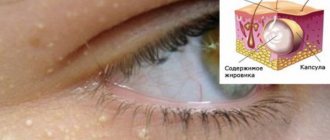
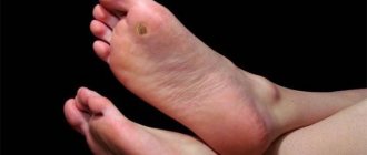
By the way. Most often, this formation appears on the skin, at any point. But in 20% of cases, the tumor can localize itself to internal organs, muscle and even bone tissue.
The neoplasm looks like this:
- a thickened tumor-like spot of various sizes and degrees of elevation above the surface of the skin or depression;
- the color is either pale burgundy, closer to pink, or bright burgundy;
- the edges are uneven;
- the structure is dense;
- composition – atypical and normal cells.
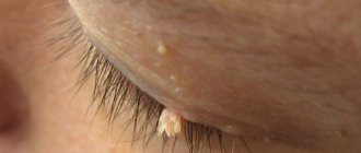
New growth on a child's cheek
Important! The key point is that although this is a full-fledged tumor, it is always benign. Therefore, there is no need to do anything. Over time, in most cases, the hemangioma subsequently resolves on its own, without a trace.
In children, quite often hemangioma simply resolves over the years.
Of course, exceptions, deviations and anomalies occur, since each person’s body is individual, including a newborn baby. The most important thing for parents when a neoplasm is detected is not to panic and carefully study the issue of how the local pediatrician and this material can help them.
Answers to frequently asked questions
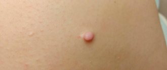
What does a hemangioma look like?
Externally, the tumor is a red or bluish spot that merges with the skin or rises above its surface. The size of the spot in diameter ranges from 1-2 cm to 10-20 cm. The tumor has different shapes. In children, temperature asymmetry appears; the defect is always warm to the touch.
Is it necessary to remove the hemangioma?
A skin hemangioma must be removed if it causes discomfort, itches or bleeds. It is necessary to remove the tumor if it is located in difficult places that are constantly rubbed by clothing.
Can a soft tissue tumor go away on its own?
It is not always necessary to remove hemangioma surgically. The tumor can regress on its own. There are several stages in the extinction process. It begins with the formation of a pale spot in the center of the tumor, then the shade changes and becomes normal towards the periphery. The process of disappearance is long, up to several years.
Why is hemangioma dangerous?
If treated incorrectly and untimely, complications may occur that pose a threat to human life and health:
- germination with subsequent destruction of nearby organs;
- destruction of bone and muscle tissue;
- spinal cord compression;
- ulceration of the tumor, infection;
- malignancy;
- development of pathologies of the vascular system;
- cosmetic defect.
How does a tumor occur?
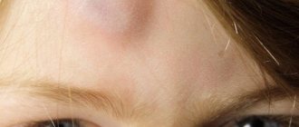
Statistically, hemangioma appears in ten percent of infants. The percentage is not so small if you think about it. Most often, formations found on the baby’s skin are located at such points that the tumor does not pose a danger to vital organs and does not cause damage to the mucous membranes. The only problems that education can cause are aesthetic ones. But they are precisely the most significant. Agree, a rough red “blot” in the middle of the forehead or on the child’s face, reminiscent of an exaggerated wart or depression, will bring few parents into a relaxed state.
Hemangioma on the forehead. Unsightly stain
By the way. Not every formation found on the skin of a baby is a hemangioma. Therefore, those parents who, at the sight of a spot, begin to sound the alarm and go to the doctor are right.
Signs of hemangioma.
- The most common type of tumor among newborns.
- Benign education.
- Appears only in children from age zero (immediately after birth) to the age of 60 days.
- Affects every tenth child on the planet.
- It is three times more common in girls (for every three newborn girls with this type of tumor, there is one male child).
- May appear as a flat spot.
- May resemble a depression (grow deep).
- Forms a bulge (most often).
- It has any size - from a “grain” to a large spot.
- There may be several or even many formations.
- If more than three hemangiomas are found on the skin, they are also present inside the body.
- Goes away on its own (in the vast majority of episodes).
Hemangiomas
Combined hemangioma in the breast area
Important! The most important sign that this is not a malignant formation, but a “harmless” benign hemangioma is that it contains elements of endothelial cells (the inner surface of blood vessels).
Of course, we are talking about “harmlessness” only when the formation is located outside, does not block physiological openings, does not damage the mucous membranes and does not increase in size.
There are several types of hemangiomas based on their location and structure.
Table. Types of hemangiomas.
| Variety | Description |
| It is formed from dilated vessels and cavities filled with venous or arterial blood. Does not penetrate deep into the skin, is located on the surface. It has the softest structure of all varieties. Amenable to basic therapy. |
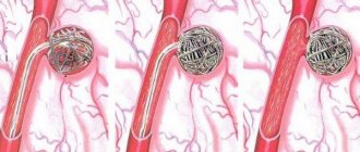
| Located in internal organs, the blood supply to which is enhanced. These are the spleen and liver, adrenal glands, kidneys and brain. Diagnosed using ultrasound. It is considered dangerous, especially if localized in the liver, spleen and brain. Education is asymptomatic, and this poses the greatest danger. In case of injury, the spleen and liver rupture, filling the abdominal cavity or liver capsule with blood, which in more than 80% can be fatal. Rupture of a cerebral hemangioma leads to hemorrhage in the brain, which plunges the patient into a coma or causes death. |
| It consists of a combination of simple capillary and cavernous parts. And this is the greatest danger. The formation can go deep into the skin, and it is often mistaken for capillary, while the hidden cavernous part is capable of ruptures and hemorrhages. |
| Mixed | This is no longer quite a hemangioma, since, along with the vascular formation, in parallel with it, a tumor of other tissues develops - nervous, connective, and so on. |
| Capillary | It is considered the simplest formation consisting of blood vessels of the dermis. Does not enter the lower skin layers, usually has a convex shape. These are capillary vessels intertwined with each other, which in very rare cases are capable of hemorrhage. As a rule, its size is no more than a centimeter, and its color is light, which indicates its involutionary development. |
Diagnostic methods
When young parents go to a medical facility, suspecting that their baby has a hemangioma, the child should be examined by three specialists:
- pediatrician;
- surgeon;
- dermatologist.
Venous hemangioma
In the future, if it is determined that not only the skin, but also other systems and organs of the body are affected, the help of ophthalmologists, neurosurgeons and other doctors may be needed. To understand how widespread the tumor is, the following medical tests are done:
- ultrasound diagnosis of tumor;
- radiography, during which the affected area is examined by injecting a contrast agent into nearby vessels;
- computed tomography of the area where the hemangioma is located (vertebral column, head or eye sockets).
Computer tomograph
If the hemangioma has developed as a consequence of some other disease or pathology in the functioning of any internal organ, then it will be necessary to take blood from the child to determine its coagulability and platelet counts.
Education mechanism
Medicine has not found any exact reasons why this type of tumor forms on a baby’s body. There are more hypotheses than facts, despite the fact that many years of research into infant hemangiomas have been conducted.
Doctors are convinced of only one thing - the reasons are not genetic, therefore heredity in the formation mechanism is excluded by almost all specialists.
Development of hemangioma
However, there are aspects whose participation in the formation of hemangiomas is assumed with a high degree of probability.
- ARVI suffered by a pregnant woman in the early stages, between the third and sixth week. It is during this time period that the formation of the cardiovascular system of the embryo occurs.
- Rhesus conflict, when positive red blood cells from the fetus enter the maternal bloodstream.
- Pregnant women taking medications not authorized by a doctor, smoking during pregnancy, drinking alcohol.
- The presence of hormonal imbalances and disorders in both the fetus and its mother.
- Low level of ecology where the pregnant woman lives.
- Heredity aggravated by any vascular disease.
- Multiple pregnancy (starting with twins).
- The age of the woman giving birth is over 38 years.
- Birth of a child prematurely.
- Convulsive seizures in a pregnant woman.
In multiple pregnancies, there is a risk of developing hemangiomas in children
Bad habits increase the risk of developing fetal pathologies
If we briefly characterize the hypothesis of the mechanism of formation, it will sound like this. At the stage of bearing a child, when its cardiovascular system is formed, endothelial cells, under the influence of some (not precisely established) factors, are sent to the “wrong address”. Instead of forming the inner surface of the blood vessels, they form a tumor.
Bandages for pregnant women
If you want to find out in more detail what symptoms spinal hemangioma has, and also consider alternative treatment methods, you can read an article about this on our portal.
Why choose the clinic “Mama Papa Ya”
The network of family clinics “Mama Papa Ya” offers medical services for hemangiomas to patients of any age. The clinic's branches are located in Moscow and other cities. Advantages of visiting this medical institution:
- the opportunity for children and adults to get advice from doctors of various profiles - dermatologist, cosmetologist, surgeon, neurologist, pediatrician;
- individual decision on the need to remove hemangioma;
- the use of low-traumatic methods of tumor removal;
- affordable prices for diagnostic and therapeutic procedures.
To obtain a doctor’s consultation, we invite you to call the clinic’s phone number listed on the website and make an appointment at a convenient time. You can also leave a request by filling out a special form on our website.
Reviews
Good clinic, good doctor!
Raisa Vasilievna can clearly and clearly explain what the problem is. If something is wrong, she speaks about everything directly, not in a veiled way, as other doctors sometimes do. I don’t regret that I ended up with her. Anna
I would like to express my gratitude to the staff of the clinic: Mom, Dad, and me. The clinic has a very friendly atmosphere, a very friendly and cheerful team and highly qualified specialists. Thank you very much! I wish your clinic prosperity.
Anonymous user
Today I had a mole removed on my face from dermatologist I.A. Kodareva. The doctor is very neat! Correct! Thanks a lot! Administrator Yulia Borshchevskaya is friendly and accurately fulfills her duties.
Belova E.M.
Today I was treated at the clinic, I was satisfied with the staff, as well as the gynecologist. Everyone treats patients with respect and attention. Many thanks to them and continued prosperity.
Anonymous user
The Mama Papa Ya clinic in Lyubertsy is very good. The team is friendly and responsive. I recommend this clinic to all my friends. Thanks to all doctors and administrators. I wish the clinic prosperity and many adequate clients.
Iratyev V.V.
We visited the “Mama Papa Ya” Clinic with our child. A consultation with a pediatric cardiologist was needed. I liked the clinic. Good service, doctors. There was no queue, everything was the same price.
Evgeniya
I liked the first visit. They examined me carefully, prescribed additional examinations, and gave me good recommendations. I will continue treatment further; I liked the conditions at the clinic.
Christina
The doctor carefully examined my husband, prescribed an ECG and made a preliminary diagnosis. She gave recommendations on our situation and ordered additional examination. No comments so far. Financial agreements have been met.
Marina Petrovna
I really liked the clinic. Helpful staff. I had an appointment with gynecologist E.A. Mikhailova. I was satisfied, there are more such doctors. Thank you!!!
Olga
Therapy
The presence of hemangioma is not always an indication for any specific therapy. In many cases, on the contrary, it is not recommended to touch it - just wait until it resolves on its own. But sometimes doctors and parents note the presence of indications for treatment. The absolute indications for the fight against hemangiomas are:
- periodic bleeding from the tumor;
- ulceration of hemangioma;
- the tumor is located in a place that increases the risk of injury (in the area of underwear, on the back, etc.);
- tumors that interfere with the normal functioning and development of the newborn;
- very rapid tumor growth.
How are hemangiomas treated?
In cases where there is a risk that the hemangioma will rupture, it must be urgently removed. When a rupture occurs, heavy bleeding occurs due to damage to blood vessels. Internal hemangiomas can also be traumatized, resulting in internal bleeding affecting the spleen, liver, and even the brain. In this case, the danger from leaving the hemangioma is higher than from surgical intervention.
Results of laser hemangioma removal
If we are talking about a hemangioma that has a simple shape and is not too large in size, then there is no need to touch it - it will go away on its own over the next 2-5 years.
The fight against tumors of this type is carried out: