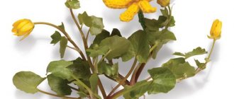
Lichen (mycosis) is an infectious disease of the human skin. Appears as a result of contact of microspores of various types of fungi on the skin. It is a mistake to think that lichen planus is an exclusively childhood disease. Adults are no less susceptible to it than children.
The main factors that influence the appearance of lichen are visiting crowded places with high humidity, for example, swimming pools, saunas, baths, gyms. Domestic and street animals are also often carriers of infections. Often, pathologies of the thyroid gland, diabetes mellitus, and hormonal imbalances in the body can contribute to infection of a person’s skin with lichen.
What is pityriasis versicolor
Pityriasis versicolor, or pityriasis versicolor, is a chronic skin disease of a fungal nature. It appears in the form of specifically pigmented spots of various sizes and shades, without signs of inflammation. Most often, the areas affected by pityriasis versicolor are located on the body (back, abdomen, neck, shoulders) and scalp. Representatives of both sexes are most susceptible to the disease at a young age, and children under 7 years of age are the least susceptible.
External manifestations of the disease usually do not cause concern, so they are often perceived as harmless cosmetic defects. For this reason, the pathology becomes chronic: spots periodically appear, then disappear, and after some time, under the influence of provoking factors, the disease worsens again.
Using effective strokes we “kill” the fungus
If you choose among the many antifungal drugs widely represented on the modern Russian market, then preference should be given to those that, firstly, are able to accumulate in those layers of the skin where the fungal process develops, and secondly, do not penetrate into those layers where fungal activity is impossible. Usually, treatment of lichen versicolor is carried out with local drugs, and in severe cases with the use of systemic antimycotics, which can significantly reduce the treatment time and prevent the frequency of relapses. But you should always remember that if you find signs of a disease in yourself, under no circumstances try to treat yourself, but immediately go to a doctor who will accurately determine your diagnosis and prescribe the right treatment for you.
Symptoms of the disease
When infected with pityriasis versicolor, the pathogens multiply in the superficial layers of the skin, disrupting the normal functioning of cells. First of all, melanocytes, which are responsible for the production of the pigment melanin, are affected, thanks to which the skin has one color or another. This means that under the influence of pathogenic microorganisms, the affected areas acquire an uncharacteristic, painful shade. Hence the second name for pityriasis versicolor - versicolor.
The main symptoms of pityriasis versicolor include:
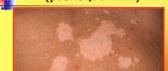
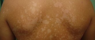
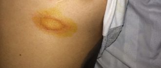
- Spots of yellow, coffee, scarlet or dark brown shades, located mainly on the back, head and neck, shoulders and stomach. In children, they also appear on the limbs, armpits and scalp. The affected areas can be of different sizes, but usually their size does not exceed 1 cm. Subsequently, the spots merge with each other, forming significant areas of the affected skin.
- No signs of inflammation in the form of redness, swelling, hyperemia and pain to the touch.
- Severe peeling of the skin in infected areas, aggravated by touch.
- No tanning on the affected areas.
Pityriasis versicolor is not characterized by itching and discomfort, so patients often mistake the manifestations of the disease for minor cosmetic defects, postponing a visit to the doctor and full treatment.
Such spots begin to itch and scratch only if a secondary infection occurs.
Publications
Versicolor (pityriasis versicolor) (Tinea versicolor) is a mycotic skin infection characterized by widespread distribution and a chronic relapsing course. The disease affects 10% of the population. In hot countries, lichen versicolor is more common.
In mid-latitudes, most cases of the disease occur in the summer. Adults and young people get sick more often; The disease is rare in children and the elderly. Transmission of the pathogen from a patient or carrier: for example, through a shared bed or clothing (underwear), is possible, however, in most cases, the source of infection is endogenous. Therefore, lichen versicolor is not considered a contagious disease. Some authors consider iatrogenic immunodeficiencies, pregnancy, and hormonal contraception to be predisposing conditions. A hereditary predisposition to the disease cannot be ruled out.
In the International Classification of Diseases (ICD-10), lichen versicolor is included in the section “Other superficial mycoses” (B36.0) and has code B36.1. Its causative agents are opportunistic imperfect yeast lipophilic fungi. In recent years, the causative agents of pityriasis versicolor have been assigned to the genus Malassezia [2,4]. Currently, the genus Malassezia has 9 described species of fungi, among which the main causative agents of pityriasis versicolor are M. globosa, M. sympodialis, M. sloolliae |6, 7|. Fungi of the genus Malassezia are imperfect yeast fungi, basidiomycetes. A common characteristic of most Malassezia species is lipophilicity: (requirement of a source of lipids for growth). Malassezia are distinguished by polymorphism, consisting in different cell shapes (round or oval) in representatives of the same genus and even species, and the ability to form mycelial forms.
The disease begins with the appearance of small spots with a clear edge, or papules slightly raised above the surface of the skin. Small spots are often located around the hair follicles. The color of the spots ranges from yellowish-pink to brown in different shades. The spots often merge, forming large lesions with scalloped edges. On the surface of the spots you can notice a gentle “pityriasis-like” peeling. Peeling intensifies with scraping (symptom of shavings, or “nail strike” of Beignets). The color of the spots differs from the color of the surrounding skin: in people with fair skin, the lesions look darker, in people with dark or tanned skin - lighter, depigmented (pseudo-leukoderma, pityriasis versicolor alba). The same patient may have both hyperpigmented and hypopigmented macules. The color of the spots is mainly determined by exposure to ultraviolet radiation: unlike the color of the surrounding skin, it does not change after tanning. In rare cases, inflammatory lesions with erythema and slight infiltration occur. Typical localization of lesions in lichen versicolor is areas of the body rich in sebaceous glands: chest, back, neck, shoulders. Less commonly, the axillary, groin, forearms and lower legs are involved. In tropical climates, lesions on the face, abdomen, and scalp are more common. The inverse form (tinea versicolor inversa) is the lesion of large, usually inguinal, folds. Hair and nails are not affected. Changes in the skin, as a rule, are not accompanied by subjective sensations. The disease is characterized by a long-term relapsing course.
The variety of clinical manifestations makes timely diagnosis of mycosis difficult [6]. A long-term recurrent course, significant contamination of the skin and scalp with pathogens, and their penetration into the pilosebaceous follicles cause certain difficulties in the treatment of patients with lichen versicolor [1]. Until recently, keratolytic agents, which have low antifungal activity, are inconvenient to use and do not reduce the number of disease relapses, have been widely used in treatment.
N.V. Kungurov et al. developed an algorithm for the management of patients with lichen versicolor. When detected in patients on the skin in typical localizations (areas rich in sebaceous glands), spots of a round or oval shape, non-inflammatory in nature, not rising above the surface of the skin, with clear boundaries, scalloped edges, of various colors (from yellowish-pink, light brown to brown) and gentle pityriasis-like peeling on the surface for the purpose of diagnosis, a Balser test is performed (when the lesions are smeared with alcohol solutions of iodine or aniline dyes, the affected skin is colored much brighter than healthy), and the symptom of “chips” is revealed - with careful scraping of the spot, a barely noticeable tender pityriasis peeling intensifies, which is due to loosening of the stratum corneum of the epidermis.
The skin of the torso and extremities is examined under a Wood's lamp in order to identify non-visual, atypical forms of the disease. Lesions occupying up to 18% of the area are limited (localized) forms, more than 18% are widespread. The erythematous-squamous form (the most typical) is recorded in 98.5% of cases and is characterized by the clinical manifestations described above. The follicular variant, found in 7% of patients, is characterized by the appearance of perifollicular papules or pustules of a yellowish color, located on an erythematous background, while in the middle of the spots, point-shaped follicular openings are clearly visible. In the inverted variant (3.5%) of the clinical course, the lesions are localized in the inguinal-femoral folds, in the pubic area, on the thighs and legs. Less commonly, rashes of pityriasis versicolor rise above the skin and to the touch create the impression of nodules the size of a lentil or smaller, located in groups (pseudo-papular form). Certain difficulties in making a preliminary clinical diagnosis arise in the presence of hypopigmented lesions on the skin that appear in place of typical spots of pityriasis versicolor, usually after irradiation with ultraviolet rays or insolation. Differential diagnosis is carried out with diseases such as Zhiber's pityriasis rosea, syphilis (primary and secondary), seborrheic dermatitis, chloasma, vitiligo, as well as other skin dyspigmentations.
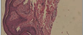
After making a preliminary clinical diagnosis of lichen versicolor, taking into account the course and extent of the process, it is confirmed in the laboratory. During microscopic examination, scales of the stratum corneum of the epidermis of the skin, obtained by scraping with a scalpel from the lesions, are treated with a 20% solution of potassium hydroxide and, after 30 minutes of exposure, are subjected to microscopic examination. The preparation shows short, slightly curved, septate, thick filaments of pseudomycelium and round spores with a double-circuited shell, located in clusters and singly.
To realize the pathogenic properties of the fungus, its transformation from a non-pathogenic form of blastospore into a pathogenic mycelial, certain conditions are necessary, therefore it is necessary to establish factors contributing to the occurrence and spread of fungal infection, analyzing anamnestic data, prescribing consultations and examinations with specialists. Pathology of the gastrointestinal tract, dysfunction of the endocrine system, diseases and functional disorders of the cardiovascular system, seborrheic processes, foci of acute and chronic infection - these conditions directly or indirectly contribute to hyperhidrosis, which results in changes in the physicochemical properties of sweat and sebum (in side of increasing the alkaline reaction), reducing the rate of physiological peeling of the stratum corneum or its loosening, which contributes to the spread of fungal colonies [3].
The choice of treatment tactics depends on the duration, prevalence and form of the fungal process, and identified concomitant diseases.
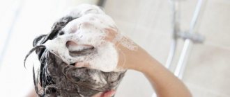
Most cases of lichen versicolor can be treated with external remedies. Patients with a limited form of lichen versicolor (the fungal process occupies less than 18% of the body skin area), whose disease duration is no more than 2 years, who have not previously been treated, as well as patients with widespread keratomycosis (affected area more than 18%), who have absolute contraindications for use systemic antimycotics, one of the external antimycotic drugs is recommended. The use of external forms that allow covering a significant surface of the body is of considerable importance in the treatment of lichen versicolor. Even if the lesion occurs in the form of separate small lesions, it is recommended to treat all areas where lichen versicolor usually develops. The shampoo is used once a day for 5 days.
Other imidazole antimycotics in the form of creams ( Fungazol "Farmakar") are used 2 times a day for at least 2 weeks. The use of modern antifungal aerosols is promising.
Lichen versicolor is characterized by relapses after treatment. As a rule, a year after treatment, a relapse is observed in every second patient, and within two years - in almost all. To reduce the likelihood of relapses, longer treatment and treatment of larger surfaces are recommended. For frequent relapses, systemic antimycotics, azole derivatives, are prescribed. Systemic antimycotics are prescribed to the following groups of patients: with the prevalence of the fungal process exceeding 18% of the skin area; with limited forms (less than 18% of the skin area), with a disease duration of more than 2 years, with relapses after treatment; with follicular and pseudopapular clinical variants of the course of keratomycosis, regardless of the area of the lesion; with the lightning-fast form of the fungal process.
Itraconazole ( Mikotrox "Farmakar") is an antimycotic with the widest spectrum of action, also effective in the treatment of lichen versicolor. It is prescribed at 200 mg/day for 1 week. Fluconazole ( Diflox "Farmakar") is prescribed once at a dose of 400 mg/day. These same drugs, with single or short-term use, can be used to prevent relapses.
We observed 24 patients with a common form of lichen versicolor, who were repeatedly treated with external antifungal drugs. To treat these patients, we chose the drug itraconazole ( Mikotrox ), which was prescribed at a dose of 100 mg 2 times a day for 1 week. All patients tolerated the therapy well, there were no side effects or cases of negative attitude of patients towards treatment. The criteria for achieving complete clinical and laboratory recovery are the absence of skin rashes, negative specific clinical tests, and the absence of fungal mycelium on microscopy.
Thus, despite the fact that lichen versicolor is a very common disease, it often causes difficulties in diagnosis and treatment, especially in recurrent forms. The use of the modern systemic antimycotic Mikotrox helps solve this problem and improve the quality of life of patients.
LITERATURE:
- Bragina E. E., Novoselov A. Yu., Stepanova Zh. V. // Esthet. honey. - 2002. - T. I, No. 5. - P. 447-453.
- Dmitriev G. A., Grammatikova N. E., Bibikova M. V. et al. // Vestn. dermatol. - 2002. - No. 2. - P. 7-9.
- Kungurov N.V., Skurikhina M.E., Budumyan T.M. etc. //Russian Journal of Dermatovenerol. - 2004. - No. 4. – P.49-51.
- Novoselov A. Yu., Bragina E. E., Stepanova Zh. V. // Immunopathol., allergol., infectol. - 2000. - No. 4. - P. 95-98.
- Potekaev II. P. // Ross. Journal dermatovenerol. - 2001. - No. 3. - P. 9-10.
- Sergeev L. Yu., Sergeev Yu. V. Fungal infections: A guide for doctors. - M., 2003. - 440 p.
- Crespo E, Ojeda M, Vera. A. et al. // JEADV. — 2000. -Vol. 14. - R. 47.
E.A. Levonchuk
Medical news, 2007 No. 13
Reasons for development
Versicolor versicolor is a type of fungal skin infection that affects the stratum corneum of the epidermis and hair follicles. Its causative agents are two types of fungi, and infection is possible only through prolonged and close contact with the patient. And in this case, provoking factors play a big role. These include:
- weakened immunity;
- hyperhidrosis;
- disruption of the sebaceous glands;
- diseases of the endocrine system (obesity, diabetes, Itsenko-Cushing syndrome, etc.);
- hormonal imbalance due to pregnancy, menopause or taking hormone-containing medications;
- vegetative-vascular dystonia;
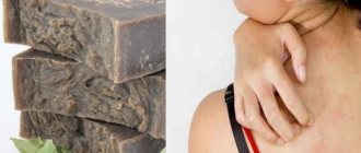
- abuse of antibacterial personal hygiene products;
- excessive exposure to ultraviolet rays (intense tanning, frequent visits to the solarium) and regular overheating of the body.
It is noteworthy that patients with pityriasis versicolor over 60 years of age are extremely rare. This is due to natural age-related changes in the skin, which make it less susceptible to pathogens.
In children under 10 years of age, the main causes of pityriasis versicolor infection are neglect of personal hygiene rules or improper skin care. At this age, with the protective functions of the skin intact, the body independently copes with pathogenic microorganisms attacking it, so the development of the disease does not occur. But closer to adolescence, when hormonal changes begin, the body’s susceptibility to bacteria, viruses and fungi increases, so children over 10 years old become infected with pityriasis versicolor just like adults.
Fungus loves heat and moisture
Pityriasis versicolor is a disease that is widespread everywhere, but it is more common in countries where the temperature and humidity are high. Young people are affected, and the disease is also registered in children. The risk of lichen versicolor increases in people suffering from excessive sweating, a certain chemical composition of sweat, favorable skin pH for the pathogen, with concomitant diseases: endocrine disorders, chronic diseases of the gastrointestinal tract, as well as immunodeficiency. In recent years, a genetic predisposition to the development of lichen versicolor has been clinically established. Often this disease develops against the background of pulmonary tuberculosis and lymphogranulomatosis, since these diseases are often accompanied by excessive sweating. Transmission of the pathogen from a patient with lichen versicolor or a carrier, for example, in a shared bed, or through shared clothing or underwear, is, in principle, possible. However, most people are carriers of the same fungi that are found on the skin (in areas rich in sebaceous glands) and do not cause disease.
Treatment of pityriasis versicolor
The diagnosis of pityriasis versicolor is made to the patient after examination by a dermatologist and dermatoscopy. Additionally, an iodine test and laboratory testing of scrapings can be used.
Treatment of pityriasis versicolor is carried out on an outpatient basis until the symptoms of the disease completely disappear. If measures were taken on time, the patient is prescribed local therapy using antifungal ointments and special agents for exfoliating dead cells.
Additionally, immunomodulators, vitamin complexes, antifungal shampoos and antihistamines are used if the patient is bothered by itching. In the most advanced cases, antimycotic agents are prescribed for oral administration. In addition, in order to avoid relapse of the disease and infection of others, the patient’s clothing and bedding are treated with disinfectant compounds.
Disease prevention also plays an important role. The following will help prevent the development of the disease:
- timely solution to the problem of hyperhidrosis (use of medicinal deodorants, creams, powders, compliance with personal hygiene rules, frequent changes of underwear, etc.);
- use of high-quality soaps and skin care products;
- regular water procedures;
- wearing clothes made from natural, hypoallergenic materials;
- avoiding stress;
- balanced diet rich in vitamins and microelements.
Experts also recommend avoiding overheating of the body and promptly seeking advice from cosmetologists and dermatologists in order to identify the problem and begin proper treatment.
Relapses
Tinea versicolor often returns after treatment, especially in the summer or while on vacation in warm, humid countries. The patient can reduce this likelihood by regularly using antifungal shampoos as a preventative measure. For example, using shampoo once a day for a few days before going on vacation can help prevent a recurrence of tinea versicolor.
If a patient develops tinea versicolor again after treatment, they can try treating it themselves with an antifungal shampoo or contact their GP for advice and other treatments. If the patient has frequent and severe episodes of tinea versicolor, the GP may consider prescribing antifungal tablets several times a month to prevent recurrence of the condition and recommend consultation with a dermatologist.
What is erythrasma
Erythrasma is a chronic bacterial disease affecting the epidermis layer in the deep folds of the skin. It is characterized by a long course - in some cases, symptoms develop for at least 10 years, without causing significant discomfort to the patient. The clinical picture of erythrasma is similar to a fungal infection of the skin, but modern dermatology classifies it as a group of pseudomycoses.
The following main stages are distinguished in the development of the disease:
- Progression. The first characteristic spots appear on the skin, their size slowly increases, and additional symptoms develop. In some cases, secondary infections occur. The spots gradually merge with each other, forming large areas of damage.
- Stabilization. New spots do not appear, and existing ones stop growing. Peeling of the skin begins. This stage is usually associated with a change in external conditions, for example, cold weather, during which the intensity of sweating decreases and the skin condition stabilizes.
- Exacerbation or relapse. Usually associated with the beginning of the warm season. But in the case of prolonged erythrasma, the disease constantly develops in waves, and after a slight decline its symptoms again actively appear.
- Remission. Occurs with a favorable microclimate, compliance with preventive measures and proper skin care. The color of the affected areas gradually returns to normal, itching and flaking disappear, and the skin is restored.
Without timely, well-chosen treatment, erythrasma can lead to the development of serious complications.
For example, it can provoke eczema and secondary infection in patients with diabetes or obesity. Also, the course of the disease is aggravated by increased humidity and contamination of the affected areas. As a result, its typical symptoms are complicated by burning, itching and pain.
Can Fluconazole be used during breastfeeding and pregnancy?
The use of the drug during pregnancy should be avoided, with the exception of severe or life-threatening systemic fungal infections, when the expected benefit of therapy for the mother outweighs the possible risk to the fetus.
Women of childbearing age should also use reliable methods of contraception during treatment with Fluconazole and for at least a week after taking the last dose. Since there is information about cases of spontaneous abortion and the appearance of congenital pathologies in children whose mothers received Fluconazole at a dose of 150 mg once or repeatedly in the first trimester of pregnancy.
When pregnant women take increased doses for a long time in the first trimester, the number of defects in infants also increases: curvature of the femurs, impaired formation of the cranial vault, brachycephaly, impaired development of the facial part of the skull, cleft palate, thinning and elongation of the ribs, arthrogryposis and congenital heart defects.
Fluconazole passes into breast milk, so use during breastfeeding is prohibited.
Signs of the disease
Externally, erythrasma manifests itself in the form of light brown, brick-red, brown or yellow-brown spots on the skin, most often round in shape and without signs of inflammation. The diameter of the lesions can reach several centimeters, and they tend to merge, forming large affected areas. First of all, erythrasma spots appear in the folds of the skin, where there is a favorable environment for the proliferation of bacteria.
In addition to spots, erythrasma is characterized by:
- Peeling of the skin on the affected areas, aggravated by touch. Usually this is where the development of the disease begins.
- Mild, irregular itching. It intensifies and begins to cause significant discomfort only if a secondary infection is added to the primary disease.
- Absence of fever, wounds, ulcers and ulcers with copious discharge. This distinguishes erythrasma from most bacterial skin pathologies.
- Getting wet. An optional symptom, the manifestation of which depends on the amount of sweating and the quality of skin care.
It is noteworthy that in children, symptoms of erythrasma appear extremely rarely. The risk group includes adults, primarily men, who are predominantly overweight and prone to excessive sweating. In this case, in men, the skin in the groin, navel and inner thighs is usually affected, and in women, the entire abdomen, armpits and areas under the breasts are affected.
Lichen versicolor or vitiligo?
Tinea versicolor can sometimes be confused with vitiligo as both cause patchy discoloration of the skin. There are the following ways to tell the difference:
vitiligo often develops on both sides of the body at the same time, while lichen versicolor can be one-sided;
skin affected by vitiligo usually has a normal texture, while areas affected by tinea versicolor are usually slightly scaly or flaky;
Vitiligo is more common around the mouth, eyes, fingers, wrists, armpits and groin, while tinea versicolor tends to develop on the back, chest, shoulders, neck and abdomen.
| Service | Price | Promotion Price | Promotion Price |
| Appointment (examination, consultation) with a therapist | 1800 rub. primary | 1500 rub. repeated | free appointment with vitamin therapy course |
| Autohemotherapy | 650 rub. 1 session | 3000 rub. 5 sessions | 6000 rub. 10 sessions |
| Ultrasound therapy procedure | 450 rub. | ||
| Plasma therapy | 8000 rub. 1 session | 13500 rub. 3 sessions | 22500 rub. 5 sessions |
| Vitamin therapy (course of 10 injections) | 4000 rub. | 3000 rub. | free appointment with a therapist |
| Recovery course of IVs after COVID-19 (Coronavirus) | 950 rub. 1 session | 4050 rub. 5 sessions | 8100 rub. 10 sessions |
| Intravenous drip administration of drugs (without drugs, 1 bottle) | 950 rub. 1 session | 4050 rub. 5 sessions | 8100 rub. 10 sessions |
| Intravenous drip administration of medications (with existing clinic medications, 1 bottle) | 1000 rub. 1 session | 4500 rub. 5 sessions | 9000 rub. 10 sessions |
| Intravenous drip administration of drugs (without drugs, 2 bottles) | 950 rub. 1 session | 4050 rub. 5 sessions | 8100 rub. 10 sessions |
| Intravenous drip administration of medications (with existing clinic medications, 2 bottles) | 1150 rub. 1 session | 5200 rub. 5 sessions | 10500 rub. 10 sessions |
| Intravenous administration of drugs (jet) | 450 rub. 1 session | 2240 rub. 5 sessions | 4050 rub. 10 sessions |
| Subcutaneous-intradermal administration of drugs (course) | 250 rub. 1 session | 1180 rub. 5 sessions | 2250 rub. 10 sessions |
| Intramuscular administration of drugs (course) | 350 rub. 1 session | 1750 rub. 5 sessions | 3000 rub. 10 sessions |
| Registration of a certificate for the swimming pool | 500 rub. | ||
| Registration of a sanatorium-resort card | 400 rub. | ||
| Examination program Men's health passport | 12350 rub. | 9999 rub. | free appointment with a therapist |
| Examination program Women's health passport | 10840 rub. | 8999 rub. | free appointment with a therapist |
| Comprehensive body cleansing program | 23660 rub. | 18999 rub. | free appointment with a therapist |
| Complete check-up for male athletes | 18500 rub. | 14999 rub. | |
| Complete check-up for female athletes | 18900 rub. | 14999 rub. |
Causes of pathology
Corynebacteria, which are the causative agents of the disease, are normally always present on human skin. Moreover, they provoke the development of pathology only under certain, favorable conditions. Corynebacteria do not penetrate deeper than the epidermis, and also do not affect nails and hair. Since the appearance of erythrasma is directly related to increased sweating, the disease most often manifests itself in the hot summer season.
Among the main reasons for the development of erythrasma are:
- hyperhidrosis;
- deviation of the normal pH of the skin to the alkaline side;
- diaper rash, constant friction and mechanical damage to the skin;
- dermatitis and other skin diseases;
- neglect of personal hygiene rules;
- wearing synthetic, overly warm clothing;
- the use of low-quality care products or the abuse of soap with an antibacterial effect, which destroys the natural protection of the skin.
Erythrasma is transmitted by contact, most often after the use of clothing, bedding and personal hygiene products of the patient. You can also become infected during sexual intercourse, when visiting a pool or bathhouse, and when walking barefoot on the ground or beach. At the same time, it is not always possible to accurately determine the source of infection, because the carrier may not have obvious external manifestations of the disease in the form of characteristic spots and peeling.
What does lichen look like in humans?
The external manifestation of the pathology is obvious - lichen is accompanied by a number of signs:
- rash in the form of spots of different colors - red, pink, brownish, yellow;
- itching, burning, unpleasant (painful) sensations;
- scales, bubble formations on spots;
- bald spots on the head in the hairline area (clipping form);
- fever (rare);
- malaise, lethargy.
Treatment of erythrasma
To diagnose a patient with erythrasma, a dermatologist first uses a visual examination. This is especially true for rashes in the groin area, which have characteristic distinctive features in the form of pronounced protrusions and bubbles along the edges. Also, the affected areas of the skin are illuminated with a Wood's lamp and a microscopic examination of the scraping is performed to exclude other diagnoses: pityriasis versicolor or pityriasis rosea, candidiasis, dermatitis or eczema.
Treatment of erythrasma is primarily based on the use of antibacterial ointments that are used to treat the affected areas of the skin.
Under their influence, corynebacteria die, and the spots gradually lighten, decrease in diameter and disappear. On average, such therapy takes at least 7-10 days. Used in parallel:
- Antiseptics. Treatment with them is carried out before applying antibacterial ointment, as well as after it, to maintain dryness of the affected areas and prevent re-infection.
- Antifungal drugs. They are prescribed together with antibacterial drugs, since corynebacteria are similar in structure to fungal micelles.
- Exfoliating ointments. They help cleanse the skin of a layer of dead cells, activating its regeneration.
- Ultraviolet irradiation. Promotes skin disinfection and restoration. Patients benefit from both natural sunbathing and physiotherapeutic UV irradiation.
If the disease has not reached an advanced stage, the use of external medications is sufficient to solve the problem. But in some cases, with multiple skin lesions, to obtain the desired result, patients are prescribed systemic antibacterial therapy.
A drop of iodine, a lamp beam, a microscope lens
In our Center, a wide variety of methods are used to diagnose lichen versicolor, one of which is the Balser iodine test: lesions and adjacent areas of healthy skin are smeared with 5% iodine tincture. Rashes due to the loosened stratum corneum are more intensely colored compared to the surrounding healthy skin. Just do not try to perform this test yourself, since only a doctor can diagnose the disease. When examined using a Wood's lamp, a yellow glow is noted in the affected area. Microscopically, in scales taken from foci of pityriasis versicolor and treated with a 15-20% KOH solution, budding fungal cells, as well as pseudomycelium, are detected. Another diagnostic method is the use of Sabouraud’s medium, on which after 3 weeks a white, creamy culture is formed, similar to yeast. Microscopic examination reveals budding fungal cells.
General provisions of therapeutic measures for tinea versicolor
The main condition for eliminating tinea versicolor is to contact a dermatologist, since self-prescription of drugs or the use of folk remedies is irrational. Incorrect selection of a course of medications during treatment or non-compliance with the dosage of medications can cause complications or relapse of the disease (repeated skin damage by the pathogen). The use of infusions, ointments, lotions and sprays, which are prepared according to folk recipes, is allowed to a person only after consultation with the attending physician and under his strict supervision.
Treatment of tinea versicolor is carried out strictly under the supervision of a specialist and consists of the use of medications, various ointments, special diets and physiotherapeutic procedures. Medicines are selected for each patient individually, depending on the cause of the fungal infection, the area of development of color pigmentation and the individual characteristics of the patient.
Depending on the degree of infection of the patient by the colored fungus, the method of therapeutic treatment is selected - outpatient (in the absence of a bacterial infection) or inpatient. Specialists use an integrated approach to treatment using local and systemic drugs. Ointments and creams cannot completely remove the causative agent of the disease, since they only have a superficial effect.
Treatment of tinea versicolor requires the dermatologist to prescribe medications and vitamin-mineral complexes to the patient that support the immune system and increase the body’s defenses. To enhance the healing effect of skin affected by lichen, physiotherapeutic techniques are used, which are carried out in the form of ultraviolet irradiation.