Herpes zoster (shingles) occurs as a result of reactivation of the varicella-zoster virus.
- Herpes zoster
- Symptoms
- Treatment
- Our doctors
- Price-list
- Analyzes
- Make an appointment
- Questions and answers
Pathogen
— DNA-containing virus of the herpes group.
It is highly contagious, but occurs in the majority of the population (more than 90%) before reproductive age. Typical skin lesions are characteristic: a spotty rash, turning into a vesicular rash, then a pustular rash with the formation of crusts and scratches. A disease in adults - reactivation of a latent viral infection, manifested by a painful blistering rash along the nerves - “shingles”, herpes zoster .
Chicken pox
is a primary infection caused by the varicella-zoster virus. In children (90% of the disease occurs before the age of 13), the disease is relatively mild; in adults it can be complicated by encephalitis and pneumonia.
Infection
Herpes zoster is transmitted by airborne droplets. The virus enters the respiratory tract, where it replicates; sometimes the virus invades the lymph nodes, causing primary viremia. The virus subsequently spreads within the body through hematogenous, lymphogenous and neurogenic routes, infecting the sensory ganglia of the autonomic nervous system, which practically ensures its lifelong persistence in the human body. The incubation period is 10-20 days (13-17). In children, fever and rash appear simultaneously. In adults, malaise and fever appear several days before the rash. The rash begins on the face and head, spreads to the torso, and rarely affects the extremities. Its development is accompanied by itching. New rashes appear after 2-5 days, all elements of the rash exist simultaneously.
Cytomegalovirus infection
Cytomegalovirus infection is characterized by the formation of giant cells and specific infiltrates in the affected organs, a latent (asymptomatic) course in individuals with a normal immune system and with clinical manifestations in immunodeficiency states, mainly in young children.
Clinical forms of cytomegalovirus infection (CMVI):>
Congenital CMV infection is infection from the mother during pregnancy and childbirth and acquired, which is often asymptomatic. The full clinical picture of CMV infection is characterized by:
- increase in body temperature;
- lethargy;
- drowsiness;
- adynamia;
- enlarged lymph nodes, liver, spleen, salivary glands;
- rash;
- other organs are involved (lungs, kidneys, central nervous system).
The disease has an undulating, relapsing course, reminiscent of the clinical picture of infectious mononucleosis.
Symptoms of herpes zoster
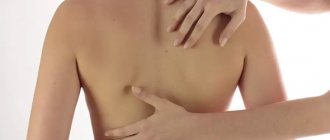
Herpes zoster is characterized by rashes along individual sensory nerves of fuzzy pinkish spots (3-5 cm in diameter), against which after 18-24 hours groups of painful vesicles form; the main feature that distinguishes them from other herpetic skin lesions is the presence of a clear demarcation zone. Most often, lesions are localized on the chest, but can also be located along any sensory nerve and, as a rule, are unilateral. The lesions disappear within 2-4 weeks, but pain (neuralgia) may persist for weeks or several months. Contagiousness: 1-2 days before the rash and until the crusts fall off. Complications include secondary bacterial skin infection, as well as encephalitis, meningitis, myocarditis, glomerulonephritis, and arthritis (rare). A very serious complication is pneumonia, which increases the risk of mortality: in adults up to 5-10%.
Prodromal period
During which pain and parasthesia appear in the area of the affected dermatome (less commonly, itching, tingling, burning). The pain can be periodic or constant and accompanied by skin hyperesthesia. The pain syndrome can simulate pleurisy, myocardial infarction, duodenal ulcers, cholecystitis, renal or hepatic colic, appendicitis, intervertebral disc prolapse, early stage glaucoma, which can lead to difficulties in diagnosis and treatment. Pain in the prodromal period may be absent in patients under 30 years of age with normal immunity.
Stages of development of the rash with herpes zoster
| Erythematous-papular stage | |
| The disease begins with the appearance of grouped erythematous papules, spreading along 1-2 dermatomes. Herpes zoster rashes have a short erythematous phase (often completely absent), after which papules quickly appear. | |
| Vesicular stage | |
| Within 1-2 days, papules turn into vesicles, which continue to appear for 3-4 days. The elements tend to merge. If the period of appearance of new vesicles lasts more than 1 week, this indicates the possibility of the patient having an immunodeficiency state. | |
| Pustular stage | |
| Pustulation of vesicles begins a week or earlier after the appearance of the primary rash. | |
| Regression stage | |
| Then, after 3-5 days, erosions appear in place of the vesicles and pustules and crusts form. The crusts usually disappear by the end of the 3rd or 4th week of the disease. However, scales and hypo- or hyperpigmentation may remain after the herpes zoster rash resolves. | |
Localization of herpes zoster by dermatome
A feature of rashes with herpes zoster is the location and distribution of the elements of the rash, which are observed on one side and limited to the area of innervation of one sensory ganglion.
The areas of innervation of the trigeminal nerve, especially the ophthalmic branch, as well as the skin of the trunk T3-L2 segments are most often affected. Skin lesions in the chest area are observed in more than 50% of cases; Most rarely, the rash appears on the skin of the distal extremities. The rash may be located in the tailbone area. In this case, a picture of a neurogenic bladder develops with urinary disorders and urinary retention (due to the migration of the virus to neighboring autonomic nerves); may be associated with herpes zoster of the sacral dermatomes S2, S3 or S4.
General symptoms
The clinical picture of herpes zoster includes skin manifestations and neurological disorders. In 20% of patients, general phenomena are observed - fever, headache, poor health, increased fatigue, enlarged regional lymph nodes, changes in the cerebrospinal fluid (in the form of lymphocytosis and monocytosis). Pain often precedes skin changes and, with appropriate localization, can simulate myocardial infarction, appendicitis , renal colic, etc. The most common localization of pain is along the intercostal nerves.
Abortive (erased) herpes zoster
| It is characterized by the appearance of erythematous plaques and papules in the foci of hyperemia, but vesicles do not develop. |
Bullous herpes zoster
| Occurs when vesicles merge. Characterized by blisters, erosions and crusts with finely scalloped outlines. |
Hemorrhagic herpes zoster
| In the hemorrhagic form of the disease, the vesicular rashes have bloody contents, the process spreads deep into the dermis, and the crusts acquire a dark brown color. |
Gangrenous-necrotizing herpes zoster
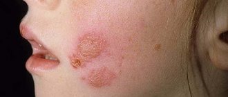
| In some cases, the bottom of the vesicles becomes necrotic and a gangrenous form develops, characterized by the formation of grouped small scabs or a solid black scab with finely scalloped outlines. After healing, scars of appropriate size remain. The duration of the course of ordinary shingles is 2-4 weeks, gangrenous shingles is 2-3 months. A particularly severe course is characterized by gangrenous herpes zoster, which develops in the area of the branch of the 1st branch of the trigeminal nerve - on the skin of the forehead, eyelids, nose and temporal region. |
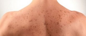
Generalized herpes zoster
| A number of patients (2-4%) experience a generalized form of herpes zoster: in addition to the usual lesion, a more or less widespread rash appears, consisting of vesicles resembling elements of chickenpox throughout the skin along with rashes along the nerve trunk. Recurrence of infection in the form of generalized rashes is usually not observed. In the presence of immune deficiency (including HIV infection), skin manifestations may appear far from the affected dermatome - a disseminated form. The likelihood of the appearance and severity of dissemination of skin rashes increases with the age of the patient. They are distinguished:
|
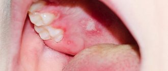
Herpes zoster of the oral mucosa
| Damage to the second and third branches of the trigeminal nerve, as well as other cranial nerves, can lead to the development of rashes on the oral mucosa (tongue, hard palate, gums), pharynx and larynx. |
Hyperkeratic herpes zoster
| A rare form of the disease, manifested in the form of continuous layers of horny masses or in the form of focally located hyperkeratic plaques, papules, nodules, sometimes resembling cutaneous horns and warts. The rashes persist for a long time within one or more adjacent dermatomes. Almost always observed in HIV-infected patients, but Cases are also rare in HIV-negative people. Also called verrucous herpes zoster. |
Ophthalmoherpes
| Ophthalmoherpes is a herpetic lesion of any branch of the optic nerve. Damage to the ophthalmic branch of the trigeminal nerve is observed in 10-15% of patients; the rash can be located on the skin from the level of the eye to the parietal region, abruptly interrupted along the midline of the forehead. Damage to the nasociliary branch, which innervates the eye, tip and lateral parts of the nose, leads to the penetration of the virus into the structures of the organ of vision. In this case, the cornea is often affected, leading to the occurrence of keratitis. In addition, other parts of the eyeball are affected with the development of episcleritis, iridocyclitis, and inflammation of the iris. The retina is rarely involved in the pathological process (in the form of hemorrhages, embolisms), the optic nerve is more often affected, which leads to optic neuritis with outcome in atrophy (possibly due to the transition of the meningeal process to the optic nerve). With herpes zoster affecting the eyes, the rash spreads from eye level to the crown, but does not cross the midline. |
Hutchinson's symptom
| Grouped vesicles are observed, localized on the wings or tip of the nose. Associated with the most serious complications of ophthalmoherpes and is a poor prognostic sign. |
Hunt syndrome
| Ganglionitis of the geniculate ganglion (geniculate ganglion of the facial nerve), caused by the herpes zoster virus and manifested by pain in the node, often radiating to the face, back of the head, neck, rashes in the external auditory canal, eardrum, sometimes on the tongue and palate, paralysis of the facial muscles and vestibular-cochlear nerve with dizziness, noise in the ear and hearing loss on the affected side. It is characterized by: pain in the area of the external auditory canal, rashes in the area of the external auditory canal, paralysis of the facial muscles, decreased taste in the anterior two-thirds of the tongue. The pain syndrome has a pronounced vegetative coloring in the form of burning, paroxysmal, sharp pains that intensify at night. Subsequently, the pain recurs and bothers the patient for many months and years, causing loss of ability to work, disrupting sleep, changing his mental and emotional status, forming a permanent syndrome - postherpetic neuralgia. The protracted, severe nature of the disease with a long-term, pronounced algic syndrome contributes to the formation of personality mental disorders. |
Herpes zoster in children
| In newborns whose mothers suffered a primary infection with the herpes zoster virus during pregnancy, intrauterine infection is possible. In these children, shingles develops without a previous history of chickenpox. Infantile shingles often leads to temporary paresis of the affected limbs. There are isolated reports of herpes zoster in young children. Risk factors for the occurrence in children include: maternal chickenpox during pregnancy or primary VZV infection in the 1st year of life. The risk of the disease is increased in children who have had chickenpox before the age of 1 year. Herpes zoster in children is not as severe as in older patients, with less pain; Postherpetic neuralgia also develops rarely. |
Herpes zoster in patients with HIV infection
The risk of developing the disease in patients with HIV infection is higher, and they are more likely to develop relapses of the disease. Additional symptoms may appear due to the involvement of motor nerves (in 5-15% of cases). The course of OH is longer, gangrenous and disseminated forms often develop (25-50%), while in 10% of patients in this category severe damage to internal organs is detected ( lungs, liver, brain). In HIV infection, frequent relapses of OH are observed within both one and several adjacent dermatomes.
Herpes zoster in pregnant women
The disease in pregnant women can be complicated by the development of pneumonia and encephalitis. VZV infection in the first trimester of pregnancy leads to primary placental insufficiency and, as a rule, is accompanied by termination of pregnancy. The presence of infection should serve as the basis for intensive prevention of the consequences of hemodynamic disorders (placental insufficiency, intrauterine hypoxia, intrauterine growth retardation).
Pain syndrome with herpes zoster
Pain is the main symptom of herpes zoster. It often precedes the development of a skin rash and is observed after the rash resolves (postherpetic neuralgia). Pain from herpes zoster and postherpetic neuralgia are caused by different mechanisms. In the early stages of the course, anatomical and functional changes are formed, leading to the development of neuralgia, which explains the connection between the severity of primary pain and the subsequent development of postherpetic neuralgia, as well as the reasons for the failure of antiviral therapy in its prevention.
The pain syndrome associated with herpes zoster has three phases: acute, subacute and chronic (postherpetic neuralgia). The acute phase of the pain syndrome occurs during the prodromal period and lasts for 30 days. The subacute phase of the pain syndrome follows the acute phase and lasts no more than 120 days. Pain lasting more than 120 days is defined as postherpetic neuralgia, which can last for months or years, causing physical suffering and significantly reducing the quality of life of patients.
The immediate causes of prodromal pain are subclinical reactivation and replication of VZV in neural tissue. Damage to peripheral nerves and neurons in the ganglia is a trigger for afferent pain signals. In a number of patients, the pain syndrome is accompanied by general systemic inflammatory manifestations: fever, malaise, myalgia, headache.
In most immunocompetent patients (60-90%), severe acute pain accompanies the appearance of a skin rash. A significant release of excitatory amino acids and neuropeptides caused by blockade of afferent impulses in the prodromal period and acute stage of OH can cause toxic damage and death of inhibitory interneurons of the dorsal horn of the spinal cord. The severity of acute pain syndrome increases with age. Excessive nociceptor activity and the generation of ectopic impulses can lead to an increase and prolongation of central responses to common stimuli - allodynia (pain and/or unpleasant sensation caused by stimuli that normally do not cause pain, such as the touch of clothing).
Predisposing factors to the development of ostherpetic neuralgia are: age over 50 years, female gender, the presence of a prodrome, massive skin rashes, localization of rashes in the area of innervation of the trigeminal nerve or brachial plexus, severe acute pain, the presence of immunodeficiency.
With postherpetic neuralgia, three types of pain can be distinguished:
- constant, deep, dull, pressing or burning pain;
- spontaneous, periodic, stabbing, shooting, similar to an electric shock;
- allodynia.
Pain syndrome is usually accompanied by sleep disturbances, loss of appetite and weight loss, chronic fatigue, depression, which leads to social maladaptation of patients.
Complications of herpes zoster
Complications of herpes zoster include: acute and chronic encephalitis, myelitis, retinitis, rapidly progressive herpetic necrosis of the retina, leading to blindness in 75-80% of cases, ophthalmic herpes with contralateral hemiparesis in the long term, as well as lesions of the gastrointestinal tract and cardiovascular systems, etc.
Wolf's isotopic response (phenomenon)
The development of a new skin disease directly at the site (anatomical area) of regressed herpes zoster. The mechanism is not fully understood, but it is assumed that herpes infection is a trigger for local immune regulation - an excessive or defective immune response. A new dermatosis can develop over varying periods of time from several days to several years. Described dermatoses with an isotopic response:
|
|
Treatment of herpes zoster
INITIAL CONSULTATION
from 2,500 rub.
Medical care for patients with herpes zoster in the acute period is provided by a dermatovenerologist, whose tasks include prescribing antiviral acyclovir-containing drugs orally in tablets. Antiviral drugs should be prescribed as early as possible from the onset of the disease, observing the single and daily dose of the drug and the duration of treatment. The second task of a dermatovenerologist is to treat the lesion. For this purpose, various drugs are used for external use, both antiviral and other mechanisms of action.
The most common complication of herpes zoster
is sympathoglioneuritis, which manifests itself as a persistent, long-lasting pain syndrome. This is postherpetic neuralgia, which is treated by a neurologist.
Epstein-Barr virus
Epstein-Barr viral infection (herpes type 4) is characterized by a variety of clinical manifestations and occurs in acute and chronic forms:
1. The acute form of EBV is infectious mononucleosis. Main clinical symptoms:
- acute onset of the disease;
- fever;
- enlarged lymph nodes, liver, spleen;
- angina;
- rash;
- difficulty nasal breathing,
- changes in the clinical blood test (increased levels of leukocytes, followed by a decrease in them, increased lymphocytes, monocytes, identification of atypical mononuclear cells).
The outcomes of infectious mononucleosis are either complete, but often long-term recovery or transition to a chronic form.
2. The manifestation of chronic EBV infection in children is:
- prolonged enlargement of lymph nodes;
- enlarged liver, spleen;
- adenoiditis;
- chronic tonsillitis;
- frequent repeated respiratory infections;
- intoxication syndrome;
- asthenic, cardiac, arthralgic syndromes;
- prolonged increase in body temperature (the so-called subfebrile body temperature - up to 38, more often it is 37 - 37.5).
Reasons for deprivation
The main reason for the formation of lichen is associated with the penetration of a viral or fungal infection into the body.
However, the infection process itself has not been fully studied. Scientists remain unknown why not all children (even those at risk) suffer from lichen. Despite this, there are factors that increase the risk of developing the disease in a child:
- excessive emotional and physical stress;
- exposure to stress;
- decreased immunity;
- the presence of infectious inflammatory processes;
- avitaminosis;
- allergic reactions;
- increased sweating;
- hereditary predisposition.
Infection of children most often occurs in crowded places: kindergarten, school, summer camp, public swimming pool.
Also, a common cause of the development of lichen is contact with domestic or street animals, after which the rules of personal hygiene were not followed. Each form of lichen has specific causes. Thus, pityriasis rosea clearly shows an infectious-allergic nature. Shingles develops as a result of various types of viruses entering the body, ringworm and multicolored fungi.