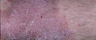
Atopic dermatitis (AD) in children is one of the most common diseases of young children. This disease is an allergic inflammation of the skin, which is characterized by itching, as well as frequent relapses and age-related characteristics of the skin rash. As a rule, dermatitis appears at an early age; if left untreated, it continues to progress into older age, which significantly worsens the quality of life of patients and their families.
For the development of this pathology, a hereditary predisposition to the development of allergies is of great importance. Therefore, this disease is often combined with other forms of allergies, for example, bronchial asthma, allergic conjunctivitis and rhinitis, food allergies. There is such a thing as “atopic march” . Atopic march is a natural progression of allergic diseases. As a rule, it all starts with a food allergy, which often gives impetus to the development of atopic dermatitis. In the absence of adequate treatment, the march progresses. Over time, the child develops allergic rhinitis. Over time, a more dangerous, in some cases, life-threatening condition arises - bronchial asthma.
Forms and complications of atopic dermatitis
Depending on your age, atopic dermatitis rashes may look different. There are infant, child and adult forms of the disease. In addition, there are currently two forms of atopic dermatitis depending on the presence of atopy - atopic and non-atopic.
In the atopic form, there is a connection between exacerbations with food, house dust allergens, and less often pollen. Other atopic diseases are often present: bronchial asthma, allergic rhinitis - both in the patient himself and in his close relatives.
It is assumed that there are other forms of atopic dermatitis, since in different patients the disease may have a different set of symptoms and proceed differently. However, clear markers that allow us to judge which scenario the disease will develop in a particular case have not yet been identified.
Atopic dermatitis can be complicated by the addition of an infection - bacterial, fungal or viral. If the pattern of rashes has changed, or you are concerned about your general condition (fever, lethargy, headache, etc.), you should consult a doctor.
Publications in the media
Contact dermatitis is an acute or chronic inflammatory skin disease caused by the irritating or sensitizing effect of exogenous factors. Incidence: 669.2 per 100,000 population in 2001.
Classification • Primary irritant dermatitis (simple contact dermatitis) • Allergic contact dermatitis (ACD) • Phototoxic dermatitis (see Photodermatitis).
Etiology and pathogenesis • Primary irritant dermatitis is associated with skin damage caused by exposure to irritating substances: chemicals (acids, alkalis, phenol), some plants (euphorbia, nettle, ash, caustic buttercup, ivy, hogweed, poison sumac), physical (UV -rays, high and low temperatures, etc.), mechanical (friction, prolonged pressure) irritants cause an inflammatory reaction in any person • ACD is caused by sensitization and the development of a type IV allergic reaction. The most common causes of ACD: salts of chromium, nickel, cobalt (jewelry, zippers, hooks, watches and bracelets); dyes (ursol, n-phenylenediamine derivatives, etc.); plants (sumac, oak, primrose); latex and various additives used in rubber production; cosmetical tools; LS • Phototoxic contact dermatitis occurs under the influence of the UV part of the solar radiation spectrum on the skin after exposure to certain substances.
Risk factors • Occupational: contact with occupational allergens • Contact with household chemicals.
Clinical picture
• A pathognomonic sign is a sharply demarcated edge of the lesion.
• The process primarily involves areas of skin with a thin epidermis (eyelids, genitals, etc.).
• The skin of the palms and soles is most resistant to irritation; The skin of deep folds is not affected.
• Forms of contact dermatitis •• Simple contact dermatitis - erythematous, vesiculo-bullous, necrotic-ulcerative •• ACD ••• Acute form: papules, blisters, blisters with surrounding erythema, oozing, itching. Initially, rashes appear only at the site of contact with an irritating substance or allergen; subsequently they can spread ••• Chronic form: thickening with lichenification, erythema, peeling, and in some cases erosion.
Research methods • If ACD is suspected, a skin patch test is performed with a standard set of contact allergens attached to a patch tape that fixes them on the skin for 48–72 hours. The reaction is assessed 20 minutes after removal of the allergen • Identification of a possible photosensitizer.
Differential diagnosis • Infections caused by HSV • Bullous pemphigoid • Seborrheic dermatitis • Atopic dermatitis.
TREATMENT
Management tactics • The influence of a possible etiological factor should be eliminated • Diet excluding spicy foods, alcoholic beverages; limiting table salt and carbohydrates.
Drug therapy
• Locally •• Cold disinfectant lotions with 2% resorcinol solution, 3% boric acid solution, Burov's fluid (1:40 dilution) •• HA ointments with high activity, for example, fluacinolone acetonide (0.025% ointment) 3-4 times a day, preferably under a compress. It is recommended to apply HA creams with lower activity to the skin of the face and skin folds. •• For secondary infection, antibiotics (for example, gramicidin, gentamicin, erythromycin).
• Systemically •• GK (only in severe forms with a large area of damage), usually prednisolone 0.5-1 mg/kg/day with gradual withdrawal over 10-14 days •• Antihistamines - hydroxyzine 25-50 mg 4 r/day or diphenhydramine 25–50 mg 4 times/day •• If a secondary infection occurs, antibiotics: erythromycin 250 mg 4 times/day.
Complications • Attachment of pyogenic, yeast infection • Malignancy in radiation dermatitis (radiation cancer) • Transformation of allergic dermatitis into eczema.
The prognosis is favorable.
Prevention. They recommend compliance with hygiene rules, rational employment with the exclusion of industrial irritants and allergens, compliance with occupational safety and health regulations, individual prevention (working clothes, protective gloves, anti-aerosol respirators, protective neutralizing creams, surfactants, etc.), elimination of household chemicals allergens.
Reduction. ACD - allergic contact dermatitis
ICD-10 • L23 Allergic contact dermatitis • L24 Simple irritant contact dermatitis • L25 Contact dermatitis, unspecified
Application. Enteropathic dermatitis is a disorder (including hereditary absence of zinc-binding factor, 201100, r) of zinc absorption (manifests with the beginning of complementary feeding of the child), characterized by blistering rashes around natural openings (eyes, nose, mouth, anus), as well as in the area of the elbows, knees, inguinal-femoral folds, on the thighs, buttocks, where scaly plaques subsequently form; accompanied by hair loss, onychopathies, photophobia and dysfunction of the gastrointestinal tract (diarrhea, steatorrhea, profuse fetid stool). The disease is often complicated by fungal and purulent infections, stomatitis, blepharitis, malnutrition, mental disorders, and developmental delays. Causes: zinc deficiency, impaired zinc absorption, hepatitis B. Treatment: zinc sulfate 30–150 mg/day orally for 3 months. The prognosis with timely initiation of treatment is favorable. Synonyms: Danbolt-Kloss syndrome, Brandt syndrome. ICD-10. E83.2 Zinc metabolism disorders.
Causes of the disease
There is no single cause for the development of atopic dermatitis. The occurrence of the disease is facilitated by a whole complex of conditions: genetic characteristics and environmental factors. These include a violation of the protective function of the skin (it becomes more vulnerable to the effects of detergents or other irritating factors), characteristics of the immune system, climatic conditions (temperature, humidity, dust, tobacco smoke and other impurities in the external environment). Possible effects of the microbiome are being studied.
General information
Atopic dermatitis is the most common dermatosis (skin disease), developing in early childhood and maintaining certain manifestations throughout life. Currently, the term “atopic dermatitis” refers to a hereditary, non-contagious, allergic skin disease of chronic relapsing course. The disease is the subject of supervision of specialists in the field of outpatient dermatology and allergology.
Synonyms for atopic dermatitis, also found in the literature, are the concepts of “atopic” or “constitutional eczema”, “exudative-catarrhal diathesis”, “neurodermatitis”, etc. The concept of “atopy”, first proposed by American researchers A. Coca and R. Cooke in 1923, implies a hereditary tendency to allergic manifestations in response to a particular irritant. In 1933, Wiese and Sulzberg coined the term “atopic dermatitis,” which is now generally accepted, to refer to hereditary allergic skin reactions.
Atopic dermatitis
Treatment of atopic dermatitis
A feature of the treatment of atopic dermatitis is a stepwise approach from simpler methods of external therapy and skin care to complex innovative techniques.
Remedies and methods for treating atopic dermatitis
When treating atopic dermatitis, the following are used:
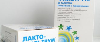
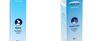
- Softening and moisturizing creams and ointments are the basis for the treatment of atopic dermatitis. These products help keep your skin hydrated and soft.
- Steroid creams and ointments are applied to the skin and help relieve redness and itching. In severe cases, steroid tablets or injections may be required, but your doctor will keep the course of treatment as short as possible, since the high effectiveness of injections comes with a high risk of side effects.
- Medicines that affect the immune system are highly effective, but have side effects and may be prescribed when safer treatments do not produce the desired results.
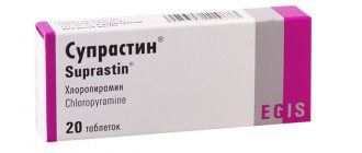
- Antihistamines may be used in patients who report decreased itching.
- To increase the effectiveness of therapy, it is possible to perform wet wraps (Wet Dressing, Wet Wrap Therapy).
Wet wraps (Wet Dressing, Wet Wrap Therapy)
Treatment with Wet Wrap Therapy helps to quickly clear skin of rashes and can be used both in a medical facility or hospital, as well as at home. The effect is associated with improved penetration of the drug used, deeper and longer-lasting hydration, and reduced water loss by the skin. The dressings provide protection from scratching, which leads to the formation of an itch-scratch cycle, preventing scratches from occurring, which further intensify the itching, which makes the skin heal better. Cooling the surface of inflamed skin by evaporating water from the dressings helps reduce inflammation, itching and soreness.
For severe eczema, wet wraps are prescribed in a medical facility. They can also be used at home to maintain good health or at the first sign of deterioration and can reduce the need to seek medical help and the likelihood of hospitalization.
There are various modifications of Wet Wrap Therapy; the essence of the method is to use external medications (emollients or steroids) under two layers of bandages. The bottom layer is warm and moist, on top of which a second, dry layer is made. For the bandage, you can use a regular bandage, special tubular bandages, or special clothing. The bottom layer must be periodically moistened with ordinary warm water, preventing it from drying out. The use of topical steroids under a wet dressing can increase the effectiveness of treatment. The procedure can be performed 1-2 times a day, every day, during an exacerbation. As the exacerbation decreases, the procedure can be done less frequently, performed 1-2 times a week during the period of remission. If well tolerated, the bandage with the medicine can be left overnight, remembering to periodically moisten the bottom layer of tissue.
The procedure takes quite a long time, and in the first stages (especially in patients with severe skin damage) may require the participation of medical personnel. After training and improvement, the procedures can be continued at home.
Complications
The main reason for the development of complications in atopic dermatitis is constant trauma to the skin as a result of scratching. Violation of the integrity of the skin leads to a decrease in its protective properties and contributes to the addition of a microbial or fungal infection.
The most common complication of atopic dermatitis is bacterial skin infections - pyoderma. They manifest themselves as pustular rashes on the body, limbs, and scalp, which dry out and form crusts. At the same time, general well-being often suffers, and body temperature rises.
The second most common complication of atopic dermatitis is viral skin infections. Their course is characterized by the formation of bubbles (vesicles) filled with clear liquid on the skin. The causative agent of viral skin infections is the herpes simplex virus. The face (skin around the lips, nose, ears, eyelids, cheeks), mucous membranes (conjunctiva of the eyes, oral cavity, throat, genitals) are most often affected.
Complications of atopic dermatitis are often fungal infections caused by yeast-like fungi. The affected areas in adults are often skin folds, nails, hands, feet, scalp, and in children - the oral mucosa (thrush). Often fungal and bacterial infections are observed together.
Prevention
There are two types of prevention of atopic dermatitis: primary, aimed at preventing its occurrence, and secondary, anti-relapse prevention. Measures for the primary prevention of atopic dermatitis should begin during the period of intrauterine development of the child, long before his birth. A special role during this period is played by toxicosis of the pregnant woman, taking medications, and occupational and food allergens.
Particular attention to the prevention of atopic dermatitis should be paid in the first year of a child’s life. During this period, it is important to avoid excessive medication and artificial feeding, so as not to create a favorable background for the body’s hypersensitivity to various allergic agents. Following a diet during this period is no less important for a nursing woman.
Secondary prevention aims to prevent exacerbations of atopic dermatitis, and, if they occur, to alleviate their course. Secondary prevention of atopic dermatitis includes correction of identified chronic diseases, exclusion of exposure to disease-provoking factors (biological, chemical, physical, mental), adherence to hypoallergenic and elimination diets, etc. Prophylactic use of desensitizing drugs (ketotifen, sodium cromoglycate) during periods of probable exacerbations (autumn, spring) allows you to avoid relapses. As anti-relapse measures for atopic dermatitis, treatment is indicated in the resorts of the Crimea, the Black Sea coast of the Caucasus and the Mediterranean.
Particular attention should be paid to daily skin care and the correct choice of underwear and clothing. When taking a daily shower, do not wash with hot water and a washcloth. It is advisable to use gentle hypoallergenic soaps (Dial, Dove, baby soap) and a warm shower, and then gently pat the skin with a soft towel without rubbing or injuring it. The skin should be constantly moisturized, nourished and protected from adverse factors (sun, wind, frost). Skin care products should be neutral and free of fragrances and dyes. In linen and clothing, preference should be given to soft natural fabrics that do not cause itching and irritation, and also use bedding with hypoallergenic fillings.