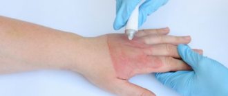
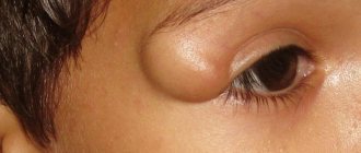
Any punctured, chopped, torn or cut wound can be complicated by the process of suppuration. Even if you cut your finger with a knife in the kitchen, you should not think that this is a trifle, because an insufficiently treated wound can fester. Everyone should know the first symptoms of wound suppuration for the reason that in most cases this problem has to be solved exclusively by surgery.
This problem is especially relevant for those who have undergone any surgery and were discharged home after rehabilitation. If postoperative sutures are not cared for at home as prescribed by the doctor, then if an infection occurs, suppuration may begin. Contrary to the generally accepted opinion of patients, suppuration after operations begins not because of the conditions during the operation, but precisely due to the fault of patients who irresponsibly approach the doctor’s prescriptions when they find themselves at home.
During surgery, it is essential to maintain absolute sterility, and this principle is never violated. Medical doctors recommend that you always fully follow the prescriptions they make to avoid such serious complications.
How does the process of wound suppuration develop?
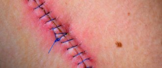
Typically, suppuration begins when a clean wound becomes infected, swelling around the wound develops, tissue necrosis and purulent discharge appear. If redness begins around the wound, which is accompanied by a tugging pain that gets worse at night, then this means that you are dealing with the first symptom of wound suppuration, and urgent measures need to be taken.
When examining the wound, dead tissue and pus are visible. The situation is dangerous because the decay products are absorbed by the body, and this leads to increasing intoxication of the body. As a result, the following symptoms appear:
- significant increase in temperature;
- chills;
- headache;
- weakness;
- nausea.
The problem of wound infection has become extremely relevant in recent years. The number of infectious complications from surgical wounds has sharply increased, resulting in severe sepsis caused by gram-negative microorganisms, as well as microorganisms resistant to almost all modern antimicrobial drugs.
Among all surgical patients, wound infection occurs in 35-45%. The number of postoperative purulent-septic infections in 1998 increased to 39% [14].
Simultaneously with the increase in bacterial infections, the frequency of candida and aspergillus fungi increases, most often due to the widespread, irrational use of antibacterial, corticosteroid, cytostatic drugs, as well as the lack of the concept of simultaneous prescribing of antifungal drugs with antibiotics for prophylactic purposes. The problem of intrahospital spread of both aerobic and anaerobic (clostridial and non-clostridial) infections remains relevant.
The reasons for the increase in the frequency and severity of purulent infections in surgery are varied and include the following factors:
— increasing the volume of surgical interventions, especially in high-risk patients;
- widespread use of instrumental examination and treatment methods accompanied by infection of the patient (intravascular and urinary catheters, endoscopic and tracheostomy tubes, endoscopic manipulations, etc.);
- traditional long-term regimens of prescribing certain groups of antibiotics for therapeutic and prophylactic purposes without regular monitoring of the dynamics of resistance of hospital strains to these drugs.
Depending on the pathogens that have invaded and the changes that occur in the wound, nonspecific (purulent, anaerobic, putrefactive) and specific (wound diphtheria, wound scarlet fever) wound infections are distinguished. In addition, a number of infectious diseases that do not have the nature of a wound infection enter the body through a wound, i.e. not accompanied by noticeable disturbances in the wound process. Some of these common infectious diseases are necessarily associated with the invasion of a pathogen into a wound (tetanus, rabies, rat bite disease); in others, the wound is only one of the possible routes of penetration of microbes (syphilis, anthrax). In practice, it is advisable to focus on the distribution of patients with wound infection into groups, taking into account the etiological and clinicopathological features:
- acute purulent diseases of the skin and soft tissues (abscessing boil, carbuncle, hidradenitis, mastitis, etc.);
— purulent post-traumatic wounds of soft tissues (with or without bone damage, with long-term soft tissue crush syndrome);
— postoperative purulent wounds of soft tissues;
— chronic purulent diseases of soft tissues (trophic ulcers of various origins, bedsores, etc.);
- hematogenous, postoperative or post-traumatic osteomyelitis;
- surgical sepsis.
Regardless of the origin of the wound process, the species composition of the wound microflora, the main methods of treatment are surgical and active complex effects on the purulent process, aimed at eliminating the tissue defect, suppressing the growth of microflora vegetating in the wound or preventing suppuration.
Along with timely surgical intervention on a purulent focus, the outcome of the disease is largely determined by adequate systemic and local antibacterial therapy, strictly focused on the data of bacteriological studies.
Only local drug treatment that is carried out strictly in accordance with the pathogenesis of the wound process can be considered reasonable, i.e. taking into account the phase of its flow [15].
Etiology of wound infections
In recent years, under the influence of various factors, primarily the powerful selective action of antibiotics, significant changes have occurred in the etiology of wound infections. Currently, the leading pathogens are:
— staphylococci (S. aureus, S. epidermidis);
- £, B, Y-hemolytic and non-hemolytic streptococci;
— representatives of the family Enterobacteriaceae (E. coli, Citrobacters
spp.,
Klebsiella
spp.,
Enterobacter
spp.,
Serratia
spp.,
Proteus
spp.,
Providencia
spp.);
- non-fermenting gram-negative bacteria (Pseudomonas
spp.,
Acinetobacter
spp.,
Moraxella
spp.,
Flavobacterium, Achromobacter).
The dependence of the species composition of wounds on their origin is clearly visible. So, for example, if in a group with acute purulent diseases staphylococcus in monoculture is detected in 69.5% of cases, then in patients with post-traumatic purulent wounds, chronic purulent diseases of the skin and soft tissues, as well as in patients with purulent wounds and developed sepsis, they are detected immediately several pathogenic microorganisms in 31.5, 48.8, 55.6% of cases, respectively. The rest are representatives of the family Enterobacteriaceae
in monoculture.
In recent years, fungi have begun to emerge from wounds much more often (9.9%), which is apparently due to insufficient attention to this problem and the lack of reliable prevention of fungal invasion (Table 1).
Obligate non-spore-forming anaerobic microorganisms, among which Bacteroides
spp.,
Fusobacterium, Peptococcus
spp.,
Peptostreptococcus
spp.,
F. nucleatum, P. melaninogenicus.
The proportion of pure non-clostridial and mixed aerobic-anaerobic microflora also depends on the location and origin of the purulent wound.
Currently, it is possible to significantly reduce the duration of systemic antibacterial therapy through the active introduction into practice of treating wounds under dressings with modern drugs focused not only on the phase of the wound process, but also on the species composition of wound microorganisms.
This tactic, with timely adequate surgical intervention and treatment with correctly selected drugs for local therapy, allows you to localize the purulent process and avoid generalization of the infectious process.
The use of modern drugs for local treatment of wounds at all stages of complex treatment makes it possible to reduce the time of systemic antimicrobial therapy, avoid the development of side effects, significantly reduce the cost of expensive antibacterial drugs, and avoid the formation of microflora resistance to the systemic antibiotics used.
Currently, several groups of drugs have been developed for local treatment of wounds in phases I and II of the wound process (Table 2).
The main groups of drugs are antiseptics, polyethylene glycol (PEG)-based ointments, modern biologically active dressings, enzyme preparations, and new antiseptics.
Antiseptics
When choosing antiseptics used for both preventive and therapeutic purposes, preference is given to drugs with a universal, broad spectrum of action, active against mixed microflora, and having a microbicidal or microbostatic effect.
Iodophors
In the practice of wound treatment, new complex compounds of iodine with polyvinylpyrrolidone (povidone-iodine, betadine, iodopirone, iodovidone, etc.), which have microbicidal and microbostatic effects, are widely used.
Drugs in this group suppress:
- gram-positive bacteria, including enterococci and mycobacteria;
- gram-negative bacteria, including pseudomonas, acinetobacter, Klebsiella, Proteus;
— bacterial spores, fungi, viruses, including hepatitis B and C viruses, entero- and adenoviruses, as well as anaerobic, spore-forming and asporogenic bacteria.
All pathogens of wound infections do not have either natural or acquired resistance to iodophors.
The activity of the complex with polyvinylpyrrolidone is not affected by the presence of blood, purulent discharge or necrotic tissue [4].
The most widely used in the practice of treating purulent-inflammatory processes are two dosage forms of complex compounds of iodine with polyvinylpyrrolidone - solution and ointment.
Ointments (1% iodopyrone ointment, povidone-iodine ointment) are used to treat purulent wounds with heavy exudation. Solutions (iodopirone, povidone-iodine) are used as antiseptics for prophylactic purposes for treating the surgical field, skin during punctures, closing surgical sutures, as well as for the treatment of wounds, trophic ulcers, bedsores, and diabetic foot syndrome in the absence of a large amount of wound separated.
Dioxidine
Dioxidin is one of two drugs derived from quinoxyline di-N-oxide, developed as a result of fundamental exploratory research in the period from 1960 to 1980 at the All-Union Scientific Research Chemical-Pharmaceutical Institute (currently the Center for the Chemistry of Medicines - TsKhLS VNIHFI , Moscow).
A number of drugs of this class of substances with high antimicrobial activity and a wide antimicrobial spectrum (quindoxine, mequidox, carbadox, temadox, olaquindox) have been developed abroad.
The drug is intended for the treatment of patients with wound infections caused by multidrug-resistant flora, Pseudomonas aeruginosa and non-clostridial anaerobic pathogens. This drug is most active against anaerobic bacteria (Clostridium
spp.,
Bacteroides
spp.,
P. melaninogenicus, Peptococcus
spp.,
Peptostreptococcus
spp., as well as aerobic gram-negative bacteria -
Ps.
aeruginosa, E. coli, Proteus spp.,
Klebsiella
spp.,
Serratia
spp.) [1-3].
It should be noted that strains of Pseudomonas aeruginosa, as well as gram-positive bacteria (staphylococci, streptococci), are more resistant to the drug. That is why, if the clinical situation allows, a 1% dioxidine solution without dilution is used for local treatment.
In the 70-90s, a solution of dioxidine in monotherapy and combination with other antibacterial drugs was considered as the drug of choice for the treatment of patients with sepsis, diffuse and local peritonitis, for the prevention and treatment of purulent-inflammatory processes in the liver and biliary tract, lungs, stomach, kidney allotransplantation, cardiac vascular prosthetics and coronary artery bypass grafting, under conditions of artificial circulation [11-13].
Currently, in Russian clinics for more than 25 years, various dosage forms of dioxidine have been used to treat various forms of purulent infection:
a) for local treatment
— 5% ointment, “Dioksikol” ointment containing 1% dioxidine, dioxidine aerosol (“Dioxisol”), polymer compositions with dioxidine (“Diovin”, “Diotevin”, Anilodiotevin”, “Colladiasob”, “Digispon A”, suture material ;
b) for introduction into cavities, for ultrasonic inhalations
— 1% aqueous solution in ampoules;
c) for intravenous administration
— 0.5% aqueous solution in ampoules.
Intravenous administration of dioxidine is carried out for health reasons. When justifying and determining the indications for the intravenous administration of dioxidine from the “benefit-risk” perspective, it should be taken into account that over the past 15-20 years, highly effective antibacterial agents have been created that have advantages over dioxidine in terms of toxicological properties. Therefore, dioxidin is prescribed intravenously only if other chemotherapeutic agents are ineffective or intolerable, strictly observing the recommended doses for the drug and the duration of each infusion.
Dioxidin is well compatible with other antimicrobial drugs. The clinical capabilities of dioxidin are expanded due to its ability to penetrate the blood-brain barrier, which makes it possible to use it in the treatment of patients with meningitis, brain abscesses and purulent cranial wounds.
Miramistin
The domestic antiseptic Miramistin belongs to quaternary ammonium compounds (cationic surfactants). Preclinical and clinical studies have shown that Miramistin has a pronounced antimicrobial effect against gram-positive and gram-negative, aerobic and anaerobic, spore-forming and asporogenic bacteria in the form of monocultures and microbial associations, including antibiotic-resistant hospital strains. The drug is most effective against hospital strains of staphylococcus and streptococcus. The drug has a detrimental effect on fungi, viruses, and protozoa.
Miramistin has been introduced into clinical practice since the early 90s of the last century. Currently, the drug is widely used in the complex treatment of purulent wounds and accompanying inflammatory complications in the bronchopulmonary system in the form of ultrasonic inhalations and the genitourinary system in the form of bladder instillations [5].
Lavasept
An antiseptic drug, the main active ingredient of which is polyhexanide, considered by experts as the drug of choice for the treatment of contaminated and infected wounds [16]. Polyhexanide belongs to the group of positively charged (cationic) polymers and contains surfactants, which reduce surface tension, which ensures easier removal of microbial biofilms.
Lavasept has a broad-spectrum bactericidal effect, is active against gram-positive and gram-negative bacteria (including Pseudomonas aeruginosa
), fungi, and MRSA.
Prontosan
A particular danger for the patient is a “dormant” infection, the aggressiveness of which is determined by the variability of the microflora, the reactivity of the body, the loss of activity of traditional systemic antibiotics and drugs for local medicinal treatment of wounds. This threat persists to a particular extent if the patient has foreign bodies, implanted devices, in patients with long-term non-healing wounds, trophic ulcers, diabetic foot syndrome, with post-traumatic and postoperative osteomyelitis, chronic post-traumatic and postoperative wounds.
It has been established that microorganisms and fungi, when they remain in a wound for a long time, thanks to the polymers they secrete, form a thin layer - a biofilm. Biofilm formed by bacteria and fungi provides reliable protection for pathogens from ultraviolet radiation, antibiotics, phagocytosis and other factors of the body’s immune system. Microbes in the biofilm can withstand concentrations of antibiotics 100-1000 times more than suppressive planktonic cells. Therapeutic effects on biofilms can be aimed at the mechanisms of initial adhesion of bacteria to the surface, blocking synthesis or destruction of the polymer matrix.
Currently, the attention of researchers on this problem is drawn to the possibility of using various medications that destroy the biofilm formed by bacteria and fungi. One of these drugs is prontosan, which contains polyhexanide, a polymerized biguanide derivative that acts as a local cationic antiseptic. The antimicrobial effect of polyhexanide is due to nonspecific affinity for the cell membranes of the microorganism, which contains a large amount of acidic phospholipids. Polyhexanide acts on bacterial cell membranes and increases their permeability.
Two dosage forms of Prontosan have been introduced into practice - 0.1%, 0.2% solution and gel, which contains 0.1% polyhexanide, 0.1% undecylenic amidopropyl betaine (surfactant), glycerol (humectant), hydroxyethylcellulose ( gelling agent), water.
PEG-based sorbents and ointments
It should be remembered that with abundant purulent exudation, the use of antiseptic solutions for local treatment of wounds in the form of gauze tampons is considered a flawed method, since tampons placed in the wound dry out quickly and do not have the long-term osmotic activity necessary to remove pus. In extreme cases, the wound can be filled with a combined tampon - a silicone tube is placed in the center of the gauze tampon, through which 10-20 ml of antiseptic is injected into the wound with a syringe 3-6 times a day.
For the treatment of superficial infected, purulent and purulent-necrotic wounds of various etiologies in the inflammatory phase, biologically active draining sorbents (anilovine, diovin, anilodiovin, diotevin, anilodyotevin, collasorb, colladiasorb), the main component of which is helevin, are successfully used. Dioxidin is used as an antimicrobial drug. The proteolytic effect of sorbents is due to the introduction of proteolytic enzymes into their composition (terrylitin, collagenase from hydrobionts). The analgesic effect is achieved due to the presence of a local anesthetic, anilocaine, in the composition [11].
Targeted use of biologically active dressings with a differentiated effect on the wound process, taking into account its phase and characteristics of the course, providing for sorption-application therapy in phase I of the wound process using biologically active sorbents or gel dressings with antimicrobial, analgesic and proteolytic effects, followed by wound treatment in phases II and III of the wound process with biologically active stimulating coatings with a specific effect on the processes of regeneration and epithelization.
In recent years, it has become possible to more successfully treat wounds using a new combination drug Baneocin, containing two highly active bactericidal components - bacitracin (a polypeptide antibiotic that inhibits the synthesis of bacterial cell walls) and neomycin (an aminoglycoside that inhibits protein synthesis), between which there is synergy. The results of numerous studies indicate the high activity of baneocin against gram-negative, gram-positive flora, aerobes and anaerobes. The components of the drug exhibit synergism against highly resistant hospital strains of Ps. aeruginosa, E. coli, S. aureus.
Baneocin is characterized by the ability to create high bactericidal concentrations in a purulent focus and not have a systemic effect. Numerous studies have shown that when treating wounds under bandages with Baneocin, complete eradication of pathogenic pathogens and reliable prevention of reinfection of the wound surface by hospital microorganisms is achieved in a short time without the traditional systemic antimicrobial therapy in such cases, prescribed for both therapeutic and prophylactic purposes.
The drug Baneocin is available in two dosage forms, focused on the phases of the wound process. For example, for the treatment of purulent wounds in the first phase of the wound process, Baneocin powder is used, which actively absorbs wound discharge within 5 hours.
For the treatment of wounds in the II phase of the wound process, Baneocin ointment is used, which exhibits a local bactericidal effect necessary to prevent reinfection of granulating wounds with hospital strains. The resulting thin film of the drug protects the thin layer of young epithelium from damaging factors.
The positive properties of two dosage forms of Baneocin - powder and ointment - are successfully implemented both at the outpatient and inpatient stages of complex treatment of patients of different groups:
- with focal skin infections (hidradenitis suppurativa, paronychia, boil, carbuncle);
- with burns, frostbite;
- with trophic ulcers;
- for the prevention of suppuration of household, sports and industrial wounds, abrasions (violations of the integrity of the skin);
— to prevent the development of an infectious process in the area of donor wounds when collecting skin grafts in traumatology, surgery, and cosmetology.
Painless and non-traumatic application, deep penetration into tissue, and good tolerability of Baneocin make it possible to successfully treat patients with trophic ulcers even in cases of aggravated allergic history (intolerance to traditional local drugs) or the identification of highly resistant strains of Ps. aeruginosa
[6].
For the treatment of extensive and deep wounds with a purulent process in the first phase
over the past 25-30 years, PEG-based ointments have been successfully used (levosin, levomekol, 5% dioxidine ointment, dioxykol, 1% iodopyrone ointment, 1% povidone-iodine ointment, 0.5% miramistin ointment, iodometricylene, nitacid, streptonitol , 10% mafenide acetate ointment, streptolaven, stallanin-PEG 3%, oflomelide, etc.). The listed drugs have different osmotic activity for the differentiated treatment of wounds in the first phase of the wound process with copious or moderate amounts of wound discharge (boils, carbuncles, hidradenitis, mastitis, abscesses, phlegmons, suppurating lipomas, atheromas, purulent postoperative and post-traumatic purulent wounds, venous trophic ulcers , “diabetic foot” with a local purulent-necrotic process, etc.
These drugs have a fairly wide spectrum of activity against both aerobic and non-spore-forming anaerobic microorganisms.
Despite many years of intensive use of ointments containing chloramphenicol or dioxidin, their high antimicrobial activity remains against the main causative agents of surgical infection.
If there are gram-negative bacteria in the wound, in particular Pseudomonas aeruginosa, it is recommended to use 10% mafenide acetate ointment, 5% dioxidine ointment, dioxicol ointment, nitacid ointment.
For the treatment of non-sporogenous anaerobic infection in combination with aerobic infection, it is advisable to use the following drugs:
- with nitazol (Streptonitol and Nitacid ointments);
— foaming aerosol Nitazol;
- 5% dioxidine ointment, dioxikol ointment.
When using PEG-based ointments, side effects (clinically significant) are observed in 0.7% of cases, clinically insignificant - in 2.3% of cases. Most often they manifest themselves in the form of local symptoms of drug-induced dermatitis. In cases of intolerance to chloramphenicol, dioxidin, treatment can be carried out with oflomelid ointment or 5% miramistin ointment, which has not only a wide spectrum of antimicrobial activity, but also antiviral and antifungal effects, which is extremely important in the treatment of patients with trophic and long-term non-healing wounds.
Of the new ointments, the domestic drug streptolaven deserves special attention, which includes an enzyme of microbial origin (streptolysin), an antimicrobial drug miramistin and a base balanced in osmotic action that does not cause drying of wound tissue. This is the only ointment in the country with a necrolytic effect and is currently successfully used in the treatment of patients with diabetic foot syndrome, extensive burns, trophic ulcers, and bedsores [9].
The possibilities for successful treatment of patients with purulent wounds, trophic ulcers, bedsores, infected burns have significantly expanded with the advent of the new ointment Stellanin-PEG 3%, containing 1,3-diethylbenzimidazolium triiodide, low molecular weight polyvinylpyrrolidonium, dimexide, polyethylene oxide 400 and 1500. The bactericidal effect of the drug is due to the incoming it contains active iodine [9].
As can be seen from table. 3,
Stellanin-PEG ointment 3% is not inferior in antimicrobial activity to well-known drugs (levomekol, 5% dioxidine ointment).
Stellanin-PEG has high antimicrobial activity against both gram-positive and gram-negative microorganisms, including methicillin-resistant staphylococci (MRSA), E. faecalis, E. faecium,
as well as
E. coli
and
Klebsiella
spp., producing extended-spectrum beta-lactamases, and is also able to suppress the vital activity of fungi
(C. albicans).
In the treatment of wounds and trophic ulcers with severe pain, a high clinical effect is achieved using the new PEG-based ointment oflomelid, which contains lidocaine as an analgesic component and ofloxacin as an antimicrobial component.
Particular severity of the wound process occurs when the patient develops a polyvalent allergy. The solution in such clinical situations is the use of drugs containing silver (Argosulfan ointment - for light wound discharge) or Actisorb Plus dressings (non-woven nylon fiber with activated carbon and silver ions).
For the treatment of wounds with a moderate amount of wound discharge and a slow regeneration process (long-term non-healing wounds, trophic ulcers, diabetic foot syndrome, bedsores, etc.), the use of the original domestic drug 15% Dimephosphone, which has membrane-stabilizing, anti-acidotic, antimicrobial, anti-inflammatory activity, is indicated [10 ].
Treatment of wounds in phase II of the wound process
For the treatment of moderately or slightly exuding purulent wounds in the stage of transition to phase II of the wound process, as well as in the treatment of donor wounds during free skin grafting with autodermograft, the use of biologically active gel dressings Apollo PAK and Apollo PAA, which include iodovidone or miramistin, is indicated. also local anesthetic anilocaine. The hydrogel is based on a copolymer of acrylamide and acrylic acid.
If signs of a regenerative process are detected in the absence of profuse suppuration and mild symptoms of inflammation remain, it is possible to treat wounds under bandages using iodine-containing solutions: 10% Iodopirone, 1% Iodovidone, 1% povidone-iodine, Sulyodopirone.
After the acute purulent process has stopped and symptoms of intoxication have disappeared, confirmed by both clinical and laboratory tests, it is possible to cancel general antibacterial therapy. In these cases, local treatment of wounds at the stage of preparation for final closure with sutures or plastic surgery is carried out under bandages with drugs:
- biologically active stimulating wound coverings with antimicrobial and local anesthetic effects (Digispon-A, Algikol-FA, Kollakhit-FA, Anishispon);
— collagen-containing wound coverings (Gentatsikol);
— wound coverings based on alginates (Algipor, Algimaf);
— wound covering with silver based on collagen and regenerated cellulose (Promogran-Prisma);
— foaming aerosols (dioxysol, gentazol, nitazol);
- ointments with slight osmotic activity (Methyldioxylin, Argosulfan, Fuzimet, Streptonitol, Biopin 5%, 10%; Baneocin);
- oils (Miliacyl, sea buckthorn oil, rosehip oil).
The appearance in hospitals of a new Fuzimet ointment (a combination of fusidine sodium with methyluracil) makes it possible to successfully treat patients with methicillin-resistant S. aureus
(MRSA), without including vancomycin or linezolid in complex therapy [7, 8].
Prevention and treatment of fungal infections during systemic and local antimicrobial therapy of wound infections
Excessive enthusiasm for various antibacterial drugs in the treatment of infectious processes without mandatory simultaneously prescribed antifungal therapy has led to the fact that the problem of deep mycoses in recent years has become extremely urgent and difficult to solve. For example, the mortality rate for candidal pneumonia is 65-70%.
Disseminated candidiasis accounts for up to 10-15% of all nosocomial blood infections. With disseminated candidiasis, fungal infection of the skin is possible with the formation of subcutaneous abscesses, cellulite or individual muscle groups (candidal myositis). Fungal infections of the kidneys and lungs, nervous system, heart, spine, costal and sternoclavicular cartilages, stomach, intestines and bile ducts are extremely difficult to treat.
Candidaemia can be the direct cause of death in 75% of cases, and candidal sepsis as a cause of death can reach 88%.
Diabetes mellitus, antibiotic treatment, the presence of indwelling catheters, and severe neutropenia predispose to colonization of a fungal infection. Disseminated candidiasis is possible as a result of infection during surgical operations, invasive diagnostic procedures (puncture, biopsy, endoscopy), hemodialysis and peritoneal dialysis. Up to 40.2% of medical personnel may be carriers of Candida
spp., with 31.3% of them in association with
S. aureus,
43.7% with coagulase-negative staphylococci, and 25% in monoculture.
Prevalence of oropharyngeal carriage of fungi of the genus Candida
spp. among the medical staff of the general surgery department is 61.5%.
According to a study carried out at the Institute of Surgery named after. A.V. Vishnevsky, frequency of Candida albicans
from various biological media and catheters was detected in 8.6-12% of cases. The most dangerous source of the spread of fungi was a solution of furatsilin, from which fungi were detected in 58.8% of cases.
Traditionally, to prevent fungal infection, along with antibacterial drugs in such cases, ketoconazole or (in case of generalized infection) fluconazole, flucytosine or amphotericin B are prescribed. In case of generalized fungal infection, the duration of etiotropic therapy usually ranges from several weeks to a year.
Timely abandonment of long-term systemic antibacterial therapy, abandonment of traditional antiseptics (furatsilin solution), traditional absolutely ineffective fat-based ointments and active use of drugs such as 1% povidone-iodine solution, solutions of Prontosan, Lavasept (0.1%, 0. 2%) or 0.5% Miramistin ointment allows not only to avoid serious complications of the wound process, but also to successfully combat fungal invasion without resorting to systemic antifungal therapy.
Thus, the pharmaceutical industry currently has the opportunity to produce a fairly complete range of modern, highly effective drugs for successful local medicinal treatment of wounds, trophic ulcers, bedsores of various locations and origins. Timely active introduction of these drugs into the daily practice of practical healthcare can significantly reduce the cost of purchasing systemic expensive foreign drugs (there are no domestic drugs).
However, I would like to draw the attention of leading pharmaceutical manufacturers to the unacceptable cessation of production of such drugs as 5% dioxidine ointment, dioxicol, nitacid, streptonitol, 1% iodopyrone ointment, 0.5% quinifuril ointment, foaming aerosols (dioxysol, gentazol, nitazol, domestic hydrocolloids galagran and galactone, collagen coating with gentamicin gentacycol, wound dressings containing sodium alginate (algipor, algimaf).
The current situation requires an active position of all specialists involved in the treatment of surgical infections. It is extremely important to review the standards of wound treatment using modern drugs, since from the standpoint of evidence-based medicine, such standards can reduce mortality in patients with sepsis to an average of 26.5%, which is of extreme social significance.
Timely equipping of emergency medical teams, teams providing assistance to victims of natural and man-made disasters, military conflicts, outpatient clinic doctors, trauma centers and hospitals, specialized purulent surgery departments with modern drugs for local treatment of wounds will undoubtedly help reduce the development of severe purulent wounds. complications and diseases, will reduce the cost of antibacterial drugs by 2-3 times.
Causes of wound suppuration
Any wound, small or large, is considered infected because bacteria will get into it anyway. However, this does not always lead to suppuration. In order for this destructive tissue process to develop, additional conditions are required:
- sufficient tissue damage;
- the presence of non-viable tissue in the wound cavity;
- the presence of foreign bodies in the wound cavity;
- the presence of bleeding blood in the wound cavity;
- high concentration of pathogenic microorganisms.
Therefore, the first symptoms of wound suppuration can appear even after an ordinary splinter, provided that a piece of it remains in the body tissue, and at the same time there are pathogenic microorganisms on the foreign body itself. The latter include those that are responsible for the development of the purulent process: staphylococci, streptococci, E. coli and similar microorganisms.
In addition, there is a high risk of suppuration when the patient has a history of diabetes mellitus, vascular and somatic diseases, excess weight and old age.
The nature of the wound also matters for the development of this process. Thus, a puncture wound can fester due to the wound channel being too narrow and due to which there is no normal outflow. In the case where the wound is accompanied by crushing of surrounding tissues, suppuration occurs due to too much dead tissue in the wound and excessive contamination. At the same time, wounds on the head and neck heal faster and better, and worst of all – on the feet.
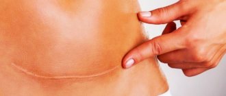
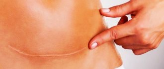
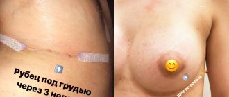
Types of postoperative scars
Keloid.
A lumpy scar made of overgrown connective tissue that is red or bluish in color. It looks like a tumor or a mushroom cap. The size of the keloid scar is larger than the wound itself, it hurts, itches, and sometimes pulsates.
Hypertrophic.
Outwardly similar to a keloid, but its size does not extend beyond the wound. A hypertrophic scar does not cause any discomfort and lightens over time.
Normotrophic.
A well-healing scar is initially convex and reddened, after 3 months it becomes almost invisible, there is no sensitivity.
Diagnostics
Determining the presence of the fistula itself is not difficult. To assess its depth and diameter, it is possible to use a special probe inserted into the resulting oroantral tract. To assess the extent of inflammation and the presence of additional passages, it is possible to use radiography of the upper jaw with the introduction of a contrast agent.
Evaluation of the presence of a foreign body or apex of the tooth root is carried out during radiography or directly during the operation to close the fistula. If there is no effect from antibacterial therapy, it is possible to culture the microflora to determine its resistance to antibiotics of various types of action.
Why does postoperative pain occur?
Postoperative pain according to ICD-10 (International Classification of Diseases) refers to unspecified types of pain and does not carry any signaling information for either the patient or the doctor, since the causes and mechanisms of its occurrence are clear. Therefore, modern principles of patient management after surgery provide for maximum relief from unpleasant pain. Moreover, pain has a negative impact not only on healing, but also on vital processes in the body: the functioning of the cardiovascular, respiratory, digestive, central nervous systems, as well as blood clotting4.
The formation of painful sensations is ensured by a multi-level reaction that connects the immediate area of damage (wound surface) and the central nervous system. It begins with mechanical stimuli in the area of the incision and the release of biologically active substances (prostaglandins, bradykinins and others)4, and ends with information processing in the cerebral cortex and the connection of emotional and psychological components.
Pain syndrome develops as a result of the emergence of areas of increased pain sensitivity (hyperalgesia). Primary hyperalgesia is associated directly with damage and forms near the wound. The area of secondary hyperalgesia covers a larger area and occurs later, over the next 12-18 hours4, as it is associated with stimulation of other types of receptors. It is she who is responsible for the preservation and intensification of pain on the second or third day after surgery, and subsequently for the development of chronic pain syndrome7.
Up to contents
Symptoms of appearance
The process of fistula formation is difficult to track; it can take several days, as happens with acute pancreatitis, or several months, as with post-radiation tissue changes.
Manifestations at the initial stage of fistula formation are due to its root cause, as a rule, a local inflammatory process resulting in purulent melting of tissue with pain and infiltration, often intoxication and fever.
Without an exacerbation of the inflammatory reaction, the fistula can be felt as a cord. The size of the compaction around the fistula tract is due to inflammatory infiltration and branching of the fistula tracts themselves, cicatricial changes in the surrounding tissues, previously involved in the inflammatory conglomerate.
A formed fistula has an entrance and, sometimes, an exit, the tissues around it are compacted, and discharge can be squeezed out of the hole: pus, bile, pancreatic juice, and so on. With a fistula, feces may leak from the intestine into the vagina from the genital organs; when there is an anastomosis of the intestine with the bladder, urine leaks from the anus. The discharge from the intestinal fistula has a fecal smell, and the purulent secretion from the vagina also has a specific smell. The smell of a discharged fistula, leading from the zone of disintegration of a malignant tumor, seems especially heavy to others.
Inflammation causes pain ranging from slight discomfort to unbearable pain. Tumor fistulas do not hurt because they form inside a disintegrating tumor.
When the infection activates with the formation of streaks of purulent contents, a general reaction occurs: intoxication, high temperature, sweating and pallor, palpitations and rapid breathing.
Treatment
To identify the pathogen and prescribe the correct treatment, it is necessary to examine the wound discharge and identify the sensitivity of bacteria to different groups of antibiotics. The usual infiltration is eliminated by probing the wound and removing purulent fluid. Sutures may be removed to relieve pressure on the tissue.
The patient is prescribed bed rest. Treatment of purulent inflammation involves the mandatory removal of suture material, cleaning the wound and installing drainage. Anaerobic infection requires excision of the affected tissue, and in some cases, additional surgical intervention.
For any form of inflammation, the wound is drained daily and cleaned of pus. As healing progresses, stitches may be repeated and gauze bandages with wound-healing ointments may be applied. To prevent suppuration of sutures, it is necessary to strictly follow medical instructions and follow a number of antiseptic measures.
Patient hygiene
Hygiene rules will help to avoid the addition of a secondary infection, they are as follows:
- When using antiseptics, solutions are used regularly until the damage is completely healed. Antiseptics stop the growth and reproduction of pathogenic microorganisms.
- While washing. It is recommended to carry out them at least 2 times a day, using warm water. You can give preference to herbal decoctions, use: chamomile, oak bark, plantain.
- In changing bed and underwear, which is carried out daily. Preference is given to natural fabrics.
Brief anatomical summary
The upper jaw contains the maxillary sinus (maxillary sinus), the alveolar process located under the sinus and a number of other anatomical formations. In the absence of pathology, the roots of the teeth of the upper jaw are tightly fixed in the alveolar process. Normally, between the tips of the roots of the teeth and the sinus cavity there is from 3 to 15 mm of bone tissue.
A fistula is a pathological course formed as a result of an infectious-inflammatory process. Due to the fact that during sinus lifting the amount of bone tissue is reduced, when an infection enters the surgical wound, the soft and hard tissues of the alveolar process and the bottom of the maxillary sinus melt. An oroantral fistula is formed, a fistula connecting the sinus cavity with the oral cavity.
Prevention of complications
If a person recently underwent surgery, the following recommendations will help eliminate the likelihood of complications:
- Do not break your diet, strictly follow your doctor’s recommendations, treat the wound, and if problems arise, consult a doctor as an emergency.
- Monitor the temperature, monitor changes; an increase indicates an inflammatory process in the body. You can keep a diary in which you should record your body temperature. You will have to measure it 3 times per knock.
- Try to avoid problems with stool and digestion; constipation and bouts of diarrhea are unlikely to help you recover faster.
- Follow the drinking regime, consume a sufficient amount of liquid every day. This will help normalize the functioning of the gastrointestinal tract.
- Take a course of antibiotics. If treatment is stopped, even with significant improvement in condition, the risk of complications increases. Because the inflammatory process was not completely stopped.
- Maintaining an active lifestyle is also considered a preventive measure, but do not forget about the doctor’s instructions. If the body is weakened, then additional stress will not bring benefit.
- Taking vitamins and immunostimulants should also be regarded as preventing complications.
- You will have to avoid systematic hypothermia of the body, stressful situations, and inflammatory diseases of the nasopharynx.
- During the off-season, increase immunity and, if it occurs, eliminate imbalance in the intestines.
- After complete recovery, prevent parasitic diseases 2 times a year.
A passive lifestyle, bad habits, frequent stress - all this weakens the body and reduces its protective functions. For this reason, you will have to lead a measured lifestyle, play sports, and give up alcohol and smoking.
Recommended video:
Paraproctitis after surgery can be treated in various ways, but an integrated approach is considered optimal. This way you can achieve better results. Correctly planning therapy helps to avoid relapse of the disease and repeated surgery. It also significantly reduces the risk of complications.
Diet after surgery
It is considered a component of treatment because it speeds up recovery and reduces the risk of complications. It involves giving up certain foods, which normalizes the functioning of the digestive system and reduces the unpleasant symptoms that are often observed in patients during the postoperative period.
The table shows the products that are not prohibited, as well as those that will have to be avoided:
| What is prohibited: | What is allowed: |
| meat broths; | vegetable broths; |
| cabbage in any form, due to the presence of coarse fiber; | green onions, spinach, beets (preferably boiled), carrots (can be eaten raw), cucumbers and radishes; |
| tea, coffee, sweet carbonated drinks; | compotes from dried fruits, fruit drinks from berries, vegetable and fruit juices, it is not forbidden to drink water, including mineral water, only without gas; |
| you should not eat rice, oatmeal, cruise cereal, as well as beans and peas; | you can eat buckwheat, semolina, barley and wheat porridge cooked in water; |
| sweet baked goods, various products made from refined flour: gingerbread, cakes, cookies; | do not exclude bread from your diet; |
| pork and beef; | you can eat boiled chicken, turkey or rabbit meat - these are dietary products; |
| fatty fish: herring, salmon. | low-fat fish varieties such as hake, cod, flounder. |
Despite the fact that it is not prohibited to drink fruit juices, you still have to give up fruits. Patients can only eat baked apples.
In this case, the food must meet certain characteristics:
- Easy to digest and does not burden the stomach.
- Do not provoke increased gas formation in the gastrointestinal tract, do not cause problems with stool.
- Completely digested in the intestines.
Fatty, salty, sour, spicy, smoked and hot foods are prohibited. You should not drink alcohol and its derivatives. You will have to give up fast food, instant noodles and processed foods.
Preventive measures before and after sinus lift to prevent the development of a fistula
Fistula after sinus lifting and bone grafting is quite easy to prevent by taking certain groups of medications before and after surgery, as well as observing the rules of oral hygiene.
Rules of conduct after surgery
The protective regimen should last from 5 to 7 days after the sinus lift. It includes a number of behavioral features:
- When brushing your teeth, special dental pastes with antibacterial and anti-inflammatory properties are used: Solcoseryl, Lakalut, Sensodyne. Oral hygiene procedures should be carried out twice a day (in the morning and before bedtime). It is necessary to brush your teeth on the operated side with great care so as not to disturb the condition of the tissues and implant after surgery.
- When eating, chewing movements should be carried out exclusively on the healthy side. Drinking all kinds of liquids through a straw is strictly contraindicated. You should also give up sweet foods for a week.
- During the first two weeks, you must stop playing any sports. This is especially true for athletics, gymnastics, cycling, swimming and martial arts. You should avoid the latter for a month.
- Try, if possible, not to sneeze or cough, avoid head injuries and bruises, and shaking.
- When sleeping, do not lie on your operated side.
