The first and, perhaps, the most important step towards curing psoriasis is diagnosing this complex disease. Modern research methods and the experience of dermatologists make it possible to quickly diagnose psoriasis and begin its treatment.
If you notice any strange rashes on your skin, see a dermatologist as soon as possible. The sooner you diagnose the pathology and begin its treatment, the greater your chances of significantly extending periods of remission and reducing the intensity of psoriasis exacerbations.
Methods for diagnosing psoriasis
The main symptom of psoriasis is a skin rash that can appear on different parts of the body. Small pink lumps appear on the patient’s arms, legs, back, head or other parts of the body, which quickly become like tubercles.
These spots become bright pink bumps and are covered with loose silvery-white scales, so-called plaques are formed. These plaques increase in size and reach 7-8 centimeters in size.
To accurately diagnose psoriasis, the doctor refers the patient to the following tests:
- General and biochemical blood tests and urinalysis;
- stool analysis for worm eggs;
- rheumatic tests - for joint damage;
- skin biopsy – if the picture of psoriasis is unclear. During the examination, a small area of skin is cut off. A biopsy allows, under a microscope, to differentiate psoriasis from many other diseases with similar symptoms.
During the examination, the doctor must question the patient in detail about concomitant diseases, lifestyle, and the presence of lichen planus among the patient’s relatives. An examination is also carried out for the so-called psoriatic triad - spots, film and pinpoint hemorrhage.
On the body's defense
The immune system is a system of organs and cells that protect the body from pathogens: viruses, bacteria, protozoa, helminths, and so on. Every day the body encounters millions of microbes, many of which are pathogens, but due to immune responses, diseases rarely develop. The reason the immune system fights enemies so well is that it can identify them very precisely: it can recognize the characteristic pieces of molecules of the pathogens that attack it, mainly the substances with which they are coated. Such pieces are called antigens.
In order to communicate with each other, immune cells release special information molecules - cytokines.
Cytokines can be either stimulating (“we’ve been attacked, call everyone up!”) or inhibitory (“false alarm, let’s return to peaceful life!”). In principle, cytokines can be released by all cells of the body, but immune cells communicate in the language of cytokines especially actively. The table below lists the main groups of cytokines and their functions. Table. Cytokines are molecules produced by immune and non-immune cells that play an important role in regulating the immune response.
| Name | Function | Some representatives* | |||||
| Interferons | A group of molecules released by cells in response to viral invasion; have similar properties and antiviral effects | Interferons alpha (IFN-α), beta (IFN-β), gamma (IFN-γ) | |||||
| Chemokines | Small proteins produced in cells and tissues when a pathogen appears; stimulate cell migration, adhesion and activation | CXC, CC, CX3C, XC | |||||
| Tumor necrosis factors | Proteins synthesized mainly by monocytes and macrophages; cause necrosis of tumor cells and enhance the immune response | Tumor necrosis factors alpha (TNF-α), beta (TNF-β, also known as lymphotoxin-α) | |||||
| Interleukins | Soluble peptides synthesized mainly by T lymphocytes; in isolation stimulate groups of cells to divide or differentiate. Have pro- and anti-inflammatory effects depending on the interleukin and the cell | IL-6, IL-17, IL-23 | |||||
| Hemopoietins | A group of molecules with similar properties; stimulate the reproduction and differentiation of blood cells | Colony-stimulating factors (CSF) | |||||
| Transformative growth factors | Proteins that control proliferation, cell differentiation and other functions in most cells, including immune cells | Transforming growth factor alpha (TGF-α), beta (TGF-β) | |||||
| * Some cytokines have multiple effects and can belong to several groups at once (for example, IL-8 is the chemokine CXCL8, and IL-2-4 affect hematopoiesis) | |||||||
There are two types of immunity: innate and acquired.
- Innate immunity is the sum of all evolutionarily ancient immune cells (among them macrophages, dendritic cells, blood granulocytes, etc.) and molecules (antimicrobial peptides, lysozyme, a special group of proteins called the “complement system”, etc.). Innate immune cells are able to nonspecifically (by antigens common to different groups of foreign agents) recognize pathogens and, attracting other immune cells and molecules, immediately destroy them. Two types of innate immune cells are especially important in the pathogenesis of psoriasis: dendritic cells and neutrophils . Below we will talk about these cells in more detail. Although innate immunity is indispensable for a quick reaction to an enemy, its work lacks specificity: if, for example, one bacteria multiplies very strongly in the body, then innate immunity will lose in the fight against it - it will win in numbers. On the other hand, if the body reacts equally actively to the plague and to a common cold, this will also not lead to anything good. Therefore, the next stage in the evolution of the immune system was the emergence of acquired (adaptive) immunity.
- Cells acquired immunity recognize much more specific antigens. If the cells of the innate immune system are able to understand: “our enemy is some kind of bacterium,” then the cells of the acquired immunity notice: “we were attacked by a bacterium, on the surface of which there is such and such an antigen,” - which, you see, where more useful for defeating the enemy. In addition, acquired immunity has a so-called immunological memory, a “black list”, where the antigens of all pathogens that have ever encroached on the integrity of the body are recorded (by the way: the effect of vaccinations is based on the phenomenon of immunological memory). The combat unit of acquired immunity is special immune cells, lymphocytes. There are two types: B- and T-lymphocytes. B lymphocytes - these are, one might say, the artillerymen of immunity. One of the subtypes of B lymphocytes is plasma cells, which are able to produce special proteins against antigens - antibodies. Antibodies stick to antigens, stick around pathogens from all sides and make them more vulnerable - first of all, attracting innate immune cells, macrophages, which recognize antibodies and safely eat such pathogens. As for T lymphocytes, then they are divided into three main types:
- T-killers kill those cells that have been labeled by the body as “wrong” - for example, infected with viruses or tumor cells;
- Helper T cells give instructions to other immune cells about how to work. There are different groups (populations) of T helper cells: for example, T helper type 1 (Th1) spur T killer cells. T helper cells type 2 (Th2) trigger B lymphocytes to fight; in the case of psoriasis, Th1 is of great importance, as well as another subtype of T helper cells - Th17.
- T-suppressors are needed to calm overly enraged colleagues. They regulate the balance between activation and inhibition of the immune response. Suppressor T cells either directly interact with other cells or use cytokines to send them an inhibitory signal and thus slow or stop the immune response. The importance of T-suppressors is demonstrated by the fact that the development of chronic infections and tumors is associated with their increased activity, and autoimmune diseases (when the native immune system rebels against healthy cells) with reduced activity.
The life of each lymphocyte consists of several stages. At first, when the lymphocyte has not yet encountered an enemy, it is called a naive lymphocyte. Having encountered a pathogen, lymphocytes learn to recognize it and become mature. Mature lymphocytes are divided into “fighters” (T-killer cells, T-helper cells, plasma cells, etc.) and memory cells. Memory cells do not participate in battles, but they remember the antigens of all pathogens that have ever encroached on the body: each antigen has its own clone of memory cells. And if later some pathogen attacks again, memory cells will quickly resume populations of “fighters” focused specifically on it, and will defeat the pathogen even before it has time to cause significant harm to the body.
The connecting link between innate and acquired immunity is special innate immune cells, dendritic cells. They encounter a pathogen, capture it, “disassemble” it into antigens and migrate with them to the nearest lymph node, where they transmit information about them to other immune cells. The immune response is launched, T-helpers call T-killers and B-lymphocytes into battle, populations of Th1, Th2 and Th17 mature from naive T-cells, and the immune reaction unfolds to its fullest. And, as already mentioned, dendritic cells play a major role in the pathogenesis of psoriasis.
Laboratory diagnostics
Laboratory diagnosis of the disease is considered the most complex and time-consuming research method for psoriasis. However, these research methods are rarely used in cases where diagnosis is difficult.
The most common tests for psoriasis in this case are:
- biopsy;
- culture to determine microflora;
- examination for syphis;
The dermatologist pays special attention to the presence of the Koebner phenomenon (the appearance of plaques in areas of injured skin). This phenomenon manifests itself exclusively during exacerbations of psoriasis.
The following will help clarify the diagnosis of psoriasis:
- X-rays and ultrasound of the affected joints are performed if psoriatic arthritis is suspected
Mandatory studies
A general blood test allows the doctor to identify the general condition of the patient and determine the presence of inflammation in his body, as well as diagnose pathologies such as anemia or leukocytosis, find out the erythrocyte sedimentation rate (ESR) and other important indicators.
A general urine test provides information about the water-salt balance of the body.
To determine the degree of activity of the articular process, the patient is prescribed rheumatoid tests. When the level of protein in the blood increases, the doctor concludes that there is a chronic inflammatory process in the patient’s body.
Differential diagnosis
Many forms of psoriasis are similar in their external manifestations to other common dermatological diseases. To clarify the diagnosis, the doctor may prescribe additional studies that will determine the real cause of the skin pathology.
- In contrast to the papular form of syphilide, psoriatic plaques appear more clearly, the psoriatic triad is positive. If syphilis is suspected, the patient is prescribed a serological blood test.
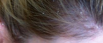
- Unlike seborrheic eczema, scalp psoriasis is accompanied by noticeable yellow flaking. Plaques can appear not only along the border of hair growth - the “psoriatic crown”, but also throughout the entire scalp.
- Unlike limited neurodermatitis, psoriasis has silvery scales, and lichenification (increased skin pattern) is absent.
- Examining the affected areas for fungi and bacteria will help distinguish psoriasis from fungal or bacterial skin lesions.
- Unlike pityriasis rosea, psoriasis does not have medallion-type elements with a pleated center and blisters along the periphery.
Properly prescribed and performed tests make it possible not only to confirm psoriasis, but also to identify the causes of its occurrence. Do not neglect laboratory tests: a complete clinical picture of the disease will help you quickly stop it.
We hope our article helped you understand what tests you should take for psoriasis. If you have already been diagnosed with this disease, during periods of exacerbations we recommend that you pay attention to Rederm ointment. This product with salicylic acid and betamethasone has an anti-inflammatory, antipruritic, anti-allergic and exfoliating effect, quickly soothes the skin and gives it a healthy appearance.
The skin I live in
Figure 1. Skin structure.
website bono-esse.ru
Skin is the largest human organ. Taking into account subcutaneous fat tissue, it amounts to up to 17% of body weight.
The skin consists of three main parts (Fig. 1).
- The epidermis is precisely that superficial part of the skin, accessible to our eyes, that peels off after a sunburn or forms calluses after mechanical stress. Thanks to the epidermis, the body's protective barrier is formed from pathogenic bacteria, fungi, viruses, temperature, ultraviolet radiation, water loss and mechanical damage. The epidermis is formed by several types of cells, but the most common type (up to 90% of the total) is keratinocytes . They are called so because they produce the structural protein keratin in huge quantities. The epidermis consists of five layers: basal, spinous, granular, shiny and horny (Fig. 2). Keratinocytes of the basal layer actively divide, displacing older cells to the surface of the skin. On the way to the surface, the cells differentiate - they flatten, become keratinized (produce huge amounts of keratin) and, gradually dying, lose their nuclei. At different stages of differentiation, keratinocytes look different under a light microscope; their appearance gives the name to the layers of the epidermis: basal - cells located on the basement membrane; spinous - cells with spine-like processes; granular - cells containing accumulations of proteins and fats in the form of grains in the cytoplasm; shiny - cells with shiny protein eleidin; horny - horny plates (nuclear-free, keratinized cells). melanocyte cells , which synthesize the pigment melanin, which is responsible for protecting the skin from UV rays, and immune cells (primarily dendritic cells), which are the first to encounter infectious and non-infectious agents that have penetrated the skin.
- The dermis is the deeper part of the skin, separated from the epidermis by a thin layer of basement membrane, formed by blood vessels and structural proteins (primarily collagen and elastin). The dermis consists of two layers: the papillary layer, rich in nourishing vessels, and the reticular layer, rich in proteins responsible for the strength and elasticity of the skin. The main and most numerous cells of this part of the skin are fibroblasts , which secrete collagen and elastin. Also present in the dermis are single melanocytes and some immune cells, primarily macrophages .
- Hypodermis is subcutaneous fatty tissue formed by adipocyte . Since fat is an excellent heat insulator and also an energy depot, the hypodermis performs thermoregulatory and energy functions. In addition, the fiber layer serves as additional protection for underlying tissues from damage.
Figure 2. Five layers of the epidermis.
website
Also in the skin there are sebaceous and sweat glands that open their ducts on its surface, hair follicles, nerve fibers and nerve endings.