Suppurating atheromas are a disease represented by the presence of a cystic formation on the surface of the skin.
In the absence of an infectious process, the cyst looks like a small round wen with clear edges.
Any mechanical injury can lead to damage to the epidermis and, accordingly, to the development of atheroma. In this case, the disease progresses and affects nearby tissues.
Festering atheromas can reach the size of a chicken egg, causing considerable discomfort to the owner.
Complications of the disease
Suppurating atheromas lead to various complications: the development of an inflammatory process, the formation of phlegmon or an abscess.
Inflammation occurs when an infection enters the cavity of the cyst - it increases in size, becomes hyperemic and painful.
Phlegmon is a disease in which the purulent contents of the atheroma do not come out, but remain under the skin.
An abscess occurs when the capsule ruptures. There are cases when the disease took on an oncological character.
Atheroma
22838 December 23
IMPORTANT!
The information in this section cannot be used for self-diagnosis and self-treatment.
In case of pain or other exacerbation of the disease, diagnostic tests should be prescribed only by the attending physician. To make a diagnosis and properly prescribe treatment, you should contact your doctor. Atheroma: causes, symptoms, diagnosis and treatment methods.
Definition
Atheroma is a cyst (pathological cavity) of the sebaceous gland, resulting from difficulty or complete cessation of the outflow of secretion (sebum) from the gland. The sebaceous glands are exocrine glands and are located on almost all parts of the skin, with the exception of the palms, soles and back of the feet. The secretion they produce is part of the water-lipid mantle of the skin. If the gland duct becomes clogged, then the secretion begins to accumulate in it, stretches it to form a cavity lined with the epidermis and containing secretion products of the sebaceous gland, cholesterol crystals, keratinized epidermal cells and detritus (decomposition products) - this is atheroma.
Atheromas occur in 5-10% of the population, mainly forming at the age of 20-30 years, with the same frequency in men and women.
Causes of atheroma
Atheroma occurs as a result of disruption of the sebaceous glands, which is manifested by increased production of sebum and blockage of the duct. A number of factors predispose to this: increased sweating, poor hygiene, narrow ducts of the sebaceous glands, individually determined high viscosity of sebum, chronic trauma to the skin, hormonal disorders, frostbite and burns. Hereditary factors also influence the development of atheroma.
The use of antiperspirants can contribute to blockage of the duct.
Classification of the disease
There are true and false cysts. True sebaceous cysts are a hereditary disease and are extremely rare. They develop as a result of a genetic defect that affects the formation of the gland. Typically, such cysts occur in newborns and are small in size. False cysts are actually atheromas that arise due to a violation of the outflow of sebaceous gland secretions.
Symptoms of atheroma
Atheromas most often occur in areas of the body where there are many sebaceous glands, for example, on the scalp, face, neck, and in the interscapular space.
Atheroma is a mobile formation of a round or slightly elongated shape, dense or elastic consistency, covered with unchanged skin. A characteristic sign of atheroma, which distinguishes it from other formations, is the presence of a point retraction of the skin (crater) in the area of the excretory duct of the gland and the adhesion of the skin to the cyst shell in the same place. Sometimes there is a hole in the middle of the atheroma through which its contents are released - cheesy masses with an unpleasant odor.
The size of atheroma can vary from a pea to a chicken egg and even more, reaching 10 cm. Atheroma always rises above the skin level, grows slowly, and is usually painless.
Diagnosis of atheroma
To make a diagnosis, the doctor performs a clinical examination; in some cases, an ultrasound examination may be required.
Diagnosis and treatment
At the first signs of suppurating atheroma, you must contact a surgeon who will conduct a visual examination.
At this stage, it is important to distinguish a cystic formation from a lipoma, hygroma and granuloma.
An ultrasound is performed to get a picture of the cyst cavity.
The treatment for atheroma is surgical removal. Laser and radio wave therapy are not performed in this case.
The operation on the inflamed area consists of two stages:
- opening the abscess, cleaning the cavity from pus and gland secretions;
- removal of the inflamed capsule with formed atheroma.
This manipulation is characterized by local anesthesia (lidocaine, ultracaine). At the final stage, the patient is given a cosmetic suture, which is removed 3-5 days after the operation.
The course of therapy includes antibiotics such as Sumamed, Doxycycline, Lincomycin.
Wen removal is performed on an outpatient basis.
The resulting material is subject to laboratory testing.
Laser removal of atheroma
The procedure takes place under local anesthesia. During removal, a laser beam is applied to the tumor and it evaporates. Laser removal is bloodless, since the light beam coagulates all vessels. During removal there is no contact between the instrument and the wound, which prevents infection of the wound. Removal time lasts approximately 5 minutes. After removal, treat the wound daily with a solution of potassium permanganate, once a day after a shower. The wound heals without complications, complete healing occurs on the 10th day. During the p/o period, you can take a shower immediately the next day after removal, but exclude visits to the swimming pool, baths, and saunas.
During laser removal of large atheromas, a defect in the form of a crater (minus tissue) remains at the site of removal, so doctors more often use the surgical method.
Recurrence of atheroma
During the operation, the atheroma is completely excised. Relapse can only occur if the sebaceous gland duct is poorly forgiven. The neoplasm must be removed completely, in some cases along with nearby tissues if the capsule has melted. The cause of relapse may also be the formation of a new cyst due to proximity to the postoperative scar. In some cases, relapse occurs due to incorrect diagnosis, when a lipoma or dermoid cyst is mistaken for atheroma. These tumors can also be treated surgically, but the operation is performed in a slightly different way. According to statistics, relapse of atheroma occurs in 15% of patients who have undergone surgery, and in 10% it is the result of opening an abscess cyst, when it is difficult to remove its contents due to purulent content. Such tumors must first be cured and removed after 2-3 weeks. It is recommended to remove atheromas in the winter season, when the cyst has just appeared and has not yet become inflamed. Relapse may also occur due to hyperhidrosis or a hereditary predisposition to obstruction of the sebaceous glands. In these cases, neoplasms appear not in the place where the operation was performed, but in the nearby excretory ducts of the gland.
Only radio wave removal of atheroma completely eliminates the risk of relapse. Before deciding on this procedure, try to find experienced surgeons to avoid possible unpleasant consequences due to an incorrectly performed operation. You can find out where to remove atheroma from reviews of people who have undergone this operation.
Danger of atheroma
This dermatological disease is a tumor-like benign formation that can be located intra- and subcutaneously. Atheromas can be congenital (as a result of impaired development of the epidermis) or appear secondary due to obstruction of the sebaceous gland duct. Atheroma looks like a dense, painless, round formation, somewhat mobile and can increase in size for a long time and cause significant discomfort; it can also become infected and suppurate. Cases of malignant transformation of these formations have been recorded, but they were observed extremely rarely.
Causes of atheromas
True atheromas are usually called congenital variants of benign tumor-like formations (steatomas), which are formed from the epithelium. Acquired forms (secondary) are called false atheromas - they appear as a result of blockage of the excretory ducts of the sebaceous gland with the subsequent accumulation of sebum and other components of the secretion and the formation of a retention cyst. The appearance of secondary atheromas is promoted by improper extrusion of the acne rod, when a small amount of it remains in the duct and blocks it. Risk factors for the appearance of secondary atheromas are also various hormonal disorders, abnormal structure of the ducts of the sebaceous glands, inflammatory changes in the skin, acne, rosacea, improper use of decorative cosmetics or neglect of personal hygiene rules
The clinical picture is limited to the appearance on the skin of a dense elastic formation of any size (usually from one to several cm) with a favorite localization in the face, neck or back. The formation gradually increases in size and can cause severe discomfort. Tumor-like formations can be single or multiple and are usually characterized by clear contours, mobility, and an elastic, dense consistency. The atheroma itself is represented by a connective tissue capsule that surrounds the contents (cholesterol, sebum, desquamated cells). On the surface of the formation, the opening of the excretory duct may be visible and the contents may periodically be released. When the atheroma becomes inflamed, the color of the skin changes, local soreness and swelling appear, which can be resolved by purulent melting of its contents and the pus breaking out. There is a high probability of recurrence of atheromas if they are not treated correctly. Complications of atheroma, in addition to secondary inflammatory changes, can be the spread of pus with the development of extensive abscesses and phlegmon, recurrence if the contents are not completely removed during treatment.
Atheromas: diagnosis and treatment
A dermatovenerologist and a surgeon are involved in the diagnosis and treatment of atheromas. Differential diagnosis must be carried out with such skin diseases as boils, benign tumors (lipomas, fibromas), syphilitic gummas, hygromas, dermoid cysts. For congenital true atheromas, which are usually located in the scalp, a differential diagnosis is made with cerebral hernias.
Diagnosing atheroma is not difficult. The main method of diagnosis is an external examination with the identification of a characteristic clinical picture: there is a retention cyst on the skin, which has all the properties listed above (dense structure, clear contours, mobility, etc.), but the reference sign is the identification of the opening of the excretory duct in the central part of the formation. In diagnostically difficult cases, it is possible to perform additional examination methods: material is taken for histological examination during surgical treatment.
Removal of atheroma
All atheromas are subject to surgical treatment, regardless of their size and location, because using a conservative method is quite difficult to completely clear the cyst and prevent its reappearance. Small formations, as well as uncomplicated atheromas, can be removed using diathermocoagulation; laser and radio wave therapy techniques are also used. In the presence of inflammatory phenomena, primary conservative anti-inflammatory treatment (ointments, physiotherapeutic techniques, use of antibacterial drugs) is indicated. After the operation, the sutures are removed after 1-2 weeks, depending on the speed of healing. Postoperative complications include secondary infection of the wound during surgery, the formation of a significant postoperative scar after removal of complicated atheromas. There is always a risk of malignant transformation, especially with atheromatosis (multiple formation of atheromas), so this disease should not be taken lightly.
Diagnosis of atheroma
Diagnosis of atheroma is not difficult. This is usually done through a routine skin examination. Atheroma does not pose a danger to life. If there is no suppuration, then you can limit yourself to ordinary observation without undertaking any treatment. There is a high probability that the atheroma will open on its own. But if it has reached a very large size and begins to ache, then it is better to remove it. If the contents of the atheroma break out on their own, the inflammation may subside. Further prospects: atheroma may disappear completely, or it may continue to fester periodically. Despite the fact that atheromas are benign tumors, if you discover a neoplasm, it is best to consult a doctor. Let the doctor tell you what kind of tumor it is and whether treatment is required.
Proper care
If the operation was performed using a scalpel, the surgeon will apply stitches. When using the laser or radio wave method, no stitches are applied. Stitches will be removed approximately a week after the procedure, and sometimes sooner.
If the removal of atheroma involved a serious surgical intervention, for example, a procedure was carried out for enucleation of a giant cyst located on the head under the hairline, bandages will be required.
After removing a large purulent cyst or atheroma, the wound is treated with spruce to prevent inflammation of the scar.
For two weeks after surgery (this is how long the healing process lasts), you must follow the doctor’s recommendations:
- Do not wet the removal area for at least two days.
- If indicated, use anti-inflammatory absorbable agents topically.
- If necessary, treat the wound daily with an antiseptic.
- Wear a bandage or a clean hat (if the operation was performed on the scalp). This will prevent infection from entering the wound.
The speed of wound healing and the likelihood of a scar depends not only on the methods used by the surgeon, but also on the characteristics of the patient’s skin.
Causes of atheroma on the head and methods of treatment.
The occurrence of atheroma on the head depends on external and internal factors. In the first case, these are difficult living and working conditions, poor ecology, head trauma, in the second - hyperhidrosis, hormonal imbalance and impaired metabolism. Most experts recommend removing atheroma on the head without waiting for it to grow to a large size or for an inflammatory process to appear, since in this case the treatment is aggravated and becomes more protracted. Removal of atheroma on the head occurs surgically under local or general anesthesia. The essence of the operation is to carefully remove the atheroma without damaging its capsule. If the removal of atheroma occurs with damage to the capsule or with its incomplete removal, then re-formation of atheroma or suppuration of the wound is possible at this site.
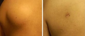
Rice. 3. Removal of atheroma on the head should be carried out only with a capsule.
Surgical intervention on atheromas of the head can be carried out in three ways:
- Laser. This method does not require hair removal, as is usually done. But this is not the only advantage, there are a number of others: relapse is excluded, no scars are left, and rapid recovery is noted.
- Traditionally. In the traditional method of removing atheroma on the head, a small puncture is made in the atheroma, which allows the contents of the cyst and its membrane to be removed. This method is suitable for atheromas of any size. The operation time is approximately 10 minutes.
- Radio wave technology. This method is used at the initial stage of atheroma formation and, as a rule, eliminates the appearance of new cysts. The wound heals in a few days.
You can avoid the reappearance of atheroma on the head by adhering to preventive measures: a balanced diet and personal hygiene. And a timely visit to the doctor is the key not only to proper treatment of atheroma, but also to a quick recovery with minimal consequences for health.
The main types of treatment for atheroma without surgery
Atheroma is a benign tumor formed as a result of blockage of the sebaceous glands.
Atheroma forms mainly on the neck, face, groin area and back. The neoplasm is considered relatively safe, since it has no tendency to degenerate into cancer. Below we will tell you whether it is possible to remove atheroma of the scalp with a laser? and “is it possible for the tumor to re-form, as well as what earlobe atheroma and other forms of the disease look like. Causes of the disease:
- improper skin care;
- injuries, skin cuts;
- excessive use of deodorants;
- hormonal disorders;
- hypersweating (hyperhidrosis);
- pimples, blackheads;
- inflammation of the epidermis;
- metabolic disease.
The main method of treating pathology is surgery. Below we will talk about the types of treatment for atheroma without surgery.
Surgical removal of lipomas, atheromas, hygromas and skin fibromas
Lipomas, atheromas, skin fibromas and hygromas are benign formations of the skin and subcutaneous tissue. Having appeared once, these tumor-like formations do not disappear spontaneously and are not amenable to “traditional methods” of treatment.
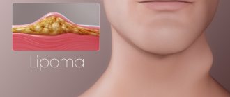
Lipoma
Lipoma
Lipoma (fat) is the most common benign neoplasm of subcutaneous and intermuscular tissue. Lipomas can appear in any area where there is a layer of fat. In the subcutaneous tissue, lipomas are most often observed on the hips and shoulders in the form of soft, jelly-like, clearly defined, painless formations of various sizes. Also, there is a disease - lipomatosis, which is manifested by multiple wen on the body, often located symmetrically.
Subcutaneous lipomas are mobile relative to the surface of the skin; lipomas of intermuscular tissue are located more deeply. Lipomas slowly increase in size; As they grow, lipomas begin to cause cosmetic (and sometimes physical) discomfort. When an infection occurs and attempts are made to get rid of the wen on your own, the lipoma suppurates.
Atheroma
Atheroma (sebaceous gland cyst) is a benign tumor-like formation of a round shape and soft consistency, enclosed in a capsule and resulting from blockage of the sebaceous gland duct. Atheroma is painless and can appear on any part of the body where hair grows, but more often it occurs on the scalp, face, back, and genital area. Once formed, atheroma may not grow for a long time, not bother you, and remain on the skin for many years; however, when an infection occurs, a subcutaneous abscess develops, accompanied by redness, pain, swelling and fever. If the atheroma has reached a large size, then it causes inconvenience and there is a high risk of infection; it must be removed.
Removal of atheroma in our clinic can be performed surgically. Surgical removal of atheroma is performed under local anesthesia and takes about 15-20 minutes. Removal of atheroma must be carried out together with the capsule in which it is enclosed, otherwise the disease develops again.
Skin fibroma
Skin fibroma is a common benign neoplasm that can be localized on any part of the skin or mucous membranes, but most often appears on the head and torso. Fibroma is a dense, painless formation, subcutaneous or intradermal, consisting of fibrous connective tissue. Skin fibroids may not increase in size for a long time and not cause any discomfort. However, if the fibroid enlarges, pain, physical discomfort or injury to the fibroid occurs, it is recommended to immediately consult a surgeon. You can get rid of skin fibroids using surgical excision. Surgical removal of fibroids is performed on an outpatient basis under local anesthesia and takes about 10-20 minutes.
Hygroma
Hygroma
Hygroma is a benign cyst-shaped formation that originates from the joint capsule or tendon sheath (tendon ganglion). At its base, the hygroma is tightly connected to the joint capsule or tendon sheath. The skin and subcutaneous fat, while remaining mobile, stretch in proportion to the growth of the hygroma. The wall of the hygroma is a dense connective tissue membrane. The contents of the hygroma are most often a jelly-like serous fluid of a light or yellowish color. Externally, the hygroma looks like a rounded formation of soft elastic consistency, 1 to 5 cm in diameter, slightly painful and limited in displacement along with the tendon. Most often, the hygroma is located in the area of the hands (the palmar surface of the fingers, palm, dorsum of the hand, wrist); popliteal fossa (Becker's cyst). A predisposing factor in the appearance of hygroma is chronic tissue trauma associated with professional activities.
When examining a patient, the surgeon distinguishes hygroma from other tumor-like formations, such as lipoma, fibroma, atheroma. Indications for removal of hygroma are: a cosmetic defect that affects the patient’s quality of life, rapid growth of the cyst, pain in the area of formation, inflammation and suppuration of this area. Among the methods of getting rid of hygroma, some doctors still use an outdated method - “crushing” the hygroma, that is, by mechanical pressure or impact, the capsule of the hygroma is destroyed and its contents spread throughout the subcutaneous tissue. The main disadvantages of this “treatment” include pain, frequent relapses; Inflammation and suppuration in the area of capsule rupture are common. To successfully get rid of hygroma, there is only a surgical method for its complete removal. Surgical removal of hygroma most often takes place under local anesthesia. The patient leaves the clinic on the same day. The next day, the first dressing is performed and the general condition of the patient is assessed. On days 6-7 after removal of the hygroma, the skin sutures are removed. Physiotherapeutic methods of influence are good only at the stage of rehabilitation of the patient after removal of the hygroma.
Indications for removal of lipoma, atheroma, fibroma, hygroma:
- cosmetic defect
- large size and fast growth
- physical discomfort
- location of the tumor near nerve branches and large blood vessels
- development of complications: inflammation, pain, swelling, redness, suppuration
Removal of lipomas, hygromas, atheromas and skin fibromas:
To diagnose lipomas, atheromas, hygromas and fibromas, an in-person examination and palpation of the tumor-like formation is usually sufficient.
The only possible method for removing lipomas (fat), hygromas, atheromas and fibromas is surgical. You can get rid of the tumor by surgical excision with the application of an invisible intradermal cosmetic suture. The operation is performed on an outpatient basis without hospitalization and takes 15-20 minutes. Under local anesthesia, the smallest possible incision is made at the site of formation with a microsurgical scalpel. The length of the incision during removal depends on the location, size and type of subcutaneous formation.
After opening a lipoma, atheroma, hygroma or fibroma, their contents, if necessary, are sent for histological examination. In the vast majority of cases, lipomas, atheromas, fibromas and hygromas are benign in nature. However, it is impossible to completely eliminate the risk without additional research.
An infected and festering lipoma, atheroma or fibroma requires more extensive surgical intervention, so in no case do we recommend self-medication. If suppuration occurs due to improper treatment, it is necessary to urgently consult a surgeon.
Surgical removal of atheroma
The procedure also takes place under local anesthesia, most often Lidocaine, after which the patient does not feel anything, after which a careful incision is made with a surgical scalpel, the size of the incision depends on the size of the formation. Afterwards, all contents with the capsule are removed and sent for histological examination. Next, the wound is inspected for the remains of the capsule, after which cosmetic sutures are applied to the wound. Then the ass is applied. bandage with Betadine solution and Levomikol ointment. In the postoperative period, the patient takes prophylactic antibiotics, daily dressings of the wound with Betadine solution. The PO wound heals by primary intention, the PO sutures are removed on the 7th – 10th day. In most cases, no defects, scars, or scars remain.
In case of festering atheroma, an incision is also made with a scalpel, the purulent contents, along with sections of the capsule walls, are removed. A complete inspection of the wound is carried out, the wound is washed with aseptic solutions. No sutures are applied; drainage is inserted into the wound. After Ass. bandage with Levomikol ointment. The PO wound heals by secondary intention in about 10 days.
Wound care after removal of atheroma
In both cases, in the postoperative period, it is necessary to exclude active physical activity, visits to the shower, swimming pool, baths, saunas.
Contraindications for removal of atheromas are pregnancy, lactation, and should not be removed during menstruation, during a cold, flu, or during an exacerbation of herpes.
Symptoms and diagnosis of atheroma on the head.
It is not difficult to identify atheroma on the head, however, during examination you need to be extremely careful to exclude a cerebral hernia. If the doctor has doubts, then a craniography (head x-ray) is prescribed, which will clearly confirm the diagnosis. The following signs are characteristic of an inflamed atheroma on the head:
- pain on palpation;
- release of liquid with a specific odor;
- a reddened, inflamed area of the scalp around the tumor;
- intensive growth of atheroma;
- elevated body temperature.
Rice. 2. Atheroma of the scalp, scalp.