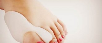
Why do growths form on the toes?
The definition of “growth” is not a specialized term, so this concept hides a variety of diseases and the causes of their occurrence.
Inflammatory formations can affect the feet and phalanges of the fingers in the following ways:
- Insufficient disinfection in baths, swimming pools, and public showers causes growths of the viral type.
- Microcracks and wounds on the legs and arms through which bacteria, for example, the human papillomavirus, enter.
- Neglect of body hygiene standards leads to the breeding of bacteria and increases the chance of infection.
Damage to bone and cartilage tissue occurs due to the following factors:
- Small shoes and too high a heel lead to deformation of the metatarsal bone.
- Flat feet contribute to the development of bunions.
- Obesity or later pregnancy increases the stress on the legs and feet.
- With age, both the immune system and the human skeleton weaken, which gives a higher percentage of neoplasms.
- Sometimes the problem of growths in a person is hereditary.
Flat feet
Reasons for education
Interdigital callus can appear in men and women of any age. The skin cannot withstand prolonged friction or compression and uses its own protective functions, actively growing the stratum corneum of the epidermis at the site of increased load.
The most common causes of calluses between the fingers:
- Wearing uncomfortable, tight shoes;
- Prolonged walking in high heels;
- Flat feet;
- Deformation of fingers;
- Overweight;
- Joint diseases (arthrosis, arthritis, gout);
- Swelling of the lower extremities.
Diagnosis of growths and which doctor to contact?
Before prescribing treatment for lumps and growths, it is necessary to find out the patient’s symptoms.
First of all, anamnesis is used - a patient interview conducted by an orthopedist or surgeon. If the problem does not lie in bone pathology, treatment is carried out by a dermatologist. During the conversation, it is important to find out complaints, possible predispositions, causes, and also how long the patient has been worried about it.
For mild forms of bacterial infections and the initial stage of bone growths, treatment is prescribed by a doctor without additional diagnostics.
But for a full-fledged study, the patient may be prescribed instrumental studies:
- X-ray stop. Three types of projection allow us to examine existing deviations and deformations of the metatarsal bones, toes, as well as pathologies in the bone processes.
- Computer plantography. A specific photo of a footprint. Allows the attending physician to identify pronounced pathologies and improper distribution of the load on the legs.
- Computer podometry. Gait studies on technical devices reveal the first stages of growths on the toes and feet.
- Biomechanical studies. Fixation of pathology expressed by reduced bio-activity of the interosseous muscles of the foot and toes, which manifests itself even in the first stages of the disease.
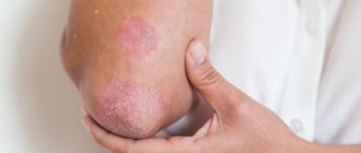
Arthrosis
Arthrosis of the big toe develops more often in women. The reason for this is long-term wearing of shoes with a narrow toe, while the big toe deviates strongly inward and is pressed against the second toe. The protruding bone receives additional trauma (rubbed by shoes) while walking and gradually becomes deformed. Later, not only the protruding bone, but also the entire joint is deformed. It becomes much wider than it was before. Movement in such a joint is sharply limited. With advanced arthrosis, the deformity usually fixes the finger in the wrong position so strongly that it is almost impossible to return it to its normal position.
Types of growths on the toes
In pathologies of the feet and toes, there are the following types of bumps, both caused by bacteria and bone deformation.
Bacterial growths and skin growths due to infection:
- Warts: common, periungual and plantar.
- Hard and soft fibroids.
- Corns.
- Nevus.
- Seborrheic and actinic keratoses.
Skeletal deformity:
- Valgus foot.
- Interdigital erosion.
- Tofus.
- Hygroma.
- Sarcomas are malignant tumors.
They all differ in appearance, method of formation, painful and tactile sensations and the severity of the pathology.
Growth on the big or middle toe
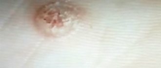
Dermatofibroma:
- A fairly common occurrence that looks like a hard pink or reddish bump. It is distinguished by a pit that appears when hardened skin is collected.
- When the field of formation becomes too wide , a tubercle appears in the middle of the growth. If not treated in a timely manner, there is a risk of developing sarcoma.
- Such a bump appears for various reasons: splinters, insect bites, stagnant pustules.
Dermatofibroma on the leg
Nevus:
- In appearance it resembles a soft brown bump , sometimes flesh-colored.
- Papillomas may appear on the surface ; less commonly, a complex nevus is distinguished by a rim around the growth.
- The reason is an excess of pigment in the skin. This often appears from prolonged and frequent exposure to the sun.
- There is a predisposition to nevus , sometimes it appears immediately after birth.
Valgus deformity:
- Expressed by a protruding bone at the base of the big toe. Here the shape of the joint changes, although in the first stages only calluses appear.
- It is detected as a result of women constantly wearing shoes with high heels or shoes with completely flat soles.
- Both cases lead to flat feet, and, as a result, hallux valgus deformity may appear.
Hallux valgus deformity of the big toe joint
A growth or callus between the toes
Interdigital erosion. It appears and multiplies between the fingers, since it is easiest for fungal infection to remain there.
Inflammations and ulcers appear at the sites of erosion, which heal and turn into a compacted callus.
The more complex and longer the fungal disease lasts, the more often and more unwanted hard calluses appear. Appears most often in people with diabetes and excess weight.
Callus and warts on the toe
Types of warts or calluses:
- Plantar warts. The growths look like compacted flat yellow calluses between the toes and on the pads of the fingers. When small in size they are called spines. When touched strongly, they hurt, which makes it difficult to walk normally. It differs from corns by the asymmetry of its location on both feet.
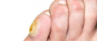
- Periungual warts. This type of growth differs in color (darker colors, from yellow to brown) and structure (more uneven). They are dangerous because they can disrupt the proper growth of nails.
- Calluses appear most often on the bend of the ring finger, middle finger, and also on the side of the little finger. A callus is a protection of the skin from constant friction.
Periungual warts on the foot
Calluses
Plantar warts on the foot
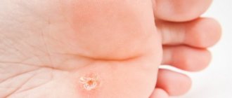
Corns
Dry calluses that feel like calluses are called corns. The cause of their occurrence is compaction of the epidermis as a result of constant rubbing and pressure. This reduces blood circulation in the legs and leads to dead skin.
The most common reason is uncomfortable or small shoes. The most common place for corns to appear is the little toe and big toe; less commonly, they occur between the toes due to flat feet.
Corns look different, the type depends on the stage of formation:
- In the first stage , a small bump appears and the skin becomes significantly redder.
- After about a week, the color changes from red to yellow-gray. To the touch, the compacted area is smooth, less often slightly ribbed.
- The last stage of corns development is manifested by cracks of varying depths.
Corns on feet
Fibroma or soft growth
A rather rare type of disease, it has the peculiarity of having a narrow base and a wide, unstable cap. This appearance evokes associations with a mushroom. Under the mushroom cap there is a large layer of fatty deposits. The lump itself is skin-colored, sometimes slightly pinkish.
Causes:
- Load on the feet due to excess weight;
- Excessive friction in shoes;
- Fungal infection.
The divided growth is called a skin polyp.
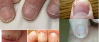
Growth on the nail and under the toenails
New growths near the nails and under them are hard, roughened, can have a round and elongated shape, and dark spots can be seen under the nail. An unnatural volume can be given to the nail by a splinter that gets inside, sometimes due to foot injuries where the toes are injured.
The cause may be:
- uncomfortable narrow shoes;
- elderly age;
- overweight.
Hygroma (ganglion) on the toe
This type of formation is distinguished by its almost invisible presence on the skin: the base, filled with liquid, is located inside the leg between the tendons. The consistency of the growth is soft and elastic. On the surface of the leg you can only notice redness and irritation of the epidermis.
A progressive ganglion leads to discomfort when walking and playing sports, aching and dull pain in the legs.
Hygroma on the toe
Tophi - a consequence of the “disease of kings”
Growths in this category occur due to gout. The base of the big toe becomes overgrown with uric acid crystals. This is also the first sign of gout. The more tophi develop, the more painful it is to touch the affected area, including moving in general.
The skin becomes very sensitive and turns red. The tophi growths themselves frame the thumb and can be either small (the size of a bead) or so large that some can create microcracks in the skin.
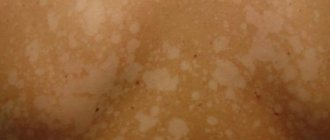
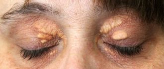
Keratoses or keratomas of different types
Keratoses on the toes are as follows:
- Seborrheic keratosis. Externally, the legs are distinguished by lumpy growths and plaques of brown and yellow shades. Most often found in the southern regions, its appearance is due to solar radiation and genetic predisposition. Usually appears on the foot, much less often on the toes and between them.
- Actinic keratosis. A particularly dangerous type of growth that can develop into skin cancer if complications arise. They appear on the skin with redness in the form of rough yellowish crusts. It is also formed due to excess exposure to direct sunlight.
Actinic keratosis
Seborrheic keratosis
Malignant formations
Just like actinic keratosis, bone sarcomas are also dangerous - malignant tumors that can return even after treatment and removal.
They are distinguished by the formation of a fairly hard lump on the leg with dilated blood vessels and redness of the skin at the site of the growth. Sometimes the epidermis does not change. The tumor grows exponentially and often forms pathological processes in the nearest lymph nodes.
IMPORTANT! Growths such as osteomas and chondromas (benign bone and cartilage formations) do not trigger malignant processes.
Classification
The blister that appears between the fingers is usually quite painful and looks like a translucent bubble filled with liquid.
Doctors know the following types of calluses:
If a watery blister appears on the little finger, this indicates that friction is occurring between it and the adjacent finger. It is necessary to get rid of it, because otherwise it will turn into a dry form. These formations are characterized by a very dense structure. Pain usually does not appear.
The callus is viral in nature and has a characteristic round shape with a small black dot in the middle. It is impossible to eliminate it at home. In a beauty salon, special equipment is used for this purpose to help get rid of the root.
Growths on a child's toe
In children, spines or warts are most often diagnosed - penetration of the human papillomavirus under the skin.
There are many causes of infections in children:
- Newborns pick up the infection during childbirth from the mother's genitals.
- Young children often run barefoot and neglect hygiene rules.
- The child often uses other people's personal belongings.
- A weak immune system allows the virus to penetrate.
The growths look like bumps with rough skin, sometimes similar to rough spots on the legs.
The main types of warts in children:
- vulgar (simple, ordinary);
- flat (adolescent, juvenile);
- plantar;
- periungual;
- molluscum contagiosum (variola virus group).
With timely treatment, the growths do not become dangerous or malignant. The bacteria themselves are killed in large numbers when using pharmaceutical products.
The specificity of the treatment also lies in the fact that often the child’s immunity copes with the growths on its own within 6-12 months. It is worth contacting a dermatologist if the child is confused by warts, but they themselves hurt and grow quickly.
Treatment of growths in children
There are several ways to remove warts and bumps:
- Over-the-counter salicylic acid-based products : ointments or alcohol. Apply every day to a wart that has been steamed and cleared of keratinized skin; the results are noticeable after a month, sometimes earlier.
- Freezing with liquid nitrogen. For use only on children over 4 years of age who are able to sit still for a certain period of time. Often one procedure is enough, but if the damage radius is large, they resort to repeated freezing.
- Electrosurgery. The lump, along with the subcutaneous infection, is burned off with electric current; it is widely used to remove simple and plantar warts. Warn your child that during the procedure there will be an unpleasant smell of burnt skin - there is nothing to worry about.
- Laser. Laser removal of warts occurs when other methods have failed. After the procedure, it is necessary to increase the child’s immunity using medications with interferon.
Pharmacy products
If a callus appears on your foot, you can try to eliminate it with medications. The choice of products for removing blisters is huge: patches, creams, special liquids - choose the form of the drug that is convenient for you and use it. Be sure to read the instructions and contraindications before doing this.
If a new wet interdigital callus has arisen, it is recommended to seal it with a special bactericidal plaster. It is applied to clean, dry skin, protects delicate tissues from further chafing and prevents the entry of pathogenic infections.
Special stickers for dry calluses are impregnated with a special composition that has an exfoliating effect and stimulates the division of new cells, due to which new smooth tissue appears on the affected area. Such patches are applied to steamed, clean and dry feet and left for 24-48 hours. Treatment of calluses between the toes is carried out until the problem is completely eliminated. The use of stickers is prohibited if the integrity of the tissue on the treated area of the foot is damaged.
Therapeutic plasters for various types of calluses from the Compeed brand are very convenient. They have an anatomical shape, do not peel off from the damaged area and act effectively on the sore spot. The Russian callus patch Salipod contains salicylic acid, which effectively eliminates dry corns.
Gels and ointments help cure interdigital calluses. They contain salicylic or benzoic acids, which soften dead tissue. They are applied pointwise, only on the sore spot in a thick layer, then sealed with adhesive tape. It is important to avoid contact of drugs with healthy skin so as not to cause dryness or damage soft tissues. After removing the compress, the damaged area must be treated with a file or pumice. Cutting off the corns is not recommended; it is not only painful, but can also lead to an inflammatory process.
How to get rid of growths on your toes?
Each type of formation requires its own treatment.
Warts and calluses:
- If there is bloody discharge from callous formations and severe pain, you should consult a dermatologist and oncologist to rule out skin cancer.
- If the source of infection is too deep, laser removal or other cryodestruction should be used.
- If the warts are periungual , cutting off the growths yourself is prohibited: this can permanently change the correct growth of the nail.
- Simple visible warts can be removed independently using pharmaceutical products.
- Hard and soft fibroids. Dermatofibromas are quite hard, making home treatment difficult. Most often, such growths are removed surgically. Soft fibroids are removed more easily, but also with the help of medical intervention, for example, with a laser.
- Corns. With such growths, first of all, they get rid of the cause of their appearance: excess weight or uncomfortable shoes. The corns themselves, depending on their complexity, can be removed using special patches or surgically.
Malignant growths on the legs are a dangerous type of growth; such growths can only be treated as prescribed by an oncologist after consultation and tests.
Treatment of other types of growths:
- Nevus. Treatment is prescribed by the treating dermatologist or oncologist, taking into account the strength of development. Treatment at home is dangerous due to possible malignant formations.
- Ganglion and hygroma. It can also be removed surgically using a simple operation.
- Interdigital erosion. You can eliminate the development with the help of ointments with an antifungal effect, special powder for shoes, as well as by adjusting your diet and losing weight.
- Keratosis. Thanks to superficial formations, seborrheic keratosis is removed using a laser. Actinic is more dangerous and can be removed by cryodestruction.
- A bone on the finger. At the first stages, the orthopedist will prescribe correction using special shoes, but neglected hallux valgus can only be removed through an expensive, complex operation.
- Tophi. First of all, gout is treated, and a decrease in uric acid in the body helps eliminate tophi.
Malignant growths on the legs are a dangerous type of growth; such growths can only be treated as prescribed by an oncologist after consultation and tests.
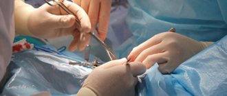
Surgical intervention
Treatment with surgery is prescribed for flat feet of 2 and 3 degrees in the absence of results of treatment by other methods.
Operations to eliminate cones are divided into:
- penetration into soft tissues;
- surgery on bone spurs;
- a combination of the first two types.
Most often, operations are performed on the bones, which either remove the lump or significantly reduce its size. Combined methods involving the removal of both bone growths and changes in the ligamentous apparatus are also common.
The operations themselves are performed under general anesthesia, with rare exceptions. the patient recovers within a week; in rare cases, rehabilitation lasts up to two months. After surgery, you must follow your doctor's recommendations and wear orthopedic bandages for a year.
Treatment with traditional methods
There are traditional medicine methods to cure growths based on the use of natural remedies:
- Baths. Soak your feet daily in a bath of hot water combined with a tablespoon of salt and 10 drops of iodine solution. Such baths will help remove and remove unwanted colonies of microorganisms and fungi on the feet.
- Apple vinegar. Wipe the growth with undiluted liquid 2 times a day.
- Celandine. Freshly squeezed celandine juice is applied to a bump or wart 2 or 3 times a day, which allows you to get rid of warts.
- Vegetable oil. Oil will help heal the tumor site. Wipe the area with dampened cotton wool until complete recovery.
- Butter and watercress. A paste of watercress juice and butter is applied to the growth several times a day.
Salt foot baths