Atheroma (sebaceous gland cyst) is a benign neoplasm located immediately subcutaneously, resulting from blockage of the sebaceous gland duct. Atheroma occurs on any areas of the skin of the body where hair grows, but the favorite place is the face, scalp, back, neck, and groin area. Atheroma has a dense capsule of connective tissue, with secretion components.
Visually, it looks like a subcutaneous ball, ranging in size from several millimeters to tens of centimeters. On palpation, this formation is round in shape, soft, elastic, freely moving under the fingers, there is no pain.
When atheroma suppurates, most often as a result of injury, the symptoms change: the formation increases many times in size, swelling and hyperemia increases around it. On palpation, the formation is denser, painful, and there is hyperthermia around it. When the process is neglected, the body temperature rises and weakness sets in. Into the flesh until an abscess forms.
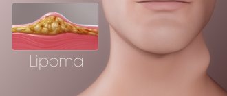
To diagnose atheroma, in addition to visual examination, ultrasound diagnostics are often used to differentiate from lipoma. On ultrasound, atheroma looks like a hypoechoic neoplasm of soft tissue, located immediately subcutaneously, at a depth of 1–10 mm. Round in shape, with smooth and clear contours, the capsule can be traced throughout. The formation is most often homogeneous, without inclusions, with distal pseudo-enhancement. With CDK there is no blood flow.
What is atheroma?
Atheroma is a cystic formation under the epidermis (upper layer of skin) filled with fat and keratin. It is formed in the ducts of the sebaceous glands and looks like a lipoma - both of these ailments are colloquially called wen.
Atheroma is quite rare - only 5% of the population, mainly in women. “Favorite” localization of formation is the place where the sebaceous glands are concentrated:
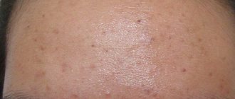
- face - lips, chin, cheeks, forehead;
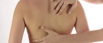
- back;
- area near the ears;
- hair growth area;
- genitals;
- armpits.
Atheroma can be recognized by the shape of an oval or a regular circle with a diameter of 0.25 to 7 cm. It occurs individually or in large quantities. Atheroma appears for a number of reasons:
- hormonal imbalance during puberty, pregnancy or menopause;
- disorders of the liver and kidneys, central nervous system diseases, sedentary lifestyle;
- dysfunction of the thyroid gland, Moon's disease, in general - metabolic failure;
- hypothermia, a course of hormonal drugs - the functioning of the adrenal glands is disrupted;
- atopic or seborrheic dermatitis;
- poor skin care with high oil content and excess ultraviolet radiation;
- overdose of medications, resulting in malfunction of the sebaceous glands;
- lack of elastin in the skin and excess calcium;
- congenital pathology.
Atheroma has two stages: cold and progressive.
Cold atheroma is safe and can remain in this state for a lifetime. Unchangeable in density and size.
The progressive stage is dangerous due to complications that can lead to death. It quickly thickens and increases in diameter. If you do not treat it, the following consequences are possible:
- the formation can break into the blood, thereby causing sepsis with a 75% probability of death;
- when compacted, the tumor can develop into a malignant one;
- if atheroma appears near the nerve endings - partial numbness of the facial muscles;
- the occurrence of necrosis around the atheroma, if it grows quickly, then there is a violation of the blood supply and, as a result, blood poisoning.
The cold stage can turn into a progressive stage at any moment, so you should not hope for a stable state. It is better to start treatment as soon as atheroma is diagnosed. This will allow you to get rid of it faster than in the case of an advanced formation, and avoid many dangerous complications.
Methods for removing atheroma
You can get rid of atheroma using laser, radio waves and surgical excision.
Laser techniques make it possible to eliminate growths in the cold and progressive stages of development. The procedure is performed under local anesthesia in several options:
- photocoagulation;
It is used if the atheroma is no more than 5 mm in diameter. It is burned with a laser beam, damaging the shell. After the procedure, a crust forms and no stitches are required. After a few weeks, the scab falls off on its own, revealing a healthy layer of skin.
- laser evaporation;
- laser excision.
Prescribed for the removal of cysts up to 20 mm in diameter. A small incision is made, damaging the shell, and the contents are squeezed out with sterile napkins. The remains of the atheroma are evaporated with a laser. After completion of the procedure, drainage is installed and stitches are applied. They are removed after 8–12 days.
Prescribed for formations ranging in size from 5 to 20 mm. An incision is made next to the atheroma, and a scalpel is used to move it upward to see where the capsule ends and healthy tissue begins. The capsule is cut out with a laser and removed. A drainage tube is placed at the operated site and stitches are applied. They are removed after 7–12 days.
Laser techniques are very effective, but have a number of contraindications:
- purulent nature of atheroma on the face;
- nerve receptors are affected;
- dry or sensitive skin.
Radio waves remove small cysts without inflammation. For the operation, as with laser methods, local anesthesia and special equipment are used. Suitable for dehydrated, dry and fair skin, but this method is not suitable for purulent inflammation.
The radio wave is directed to the top of the atheroma and burns it out, leaving a small depression at the site of formation. It is treated with an antiseptic and bandaged; stitches are not required in this case. Rehabilitation occurs in 3–5 days, and the reappearance of the growth is excluded.
Surgical excision is a traditional method that involves several options:
- with excision of the atheroma capsule;
- with preservation of the atheroma capsule.
Used for large formations. The shell is opened, the liquid is squeezed out or scooped out with a special tool, the capsule shell is removed, and the wound is sutured. If the incision was small, use medical glue.
The skin is incised near the formation, after which the entire capsule is removed through the incision. Next, the wound is sutured.
Surgical excision is prescribed if:
- atheroma more than 3 cm in diameter;
- there is a high probability of an abscess breaking through;
- degeneration into a malignant formation has begun (the nearest skin within a radius of 2 cm is also removed);
- Necrosis of the surrounding tissue develops.
Rehabilitation after surgery takes 3 weeks, and a scar always remains.
How is the operation performed?
It is carried out as follows:
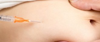
- Local anesthesia is administered.
- An incision is made on the surface of the atheroma (if there is a hole, then two).
- The cavity of the capsule is cleaned (in the case of suppuration, the surgeon must clean it more thoroughly).
- The capsule is excised.
- Stitches are applied along with wound treatment.
The suture after removing the atheroma must be removed after 7 days, for which you need to visit a specialist. Within 2-3 days it is necessary to treat the postoperative area with disinfectants. The doctor may also prescribe antibiotics.
On the ears
The procedure for removing atheroma on the ears is carried out in a medical clinic. Self-medication is strictly prohibited as it can cause complications. 3 methods are used: surgery, laser therapy and radio wave method. The most effective method for surgical removal of this type of atheroma is surgery to clean and remove the cyst. If the size of the tumor is small, laser therapy is used. All manipulations are performed under local anesthesia. For the operation, you need to go to the hospital, but for the radio wave and laser methods you don’t need to do this - sometimes, if you have the necessary tests on hand, they can be done right on the day of treatment.
In some particularly advanced cases (if the cyst becomes inflamed or suppuration occurs), you must first visit an otolaryngologist to prevent inflammation of the adjacent tissues. Removal of ear atheroma occurs as follows: the doctor opens the tumor along with its contents, after which he prescribes a course of antibiotics. Only after this can surgery be performed to remove the tumor. If the patient decides not to have surgery, the cyst will most likely reappear.
On the head
In most cases, when a cyst forms on the head, we can talk about multiple atheromas (70%). After the necessary examinations to remove the tumors, the doctor may prescribe surgery, laser therapy or surgery using a radio wave scalpel. All these methods have already proven to be effective and disease-free, and are performed under local anesthesia.
As a rule, surgical intervention is used to eliminate atheromas on the head. Despite its effectiveness, it has the longest healing period compared to other methods. After the operation, a histological examination is performed to identify the type of tumor. This procedure is not performed after the laser and radio wave method, however, atheroma of the head after removal using the above methods has a low chance of relapse.
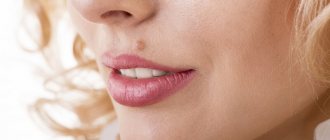
On the face
Atheromas can also form on the face - and, as a rule, these are small cysts, since if the tumor covers the face, patients seek help faster for the sake of a cosmetic effect. For this reason, in most cases, laser correction and radio wave treatment are performed to remove atheromas on the face. They help avoid recurrence of infection, are painless, and do not leave scars. In medicine, such manipulations are carried out by highly qualified specialists with sufficient experience.
On the back
Compared to other areas where atheromas appear, here they form in 45% of cases. The reason is a large accumulation of sebaceous glands and possible problems with thermoregulation.
If the cyst is small, then it may not cause any discomfort, and in this case, specialists resort to eliminating it using a laser or radio wave scalpel. If the tumor has grown to a large size, only surgery will help. It is better not to reach this state by contacting a doctor in a timely manner to remove atheroma on the back.
On the foot
As for the cyst on the leg, this location is the least common. Laser correction or radio wave method is used if the tumors are small, and surgical intervention if the atheroma has grown. You should not delay seeking medical help if atheroma appears on your leg.
Possible complications
Possible complications after removal of atheroma:
- bleeding - after surgery, laser methods and radio waves weld blood vessels together after the procedure;
- infection - due to improper care, most often - behind an unhealed suture;
- burn - occurs when a doctor makes a mistake in laser and radio wave methods;
- loose seams;
- relapse - reappearance of atheroma if the membrane was not completely removed or the conditions under which the sebaceous glands were clogged persisted;
- the formation of a rough suture is associated either with insufficient experience of the surgeon, or with the characteristics of the patient’s body.
Causes of the disease
There are many reasons for the formation of atheroma. There are two groups of etiological factors:
- The first includes congenital causes of the development of formation (anomalies in the structure and function of the sebaceous glands, genetic diseases). Such formations are also called epidermoids; they are usually multiple.
- The second group of causes includes acquired diseases and conditions that lead to the formation of atheromas. Such atheromas are called retention cysts, which arise due to blockage of the ducts of the sebaceous glands.
Scar formation
A scar almost always forms after removal of atheroma and means that rehabilitation is successful. It looks like a thin light stripe and disappears on its own over time. However, if the scar is convex or, conversely, below the skin texture, this is a reason to consult a doctor: the development of atrophic, hypertrophic or keloid scars is possible.
An atrophic scar is a pit or pockmark; it looks especially unsightly if the scar is after removal of atheroma on the face. The damaged area formed deformed collagen fibers in the dermis. If they are not destroyed and space is not cleared, healthy collagen will have nowhere to form.
Hypertrophic scar , on the contrary, rises above the skin level due to increased collagen production. It is usually red in color and occurs because abnormal scar tissue is compressing the blood vessels.
A keloid scar is similar in appearance to a hypertrophic scar, but over time it grows, affecting healthy areas of the skin and exceeding the size of the original wound. Brings discomfort, itching, burning, and a feeling of tightness. Eliminating a keloid scar is the most difficult.
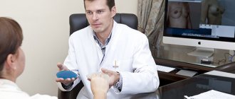
Diagnosis and treatment
After examining the patient, the doctor will prescribe a histological examination that will help differentiate atheroma from limoma, hygroma and fibroma.
The main treatment method is surgery, during which the tumor will be removed. The intervention takes place under local anesthesia in a hospital setting. The surgeon makes an incision through which the atheroma is removed. The wound is sutured and a sterile dressing is applied. The stitches are removed on the 10th day.
Laser removal of a tumor can be carried out in three ways: photocoagulation, laser evaporation of the capsule, laser excision with the shell.
Radio wave removal is indicated for small atheroma sizes. Radio waves affect tumor cells, causing them to die. A crust forms at the site of the cyst, which disappears over time. Your doctor will tell you more about how radio wave removal of atheroma on the back is carried out during your consultation.
After removal:
- The area after surgery should not be wetted for three days;
- the wound is treated daily with an antiseptic;
- You must wear a bandage for a week;
- It is mandatory to take antibiotics (as prescribed by a doctor);
- During the recovery period, a course of physical therapy is recommended.
The wound healing time depends on the location and size of the cyst.
Possible methods of dealing with scars after removal of atheroma
It is possible to remove a scar after atheroma with the help of cosmetology, physiotherapy, and home applications with anti-scar agents. In extreme cases - with surgical intervention.
Cosmetology involves traumatizing the stratum corneum of the skin in order to renew it. This could be chemical peeling, dermabrasion, etc.
Applications are made with special anti-scar creams, gels and ointments 3-5 times a day. They are effective for small, fresh scars. Their combination with physiotherapy is effective.
Physiotherapy offers a course of electro- and phonophoresis procedures - exposure of the scar to electric current or ultrasound. These manipulations stimulate metabolic processes, improve blood circulation, and when combined with anti-scar agents help to significantly speed up the process of tissue regeneration. In this case, the main task of physiotherapy is to transfer particles of the medicinal substance into the deep layers of the skin.
Surgery is prescribed in extremely severe cases, for example, when the keloid scar has reached enormous sizes. After excision of the scar, the wound should heal, and therefore a new scar will appear at the site of the skin injury. With proper rehabilitation, it is normotrophic (light and flat). However, the risk of keloid recurrence is very high.
Atheroma in a child
When examined by a specialist, it is extremely important to distinguish atheroma from lipoma. Atheroma in a child is a smooth, round neoplasm located deep in the skin. When palpated, it is soft and motionless. In most cases, the cyst does not cause any discomfort to the baby. There is no threat to health. When an inflammatory process occurs, the situation is completely different. The main symptoms include: redness of the tumor, pain when pressed, discharge of blood or pus. With such manifestations, urgent medical attention is required.
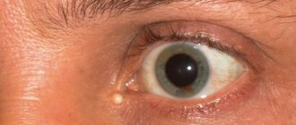
On our website Dobrobut.com you can make an appointment with a doctor and find out how to treat atheroma of the eyelid in a child. Consultations are conducted by specialists with many years of experience. If necessary, a diagnosis and course of treatment will be prescribed.