Modern approaches to the treatment of human papillomavirus infection of the urogenital tract
In recent years, in Russia, as in many countries of the world, the incidence of human papillomavirus infection has been increasing. The problem of its diagnosis and treatment attracts the attention of doctors of various specialties: dermatologists, gynecologists, urologists, oncologists, pathomorphologists, immunologists, virologists. This is explained by the high contagiousness and tendency to increase the frequency of this disease, as well as the ability of some varieties of human papillomavirus (HPV) to initiate malignant processes. The latter mostly concerns the genital manifestations of human papillomavirus infection.
Human papillomavirus (HPV) is epitheliotropic and is found in the skin, oral mucosa, conjunctiva, esophagus, bronchi, and rectum.
There is information in the literature that the introduction of HPV infection occurs at the level of immature epithelial cells of the skin and mucous membranes (basal layer). The result of this invasion is cell proliferation, but without the production of viral particles, since proliferating epithelial cells are not able to support the life cycle of viruses. Complete replication of HPV occurs only in highly specialized cells of stratified squamous epithelium: granular, spinous cells of the skin, superficial epithelial cells of the cervical mucosa. Currently, about 100 types of papillomaviruses have been described. Their tissue and species specificity should be noted. Different types of HPV are associated with different types of lesions. It has been established that certain types of HPV are associated with the urogenital area. There are varieties:
- low cancer risk;
- average cancer risk;
- high cancer risk.
Viral genome structure
Papilloma viruses belong to the papovavirus family (Papovaviridae), which infect cattle, birds, and humans and can infect basal cells of the skin and squamous epithelium. Papillomaviruses are one of the most heterogeneous groups of viruses, the differentiation criterion of which is the degree of genetic relatedness of the viruses according to molecular hybridization: it ranges from 10 to 85%. The diameter of viral particles is 55 nm. The virus does not have an outer shell. The virus capsid consists of 72 capsomeres. A detailed analysis of the HPV DNA molecule became possible after the development of a technique for DNA cleavage using endonucleases and analysis of these fragments using gel electrophoresis.
When studying preparations stained by Papanicolaou, a specific set of signs was identified that characterizes the nucleus and cytoplasm of epithelial cells (koilocytic cell atypia), caused by the cytopathic effect of papilloma viruses.
A specific cell for this infection is the koilocyte, which is an oxyphilt-stained epithelial cell with clear boundaries and a clearly defined perinuclear clearing zone and numerous vacuoles in the cytoplasm.
The term “koilocytic dysplasia” was introduced by HS Stegner in 1981. It is assumed that these changes are a consequence of the reproduction of a virus that causes disruption of cell metabolism, leading to their partial necrosis with the formation of balloon-like cells.
Cytological examination of lesions caused by HPV infection showed that the cellular material contains mostly anucleate, or orthokeratotic, cells. About 20% of scales contain nuclei - the so-called. "parakeratotic cells".
It should be remembered that morphology alone is not enough to identify HPV. In this regard, it is advisable to use the polymerase chain reaction (PCR) method and in situ hybridization. Increasingly, there are reports in the literature about the determination of HPV infection in urine using PCR as an alternative method for testing samples from the cervix. Along with this, nested PCR in one tube and type-specific nucleotide hybridization are used.
The PCR method is used for low-symptomatic or asymptomatic forms of the disease caused by HPV infection.
Using immunochemical methods, it is possible to detect HPV antigens in the tissues of genital warts in 71.4% of cases, by hybridization in 96.5%, and by PCR in 10% of cases.
The effectiveness of DNA hybridization does not exceed the effectiveness of histological examination, but it allows identifying patients with a high degree of this infection.
HPV infection clinic
The clinical manifestations of genital HPV infection are highly variable. Currently, they are divided into genital condylomas, papillary varieties of condylomas (with pronounced exophytic growth), as well as flat and inverted (intraepithelial) with endophytic growth. The last option, also known as “subclinical HPV infection,” is the most difficult in diagnostic terms, since there are no clear microscopic changes in the epithelium. In this case, special screening techniques are required to determine clear boundaries of the lesion.
A peculiar variant of genital warts is bowenoid papulosis and giant Buschke-Levenshtein condyloma. Condylomas acuminata (AC) are fibroepithelial formations on the surface of the skin and mucous membranes, with a thin stalk or a wide base in the form of a single nodule or multiple epithelial outgrowths, resembling cockscombs or cauliflower in appearance. Diagnosis of large condylomas does not cause difficulties. Genital condylomas are localized mainly in places of maceration: labia minora, vagina, cervix, urethral orifice, anal area, skin. In men, OCs are located in the foreskin, on the glans penis, in the perinatal area, and less commonly in the endurethral region. The incubation period ranges from one to 12 months (average 3-6 months).
Studies of recent decades indicate that 85% of patients with typical OC of the vulva and perineum have additional foci of HPV infection in the vagina or cervix, and almost every fourth of them has diseases associated with HPV infection - cervical intraepithelial neoplasia (CVN) of various types. degree of severity. One of the clinical types of diseases caused by HPV infection are bowenoid papules associated with HPV 16, sometimes pigmented on the skin and mucous membranes of both sexes, more often resembling common warts or seborrheic keratosis. In contrast to Bowen's disease, Bowenoid papules are benign and regress spontaneously, although they can occasionally become malignant. The course is asymptomatic.
Some authors include Lewandowski-Lutz epidermodysplasia verruciformis in this group of diseases. This disease is based on local and genetic disorders associated with chronic HPV infection.
JM Handley and WJ Dinsmore (1994), based on literature data, as well as their own studies, proposed a classification of clinical forms of HPV infection and associated diseases (Table 1).
In the vast majority of cases, manifest forms of HPV infection are combined with other sexually transmitted diseases. According to Bernard K. and Mugi K. (1996), manifest forms of HPV infection usually arise as a result of a number of factors:
- social;
- infectious, associated with associations of sexually transmitted diseases (STDs);
- associated with changes in immune status.
The most significant is the influence of urogenital tract infections associated with HPV lesions: urogenital chlamydia, mycoplasmosis, cytomegalovirus and herpetic infections, dysbiotic conditions. The result of their influence on the course of HPV infection is the chronicization of the process, the formation of persistent, usually nonspecific inflammatory changes in the genitourinary area and significant difficulties in carrying out therapeutic measures.
The significance of the presence of concomitant infection for the treatment of condylomatosis is explained by the following circumstances.
- The presence of STDs associated with HPV infection prolongs the treatment period for the latter by an average of three times.
- In most cases, relapses are associated with the above reason.
- Epithelization of cervical erosions after destruction of condylomas can be achieved only if there is a preliminary scan for concomitant STDs and bacterial vaginosis.
The possibility of a relationship between cervical neoplasia and sexually transmitted diseases has been discussed for many years. In the group of women suffering from invasive cervical cancer (CC), a higher frequency of detection of nonspecific microflora, including Trichomonas and Gardnerella infections, was noted. Examples of such effects have been discussed in relation to Treponema pallidum, Neisseria gonorrhoeae, Chlamydia trachomatis, herpes simplex virus type 2, cytomegalovirus, and human papillomavirus. Epidemiological studies have convincingly shown that genital HPV infection is an undeniable risk factor for the occurrence of precancerous changes and cervical cancer.
Principles of treatment of HPV infection
Considering the fact that specific antiviral drugs and vaccines that act on HPV are not yet available, it is generally accepted that complete elimination of the virus from the body cannot be achieved. The goal of therapy is to eliminate clinical and subclinical forms of HPV infection.
Today, practitioners have many methods for removing anogenital warts in their arsenal. Their effectiveness varies from 30 to 90%, but none of the methods is a panacea, since the relapse rate is quite high with any method of treatment. Treatment must be strictly individual: it is necessary to select the most optimal solution in each specific case, sometimes taking into account the wishes of the patient himself. The problem of relapse does not depend on the choice of therapy. Recurrences of anogenital warts are most often associated not with reinfection from a sexual partner, but with reactivation of the infection. There are three ways that events can develop in the absence of treatment:
- warts may resolve on their own;
- remain unchanged;
- progress.
At the same time, one must always take into account the possibility of persistence of the virus in the absence of any clinical manifestations.
When choosing the most optimal method in each specific case, you must be guided by four main characteristics:
- effectiveness for this pathology;
- relapse rate after treatment;
- tolerability (minimal side effects);
- ease of performing procedures.
In addition to removing anogenital warts, it is necessary to solve the following important problems:
1. Identify and treat other sexually transmitted diseases (STDs) in patients with anogenital warts (and their sexual partners).
2. Screen all women with anogenital warts for cervical intraepithelial neoplasia (CVN) using cytology and colposcopy.
3. Maintain further monitoring of CVN lesions in the early stages for timely detection of their progression or development of microinvasive carcinoma.
4. Conduct active treatment of anogenital warts, neoplasia in the early stages, occurring with a detailed clinical picture, neoplasia in the later stages and squamous cell carcinoma.
5. Provide patients with recommendations on the use of condoms and limiting casual sexual contact to prevent infection (and reinfection) with HPV infection and other STDs.
In fact, treatment of anogenital HPV lesions is aimed either at destroying papillomatous lesions by one method or another, or at stimulating an antiviral immune response; a combination of these approaches is possible.
Destructive methods
Physical destructive methods
Surgical excision. Currently used infrequently, it is mainly used in the treatment of malignant neoplasms when wide excision is necessary. This method may require hospitalization due to the fact that quite severe bleeding may occur during excision, and a long postoperative period will require special therapy.
Electrosurgical methods. These include electrocoagulation, electroacoustics, fulgation, electrosurgical excision (electroexcision) using an electric knife. Not so long ago, plasma began to be used in medicine. Our scientists have developed an original plasma coagulator (plasmaskin) EKH-1, which has no foreign analogues. Temperature measurements in plasma showed that it can reach 2000-2500°C. Such high temperature values, in turn, provide the ability to work in a non-contact mode, the operation time is significantly reduced and thereby the necrosis zone is reduced. In addition, with this effect in most cases the pain threshold is not exceeded. This temperature regime ensures almost complete combustion of tumors.
Advantages of this method:
- availability;
- cheapness;
- fairly high efficiency;
- possibility of use in outpatient settings;
- the risk of bleeding is reduced.
Flaws:
- need for pain relief
- When using this method, infectious HPV DNA is released along with the resulting smoke, so it is necessary to create adequate working conditions - vacuum extraction of smoke, the use of protective masks.
Laser excision. A fairly effective and safe method is excision of warts using a laser. Neodymium and CO lasers are used in practice. When using a CO laser, surrounding tissues are less damaged, and a neodymium laser provides a better hemostatic effect. In addition to the laser physically removing lesions, studies have shown that laser radiation has a toxic effect on HPV. The procedures require well-trained personnel. When using lasers, anesthesia is necessary - often local or local anesthesia is sufficient, which allows the procedures to be performed on an outpatient basis. Laser excision and surgical methods are approximately equally effective. Laser therapy can be successfully used to treat common condylomas that are resistant to other treatments. It allows you to stop recurrence in approximately 40% of patients. Studies have shown that such an ineffective result is due to the fact that the CO laser is ineffective when it comes to eliminating the genome from lesions that are resistant to treatment (according to the PCR method, a molecular biological cure occurs in 26% of patients).
The use of a CO laser is the method of choice in the treatment of CVI. Laser conization of the cervix is used. Relapses occur in 2% of patients. A mild method of laser therapy is vaporization, which does not cause virtually any complications. Laser vaporization has been successfully used in the treatment of low-grade CVN. Relapses are observed in 4% of patients.
Laser therapy has been successfully used to treat genital warts in pregnant women. There are reports of treatment in pregnant women at 28–35 weeks of pregnancy. In most patients, healing occurred after the first session. There were no complications during childbirth or in newborns.
Side effects include ulceration, bleeding, secondary infection, and scarring. As with electrosurgical methods, HPV DNA is released through smoke, which also requires precautions.
Laser therapy is not widely used due to the high cost of equipment and the need to train experienced personnel.
Cryotherapy. A fairly effective and safe method that involves the use of liquid nitrogen, nitrogen oxide and carbon dioxide as a refrigerant. In this case, rapid freezing of both intra- and extracellular fluid occurs, leading to lysis and death of cells upon thawing. Cryotherapy does not usually require pain relief, although local anesthetics can be used if necessary. Cryotherapy can be used to treat small warts of various locations. If the warts are multiple, then removal should be carried out in several stages. This method is characterized by the following side effects: the development of local redness, swelling, followed by the formation of blisters and their ulceration. To reduce damage to surrounding tissues, before the procedure, the surface of the warts is treated with KY-gel, which, when frozen, makes it possible to carefully lift and separate the lesion from the underlying epithelium.
The method can be used in gynecological practice.
We think the combined use of cryodestruction and plasma coagulation is extremely promising, allowing us to avoid the disadvantages inherent in the above methods separately.
Chemical destructive methods. This group of products includes solutions of acids, alkalis, and salts. Among them we can mention Feresol, hydrogen peroxide, solutions of quinacrine and hingamine, preparations of mercury and arsenic, bismuth, preparations based on salicylic and lactic acids, acetic and nitric acids, thuja and celandine juices. All these drugs are easily available, but have low, poorly predictable effectiveness, and produce numerous side effects.
Isoprinosine should be used in combination with locally destructive methods of treatment.
The effectiveness of combination treatment for PV, according to the literature, ranges from 38 to 96%.
Combined treatment methods. To treat the manifestations of HPV infection, various methods are proposed, based on the use of immune drugs in combination with laser, electrosurgical and cryodestructive effects.
The combined use of the above methods can reduce the number of relapses and thereby increase the effectiveness of treatment.
Good results have been obtained using a combined method of treating condylomas, including destruction of lesions using cryodestruction (exposure temperature from –160 to –180°C, exposure 40–120 s, twice) in combination with immune stimulation. To stimulate local immunity, the affected area was treated with an emulsion containing interferon (IF), and to stimulate the immune system of the whole body, the drug Kemantan was prescribed at a dose of 0.2 g three times a day orally for 10 days.
A combination of various destructive methods is possible. If there are manifestations of HPV infection on the skin and mucous membranes, cryospraying is first performed for 10–30 s, which makes it possible to clearly identify the boundaries of the lesion due to the characteristic papillary surface of the lesions, which turns white. Then the affected area is exposed to plasma (using the plasmaskin device).
A number of researchers recognize the best method of treating anogenital warts as surgical removal of all visible lesions followed by local administration of IF. In some cases, it is advisable to use general and local IF before surgical excision of extensive condylomas.
There is no therapeutic effect from the use of IF if the disease lasts more than one year, as well as with immunodeficiency.
Currently, there are not many remedies that can be used after using destructive methods. In particular, the drug impran has now appeared for local use in the area of lesions after destructive effects.
Specific antiviral therapy
Currently, there are no drugs that have a specific effect on HPV. Known drugs that suppress the replication of the herpes simplex virus (acyclovir, ganciclovir) turned out to be ineffective in the treatment of anogenital HPV infection.
Theoretically, vaccination is an ideal method for the treatment and prevention of anogenital warts.
There are reports of the effective use of IF inductors. Of interest is the local use of a low molecular weight derivative of imiquidaquinolamine, imiquimod, which is an inducer of cytokines and, in particular, L-IF. It is used in the form of a 5% cream three times a week or daily at night until the rash completely disappears (but not more than 4 months). Complete disappearance of condylomas is observed in 13–56% of cases. With daily use, local side effects more often developed: redness, swelling, erosion. The cream is especially indicated for the treatment of subclinical HPV infection. It is possible to use virazole.
The effect of using IF monotherapy has not been sufficiently studied and is not very high; in addition, it is necessary to take into account the high cost of such treatment. In this regard, this method is not widely used in practice.
Isoprinosine. In recent years, the new immunomodulator isoprinosine, which is a complex of inosine and the salt of N,N-dimethylamine-2-propanol and P-acetaminobenzoic acid, has attracted the close attention of immunologists. The drug can be used in the form of tablets or a solution for parenteral injection. The active substance in this complex appears to be inosine, and the amino alcohol salt stimulates its penetration through the membrane of lymphocytes and other cells.
Isoprinosine has a powerful and broad immunomodulatory effect. Numerous data and extensive literature indicate that in vitro the drug significantly enhances the proliferation of T lymphocytes induced by mitogens or specific antigens, as well as the differentiation of pre-T lymphocytes into more mature T lymphocytes, accompanied by the appearance of corresponding antigens on their surface. PI also stimulates mitogen-induced B cell proliferation. The stimulating effect of isoprinoline on the activity of natural killer cells (NK cells) in healthy people and the functional ability of cytotoxic T lymphocytes has been proven. The drug improves the CD4+/CD8+ ratio; increases the production of IL-2 by T lymphocytes; promotes the maturation and proliferation of T cells; activates the synthesis of IL-1 by macrophages. PI has an antiviral effect and prevents the use of ribosomal RNA for virus replication. It should be noted that when isoprinoline was used with other immunocorrectors, it significantly enhanced the antiviral effect of the latter.
Various treatment regimens using isoprinoline have been adopted depending on the size of condylomas, their location and the degree of malignancy.
Scheme 1: treatment of small, multiple genital warts with a low degree of malignancy.
The drug is taken in 2 tablets. three times a day for 14–28 days.
Scheme 2: treatment of multiple condylomas with individual large condylomas or flat condyloma of the cervix.
Among the chemicals used in our country and abroad that have a destructive effect are TCA and nitric acid, as well as a combined acid preparation - solcoderm.
TCA and nitric acid. TCA is used in 80-90% concentration and causes the formation of local coagulative necrosis. A solution of nitric acid has a similar effect. Due to their cheapness and availability, both methods are quite widespread to this day. Acids are effective for the treatment of condylomas of the vulva, preputial sac, coronary sulcus, glans penis, especially in cases where the use of PF and PFG is contraindicated. Cauterization is carried out once a week for 5-6 weeks. The effectiveness of using TCA and nitric acid is approximately 70-80%. In some cases, a local reaction may develop in the form of weeping and ulceration.
Solcoderm. Solcoderm is an aqueous solution, the active component of which is the interaction products of organic acids (acetic, oxalic and lactic) and metal ions with nitric acid.
acid. The solution contains nitrites in an amount of 0.02 mg/ml.
Listed below are the properties and mechanism of action of Solcoderm, which distinguish it from other drugs in this group used as part of destructive methods:
- when applied topically, solcoderm causes immediate intravital fixation of the tissue to which it is applied;
- the effect of the drug is strictly limited to the place of application;
- a sign of an immediate effect is a change in the color of the treated area;
- devitalized tissue dries out and darkens (mummification effect);
- The “mummified” scab is rejected on its own;
- The healing process is short and complications (secondary infection or scarring) are rare.
General characteristics of treatment with Solcoderm:
- the drug has a precisely limited local effect on the pathologically altered tissue to which it is applied, while the surrounding tissue is not damaged;
- the method is suitable for the treatment of various skin tumors;
- the treatment is painless;
- rapid healing, no complications;
- treatment is carried out on an outpatient basis and does not require special equipment;
- absence of any restrictions for the patient.
Indications for the use of Solcoderm: simple warts, plantar warts, anogenital warts (genital warts), seborrheic keratoses, actinokeratoses, basal cell epitheliomas (basaliomas).
Solcoderm is very easy to use and quite effective for the treatment of condylomas of any location. In most cases, a single application is sufficient.
Cytotoxic drugs
Podophyllin (PF). Pophylline is a resin obtained from the plants P.pelatum and P.emodi, which grow in North America and the Himalayas. To treat warts of the anogenital area, a 10-25% solution of PF in ethanol or benzoin tincture is used. It binds to the microtubule apparatus of the cell and inhibits mitosis, and also inhibits the transport of nucleic acids, resulting in inhibition of DNA synthesis and cell division.
The use of PF is a simple, affordable, fairly safe treatment method that can be used in an outpatient setting, as well as by patients independently. The drug is applied once or twice a week for a maximum of 5 weeks in an amount of no more than 0.5 ml per procedure. The patient must ensure that water does not enter the treated area for 4-6 hours after the procedure. PF is not recommended for use on vaginal, cervical and intraepithelial warts. According to some authors, the recurrence rate varies from 0 to 67%.
Approximately 10-15% of patients develop local adverse reactions in the form of weeping contact dermatitis. Particularly severe complications in the form of multiple ulcerations occur when used incorrectly. As a result of long-term or improper use of PF, patients may experience various adverse reactions, such as nausea, vomiting, abdominal pain, diarrhea, symptoms of damage to the kidneys, myocardium, liver, central nervous system and bone marrow.
The use of PF is contraindicated during pregnancy, since cases of teratogenic effects on the fetus and intrauterine fetal death have been reported.
Many researchers consider PF to be an insufficiently studied and crudely purified plant extract, and therefore recommend using only highly purified podophyllotoxins, and independent use of the drug by patients themselves is undesirable due to the above-mentioned complications.
Podophyllotoxin (PFT) (condylin). PFT is the most therapeutically active fraction of PF. Available in the form of solutions of 0.25, 0.3 and 0.5%, as well as in the form of cream 0.15, 0.3 and 0.5%.
It is usually prescribed twice a day for three days a week in a row for 4-5 weeks.
Although PFT is better purified than PF, a high incidence of side effects has been reported with the use of PFT, especially its 0.5% solution. The following side effects are most often observed as a result of the use of PFT: local inflammatory reactions (erythema, burning, soreness, itching, weeping and erosion in the area of application). Although systemic side effects have not been reported in the literature, it is recommended to limit the use of PFT to a dose of 0.2 ml per treatment.
The disadvantages of PFT are its high cost and long duration of treatment.
5-fluorouracil (5-FU). 5-fluorouracil (5-FU) is a pyrimidine antagonist and has the ability to disrupt the synthesis of both cellular and viral DNA. For the treatment of warts of the anogenital area, it is prescribed in the form of a 5% cream. When treating intravaginal warts, the drug is prescribed once at night for a week or once a week for 10 weeks. The degree of effectiveness of the drug, according to various researchers, is 85-90%. When using 5-FU, weeping erosions on the vaginal mucosa may occur, up to the development of severe weeping contact dermatitis. When treating warts of the terminal part of the urethra, the cream is administered immediately after urination at night for 3-8 days. Complete cure of intraurethral warts is observed in 90-95% of men. However, during treatment there are many side effects: stenosis and stricture of the urethra, dysuria, ulceration. The drug is contraindicated during pregnancy.
Immunological methods
Interferon. Since the human papillomavirus persists in epithelial cells and the use of destructive methods does not guarantee against relapses, the use of IF is promising in this regard, both as monotherapy and in combination with other treatment methods.
IFs are endogenous cytokines with antiviral, antiproliferative and immunomodulatory properties. There are three main classes of IF: leukocyte (L-IF), fibroblast (F-IF) and T-lymphocyte (T-IF). IF can be used locally, intralesional and systemically (subcutaneous, IM or IV). It has been established that when using IF in patients, the amount of viral DNA in the lesions decreases (according to PCR data), which correlates with clinical improvement or disappearance of the lesion.
There is data regarding the use of domestic IF, human leukocyte interferon (HLI), for the treatment of condylomas. It was used intralesional (under papilloma) at a dose of 100,000-500,000 IU, for a course of 3-6 procedures in combination with the application of interferon ointment with an activity of 40 IU to the lesions. PLI can be prescribed systemically and in the treatment of widespread lesions in combination with destructive methods.
L-IF can be considered the most effective drug for various methods, schedules and doses of administration. With systemic use of L-IF, complete disappearance of warts was observed in 11–100% of patients. The effectiveness of using F-IF was 45–82%. The effectiveness of T-IF, shown in different studies, is much lower than that of L-IF and F-IF, and varies from 7 to 57%.
It should be remembered that the unsystematic use of various treatment methods leads to a high percentage of relapses, however, the development of certain algorithms that take into account the gender of patients, the location and number of rashes can significantly reduce the number of relapses.
Table 1. Anogenital HPV infection and HPV-associated diseases
HPV infection
Detailed clinical forms (visible to the naked eye or invisible, but determined in the presence of appropriate symptoms):
- warts (genital condylomas, flat condylomas, vulgar warts)
- symptomatic intraepithelial neoplasia in the early stages - koilocytosis, dyskeratosis in the absence of dysplasia (flat condylomas)
Subclinical forms (not visible to the naked eye and asymptomatic, detected only by colposcopy and/or cytological or histological examination
- asymptomatic intraepithelial neoplasia (IN) in the early stages - koilocytosis, dyskeratosis in the absence of dysplasia (flat warts)
Latent forms (no morphological or histological changes when HPV DNA is detected)
back
Table 2. Diseases associated with HPV
Clinical and subclinical forms:
- VN in the early stages - mild dysplasia, /+-/ koilocytosis, dyskeratosis (VN stage 1)
- VN in late stages - severe dysplasia, /+-/ koilocytosis, dyskeratosis (VN stage 2)
- Late stage LN - severe dysplasia or carcinoma in situ /+-/ koilocytosis, dyskeratosis (stage 3 LN, or CIS)
Microinvasive squamous cell carcinoma:
- clinically visible or invisible, but in the presence of appropriate symptoms
- subclinical, not visible to the naked eye and asymptomatic, revealed only by cytological and histological examination
- latent - absence of morphological and histological changes when detecting DNA HPV infection by molecular hybridization
- intraepithelial neoplasia
Table 3. Classification of treatment methods for anogenital warts
Destructive methods
- physical
- surgical excision
- electrosurgical methods
- cryotherapy
- laser therapy
- chemical
- Nitric acid
- trichloroacetic acid (TCA)
- solcoderm
Cytotoxic methods
- podophyllin (PF)
- podophyllotoxin (PFT)
- 5-fluorouracil
Immunological methods
- interferons
- isoprinosine
Combined methods
- combined use of various methods
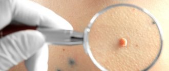
Papilloma on the eyelid
Symptoms: what papillomas look like on the eyelids
Externally, papillomas on the eyelids look like a small growth, the color of which does not differ from the color of healthy skin. The size of the neoplasm rarely exceeds 1-2 millimeters, although there are cases where the growth has reached a centimeter in diameter. The surface of the skin over the affected area is rough. The formation is painless, but if it is mechanically damaged, unpleasant sensations are possible.
The specificity of papillomas on the eyelids is that they can cause significant physical discomfort and even medical problems. Patients often complain of a feeling of “sand” or a foreign body in the eye, although the formation is located on the outer surface of the eyelid. Possible symptoms include watery eyes or dry eyes. Some patients develop conjunctivitis and blepharitis. Although these conditions do not pose a serious threat to health or vision, if papilloma appears on the eye, it is better to seek the help of a specialist as soon as possible.
How to get rid of papillomas?
Since papilloma is not just a skin growth and an aesthetic defect, but a manifestation of a chronic viral infection, solving the problem requires an integrated approach using various areas of treatment.
The patient is prescribed non-specific antiviral therapy with drugs that suppress the reproduction and activity of the virus. For these purposes, the doctor may prescribe Panavir, Acyclovir, oxolinic ointment and other antiviral agents.
The second direction of treatment is to increase the general and local resistance of the body to pathogens of infectious diseases. To strengthen the immune system, immunostimulants are prescribed. Echinacea has a general strengthening effect on the body. You can use medicinal herbs. Drugs that promote the production of interferon (Viferon) have a more specific effect.
Removal of papilloma on the eyelid is the most important condition for successful therapy. Why is it important to get rid of education? The growth is a focus of infection, and the virus is in active form. On the one hand, formations on the eyelid represent a cosmetic defect. On the other hand, it can lead to infection of family members. Finally, removing the source of infection contributes to the complete elimination of the pathogen from the host’s body.
Removal methods
How to remove papilloma on the eyelid? Because the skin on the eyelid is very thin, with the advent of modern surgical technologies, traditional surgical removal of papilloma is rarely used. In Moscow clinics, these formations are removed using microsurgical equipment - a laser, electrocoagulation or radio wave scalpel.
Laser papilloma removal is considered the most effective and safe. The procedure, which is performed using a carbon dioxide laser, has the following advantages:
- With the help of local anesthetics, removal can be done painlessly.
- The laser has stimulating properties and promotes rapid healing of the skin after surgery.
- Thanks to the coagulating properties of the laser, bleeding and damage to healthy skin areas are minimized.
- There is no risk of infection, since the laser cannot be a source of infection and has a detrimental effect on viral and bacterial flora. Thanks to these properties, the laser increases the effectiveness of antiviral therapy and helps eliminate infection.
- The rehabilitation period is short.
- There is no risk of scar or scar formation.
- There is no risk of relapse of the disease.
Removing the growth at home is a less effective (most often ineffective) tactic. Local use of traditional medicine is not justified; you should contact a specialist immediately when a formation appears.
“Effective treatment for HPV” - false or true?
It is important to understand that there are no serious and complete studies on any of the above drugs - randomized, double-blind, placebo-controlled trials. Advertising and laudatory publications and studies with non-representative samples are posted only on manufacturers’ websites and Russian-language resources. Apart from the CIS countries, these funds are not registered and are not used anywhere.
Many Russian gynecologists claim that drugs with unproven effectiveness successfully treat HPV: the test for the virus becomes negative after one or several courses. But this result is not a merit of the treatment; it means that the infection was transient and would have gone away on its own, even without the unnecessary drug and financial burden.
World practice
The US Center for Disease Control writes that specific therapy for HPV is not recommended. WHO in its fact sheet only talks about the need to treat the consequences that HPV can cause: cervical cancer, anal or genital warts. In men, oncogenic types of HPV can cause cancer of the rectum and penis.
In the modern world, there is no drug therapy, but much attention is paid to vaccination.
HPV vaccination
Two vaccines have undergone clinical trials and are registered on the market:
- Gardasil - protects against HPV 6, 11, 16, 18;
- Cervarix - protects against HPV types 16 and 18.
The drugs are very expensive (the price of one injection is about 6,000 - 10,000 rubles), so only developed countries can afford mass vaccination.
Ideally, all adolescents aged 11-12 years, regardless of gender, should be vaccinated (twice within six months) and adults under 26 years of age (three times after 14 years of age within six months). Russian vaccine instructions recommend three-time vaccination at any age.
Separately, it is worth mentioning that the American Alliance of Immunologists recommends vaccination against HPV for HIV. WHO, clinical studies of the Canadian Institute of Health Research and other publications speak about the effectiveness of vaccination of HIV-positive girls.
When should you get tested for HPV?
HPV testing is prescribed to women over 30 years of age with abnormal cytology or histology results. It’s just pointless and costly to carry it out like that. The results of the PAP test, together with the HPV test, help determine the correct interval before repeat cytological examination or colposcopy.
A positive HPV test does not mean cancer, it only allows you to make a rough prognosis; Moreover, the virus is not the only risk factor for cervical cancer.
The HPV test should not be used in large numbers - it is an additional (and expensive) screening method.
Result:
- There is no proven antiviral therapy for HPV, nor are there drugs that strengthen the immune system.
- It is not HPV that is treated, but its consequences: precancerous conditions and cervical cancer, neoplasms on the skin and mucous membranes. The treatment is not medicinal, but surgical: laser, cryodestruction, surgery.
- There is no need for everyone to get tested for HPV.