Atheromas are cysts of the sebaceous glands that are often found under the skin behind the ears and look like small whitish balls.
Atheromatous sebaceous cyst is not an oncological disease. Often atheromas go unnoticed, but sometimes they can be felt and cause discomfort upon palpation.
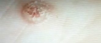
Rice. 1. Atheroma behind the ear
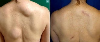
Atheromas that occur behind the ears can be located in other places: for example, on the head, back, face, chest, etc.
The most common location for atheromas is the face, neck and torso.
Atheromas can be of different sizes, large and small.
Very often, atheroma does not bother you at all, but sometimes, as a result of friction of atheroma on clothing, rupture due to pressure or injury, or infection, discomfort and pain appear. In this case, the atheroma must be removed.
Localization of wen
Where do children usually develop wen? They can also occur on internal organs, but this is rare. Tumors grow under the skin, in muscle tissue, and in places above the bone tissue. Although lipoma is called a wen, it itself, as a rule, is located where there is almost no fat. Most often, a child has only one wen. However, if more than one of them is detected, then a diagnosis of lipomatosis is made.
Most often, formations are localized on:
- head and neck;
- face and back;
- shoulders and legs.
Causes of wen appearance in children
It is believed that several factors are necessary for the formation of lipomas. Among them are hereditary causes, poor ecology, unhealthy emotional environment (stress), as well as unbalanced nutrition. However, the main factor, according to experts, is the general pathology of metabolic processes in adipose tissue. There are also versions that in older children the disease develops against the background of other diseases - liver problems, pathologies of the pancreas and thyroid gland. A typical wen in a child is a benign tumor. However, in extremely rare cases, there may also be wen in the form of malignant formations. They are called liposarcoma.
Causes of the occurrence and development of tumors:
- genetic predisposition;
- autoimmune hepatitis;
- Gilbert's disease;
- chronic gastroduodenitis;
- hereditary liver pathologies;
- improper metabolism and diseases of endocrine organs;
- hormonal disorders;
- blockage of the ducts of the sebaceous glands and others.
Isn't this dangerous?
Almost all neoplasms that appear on the head pose a real or potential danger. Wen behind the ears is no exception in this regard.
It is impossible to prevent the initial appearance of a lipoma behind the ear, so its appearance is always a surprise.
In the early stages of their development, they are painless and safe. But, gradually increasing in size, they can pose a certain threat in the following cases:
- the wen in the ear became inflamed and suppuration appeared;
- the lipoma grows in size too quickly, adding a centimeter per month;
- the wen in the auricle enlarges, closing the ear canal, which leads to partial hearing loss;
- if in an adult or child the wen becomes inflamed and painful.
If you have these symptoms, you need to go to the doctor and undergo diagnostics, after which it will be clear what to do. Most likely, the tumor will have to be removed. It makes no sense to delay any longer, because there is a high probability that the lipoma will develop into liposarcoma, a malignant tumor that forms in fatty tissue.
What does a wen look like?
The formation is not very large in size, usually no larger than a match head. However, the tumor tends to grow. Over time, as the lipoma becomes larger, it will interfere with the blood supply to the tissues and organs in the place where it is located. The wen looks like a small lump on the skin. There is a wen on a child’s cheek, on the face in general, on the head, neck, and shoulders. It can also occur on the lower extremities. For example, on the legs or thighs. There can even be a wen on a child’s eyelid, so for many parents the issue of eliminating the pathology in the most gentle way is important. Methods for treating wen will be discussed further. For now, let’s talk about the appearance of the tumor and what it resembles. It is also worth mentioning the internal structure of education.
If the lipoma is on the outside rather than the inside, it may be easier to recognize. This formation has the following properties:
- mobility;
- softness, elasticity;
- no fusion with the skin;
- if the lipoma is large, then it may have a lobular structure;
- tumor size 1.5-10 centimeters;
- education does not hurt or cause concern;
- if the wen is small, then it has a round or oval shape.
The inside of the tumor looks like this. It is made up of adult fat cells that are similar to the other fat cells around. The cells of the formation are connected into small lobules; these lobules are covered with a dense capsule.
Wen can appear where there is no adipose tissue. This phenomenon is an ectopia. This is how congenital tumors usually arise.
The appearance of a wen is a small lump under the skin, which is usually not uncomfortable. However, there are situations when you should see a specialist. Next we will talk about such cases.
Why do atheromas appear behind the ears?
Research into the mechanism of atheroma formation is still ongoing.
Today it is known that the epidermis (the surface layer of the skin) consists of layers of cells that constantly peel off from the surface during the process of growth. In the case when skin cells stop exfoliating and, accumulating and multiplying, begin to move into the tissues - a cyst wall is formed - atheroma. Inside the formed cavity, dead skin cells rich in proteins - keratin, and sebum accumulate. This is how atheroma is formed.
On the surface of the atheroma you can always find an enlarged pore through which the cyst can drain. In this case, we may notice yellowish masses with an unpleasant odor of decomposing sebum.
Other cysts, called epidermal, are very similar in appearance to atheromas. However, epidermal cysts are formed from sweat glands and their contents are very different in composition from the contents of atheromas.
When to see a doctor
At first, the lipoma does not manifest itself in any way. In principle, this state of affairs can remain for quite a long time; perhaps, even throughout life, the tumor will not bother its owner. The formation in its usual state looks harmless and is painless. Where the wen is located, there is no itching, no burning and no suppuration. However, this happens until the lipoma becomes inflamed. This situation may arise if a wen on a child’s face or in another open place was exposed to drafts or other unfavorable factors, for example, injuries, blows, etc.
When should you contact a specialist? This must be done urgently if the formation on the skin has hardened or there is pain at the site of the tumor. Wen can be similar to other tumor formations. For example, with such as atheroma, hygroma or lymphadenitis. These tumors are benign. Only a professional can determine exactly what kind of tumor a child has. At the slightest suspicion of inflammation of lymphoma, you need to make an appointment with a dermatologist. The specialist will individually prescribe a treatment regimen for the pathology, taking into account the age of your child and the location of the tumor.
Symptoms and signs of atheroma
Many patients, having atheromas behind the ears, do not even pay attention to them. However, sometimes atheromas become a serious concern.
It is worth paying attention to the following signs indicating the need to seek medical help:
- There is a small round ball under the skin;
- There is redness, swelling, thickening;
- There are signs of inflammation and infection;
- On the surface of the atheroma there is a small pinhole closed with a black plug;
- It is possible to extract thick yellow contents with an unpleasant odor from the atheroma.
You should consult a doctor immediately if the atheroma:
- Suddenly it began to grow quickly;
- Suddenly burst;
- Inflamed;
- It became sharply painful.
Diagnostics
A dermatovenerologist can recognize a wen visually, by its appearance.
Differential diagnosis can also be used. It is carried out for benign or malignant formations on the skin. We mentioned earlier that occasionally wen can be malignant. Then they are called not lipomas, but liposarcomas. To verify the diagnosis, ultrasound and computed tomography are performed.
Ultrasound examination helps to see the wen as a hypoechoic formation with a capsule, which is located among the subcutaneous fat layer or between the fibers of muscle tissue. A CT scan is needed to determine the structure of the tumor. For example, it is determined how a tumor absorbs X-ray rays in comparison with the tissues located around the tumor.
Research should be carried out only in a professional medical institution with good equipment. Then the diagnosis will be the most accurate and correct.
Removal of atheromas behind the ears.
Atheroma behind the ear can be removed in two ways:
- Laser;
- With a scalpel.
With traditional removal with a scalpel, after injection anesthesia, a skin incision is made to expose the atheroma. Then the cyst is removed along with its contents. The skin wound is sutured.
Rice. 3. Bursting atheroma behind the ear
It is clear that when working with a scalpel, a scar is left on the skin, comparable in size to the atheroma itself, and sometimes larger.
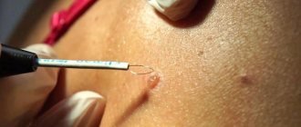
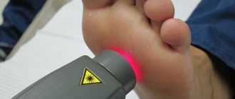
Laser technologies make it possible to remove atheroma entirely without a single drop of blood from a small puncture in the skin, which allows for the best possible cosmetic result.
Treatment
How to remove a wen from a child? In what ways does this happen? If necessary, only a qualified specialist can fix the problem.
There are different cases of lipoma formation and, accordingly, different treatment is provided for each of them. Let's look at typical cases of lipomas in children.
If a wen is on the face of an infant - how to get rid of it? In babies under one year of age, they look like pimples, white on the inside. Most often they can be seen on the baby's nose or on the child's eyelid. Such formation is usually provoked by a hormonal disorder and after some time disappears on its own. Under no circumstances should the integrity of this pimple be violated. It is prohibited to influence such a lipoma in any way. It cannot be squeezed out, pierced, etc.
The baby must make an appointment with a dermatovenerologist. If necessary, he will prescribe an appropriate course of treatment.
If the formation is located in the occipital region and is subcutaneous, only a surgeon can remove it. The same can be said about lipoma, which is located in the chest area. Surgery is also needed here. If the formation occurs on the chest, it is especially important to eliminate it in girls. In the future, the wen can interfere with the normal development of the mammary glands.
If a wen is detected in a child, treatment can also be conservative. This is practiced when the patient is still quite young in age, and the tumor has just appeared. This type of treatment is carried out therapeutically, using medications. May be assigned:
- special medicinal ointments;
- a special diet in which you should eat foods with a high content of fiber and nutrients.
Conservative treatment is also indicated after the tumor has been surgically removed. It can be prescribed for prophylactic purposes to avoid relapse of the tumor.
Surgical method for removing wen
If a child’s wen is too large, what to do? Usually in this case surgery is needed.
Surgical intervention may be prescribed in several cases:
- if the tumor is larger than 5 or 7 centimeters;
- if the lipoma grows very quickly;
- if the tumor develops rapidly in an infant in the first year of life;
- if there are white wen on the child's face.
During surgery, when it is performed on a small child, only general anesthesia is used. If the child is an older child or a teenager, then a local anesthetic is injected subcutaneously. General anesthesia is also used when operating on a lipoma on the face of a child.
The operation is performed on an outpatient basis if the lipoma is small. For newborns and children of the first year of life, surgery is performed only inpatiently. Surgery is also performed in a hospital if a tumor of significant size needs to be removed.
Before the lipoma removal procedure, certain types of tests are performed. This is a general blood and urine test. The blood is also tested for clotting and its group is determined. In addition, sometimes a test is prescribed to check for syphilis and/or HIV, etc.
Methods for eliminating lipoma tumors
The attending physician decides how best to remove the formation. The decision is made after analyzing the medical history, taking into account the child’s age, state of health and tumor size.
The methods for removing lipoma are as follows:
- radical method using a scalpel;
- mechanical method or liposuction;
- laser method.
If the tumor is removed mechanically, using a scalpel, then this operation is good because the possibility of recurrence of the lipoma is almost completely eliminated. However, this radical method has a significant drawback. After surgery, a scar is visible on the skin. Over time it may just look like a stripe. However, it can also be quite noticeable.
Laser method. It has a lot of advantages - minimally invasive, quick recovery, less pain. The possibility of infection during surgery is practically eliminated, and the risk of bleeding is low. The scar remains unnoticeable and not rough. This operation is used to eliminate small tumors, as well as if the lipoma is located on the genitals or nipples.
The liposuction method is minimally invasive and has proven itself in good aesthetic terms. Among the disadvantages of this mechanical method is the possibility of recurrence of lipoma.
In this case, the contents of the formation capsule are drawn in with a syringe. The capsule itself remains in place. There is no need to cut the skin, so the cosmetic effect of liposuction is the best.
The duration of surgery by any method is from 30 minutes to one hour. If lipoma removal is required using a laser, then you need to carefully select a clinic. Such operations are carried out exclusively with the help of high-quality expensive equipment. Doctors must have appropriate high-level qualifications and permission to perform such operations.
Advantages of laser removal of atheroma behind the ear:
- Speed (removal is possible on the day of treatment in just 15-20 minutes);
- Atheromas of any size and in any condition can be removed (even festering atheromas can be removed, since the laser has a sterilizing effect on tissue);
- Bloodlessness (the laser seals the vessels that feed atheromas and removal takes place in a dry field);
- Fast recovery;
- Almost complete absence of relapses and complications in the postoperative period.
If you have atheroma, we suggest seeking help from laser medicine specialists at the ATLANTIC Laser Surgery Center.


