Medical diagnosis of callus
Core-type calluses are fairly easy to diagnose.
After an in-person consultation, a dermatologist can provide a conclusion and diagnosis about the type of skin growth.
The only difficulty may be differentiating an ingrown callus from Verruca plantaris (plantar wart).
Because they have the same location and appearance.
You can distinguish a callus from a wart by the following features:
- An ingrown callus is not accompanied by bleeding even with strong pressure on it.
- A wart, unlike a callus, is not a single formation. Typically, upon examination, several warts that resemble cauliflower can be identified.
- The callus in its central zone has a small depression, which can be seen upon detailed examination. In the case of a wart, the HPV formation consists of thin fibers; petechiae (black dots) are observed on top, which bleed when injured.
- If a wart is present, pain occurs when the lesion itself is palpated; there is no pain when walking. In the case of calluses, pain is observed with any mechanical movement.
To confirm the diagnosis and determine the cause of the disease, the dermatologist prescribes a blood test for diabetes mellitus.
Also an analysis to determine the human papillomavirus, to exclude HPV.
Additionally, the doctor can redirect the patient to other specialized specialists.
See a podiatrist (a doctor who treats foot pathologies), a rheumatologist or an orthopedist.
An important point is to exclude Morton's neuroma.
Morton's neuroma is a benign or non-cancerous growth of fibrous tissue.
Develops in the area of the tibial nerve, most often between the third and fourth fingers.
The pathology is accompanied by thickening of the plantar nerve, pain when walking, and a feeling of numbness in the fingers.
The disease is also known as Morton's metatarsalgia, plantar neuroma, and intermetatasal neuroma.
Causes of corns on the feet
Skin renewal occurs due to keratinization of the surface layers of the epithelium. The stratum corneum thickens, the cells become denser, and then slough off. In areas of increased friction or pressure, the keratinization process occurs more actively to protect more delicate tissues from mechanical stress.
Corns on the feet occur on the foot at points that are subject to a lot of pressure. This may occur on the heels, at the base of the toes, or on the underside of the toes. BUT the process never affects the arch of the foot.
The main causes of corns on the feet are the following:
- Uncomfortable shoes - an incorrectly selected size or poorly shaped sole leads to uneven distribution of the load on the foot. In women, corns on the toes or on the toes can form when regularly wearing high-heeled shoes;
- flat feet - with pathology of the ligamentous apparatus of the foot, the load is unevenly distributed, the heel and other parts suffer. High blood pressure causes hyperkeratosis;
- valgus deformity of the foot - pathology of the joints leads to deviation of the position of the toes on the foot, the appearance of excess friction even when using the usual worn-in shoes;
- fungal infection – fungal infection of the skin of the foot increases cell division and the development of hyperkeratosis. The disease may be accompanied by the appearance of cracks in the heels, peeling of the spaces between the toes, unpleasant odor and itching;
- increased sweating - hyperhidrosis can be either an independent phenomenon or a consequence of wearing shoes made of synthetic materials that do not allow the feet to breathe. Constantly moisturized skin tolerates stress less well, and in some cases slips over the surface of the shoe, which contributes to the development of corns;
- overweight – with increased body weight, pressure on the support points increases. The feet experience increased stress, so the skin may react by forming corns.
Additional factors that contribute to the appearance of corns are diabetes mellitus, varicose veins of the legs with signs of venous insufficiency, trauma and inflammatory diseases of the joints of the legs. Stress and nervous disorders can indirectly affect the condition of the skin if they lead to sweating, changes in gait and other disorders.
Corns on the feet can form during pregnancy. A woman experiences increased stress on the musculoskeletal system, the center of gravity shifts, and her gait changes. Therefore, the consequence is hyperkeratosis in certain areas of the sole.
Methods for treating and removing ingrown calluses
The only and effective method of eliminating this problem on the legs is to remove the stratum corneum of the callus and its root.
In this case, it is important to completely remove not only the surface of the callus, but also the root.
Otherwise, the risk of relapse reaches 100%.
There are various methods for removing calluses; the optimal treatment option is selected individually.
Depending on the clinical signs, the neglect of the case and how deep the root has penetrated into the layers of the dermis.
Harvesting the plant
In order for the herb to retain its medicinal properties throughout the year, it is necessary to properly prepare the raw materials obtained from it.
Celandine is collected only during the flowering period, which lasts from May to the end of June. In this case, it is better to choose specimens growing far from roads and industrial enterprises.
This medicinal herb can be used in many ways, so it is prepared accordingly:
- in dry form - the above-ground part of the plant is cut off at a distance of 10 -15 cm from the ground for further drying,
- in fresh form - the celandine is dug up entirely, along with the roots, and washed in cool running water.
The raw materials should dry in a well-ventilated place, not exposed to direct sunlight. It should be spread in a thin layer on a surface that does not create a greenhouse effect (on paper, plywood). During the drying process, it is necessary to regularly turn the raw material to prevent it from rotting .
Fresh celandine should be used immediately or an extract should be made: the juice of the crushed plant is diluted with alcohol in a ratio of 2:1.
Workpieces can be stored:
- dry grass - 3 years,
- extract - 1 year.
Removing callus using drilling
This procedure can be carried out in a pedicure salon.
During the manipulation, a special device resembling a drill is used.
The procedure does not require the use of anesthesia.
Since it is not accompanied by pain.
Drilling/excision of callus areas is carried out using special attachments.
An antibacterial ointment is first placed in the deepening of the callus to prevent the development of the inflammatory process.
For the first few days, the patient may feel minor discomfort, which goes away on its own and does not require treatment.
Dry callus is removed using a similar method, but provided that it is not neglected.
The drilling procedure has certain disadvantages:
- If the rod is located deep enough, this manipulation may be ineffective. Often several manipulations are required to completely remove the callus.
- In the process of drilling out the keratinized area of the dermis, damage to healthy skin is possible. The procedure requires a highly qualified specialist.
- This type of callus removal is a contact type of procedure, which means it is accompanied by an increased risk of infection.
Removal of callus with liquid nitrogen (cryodestruction)
Cryodestruction is an innovative, highly effective method of removing calluses on the foot, heel or toes using liquid nitrogen.
During the procedure, the keratinized area of the skin is frozen, after which the destruction of the callus is observed.
It has been noted that the procedure is associated with an extremely low rate of callus re-development.
Many dermatologists note that no other method for removing core calluses gives results as favorable as cryodestruction.
Removing callus with liquid nitrogen takes about 10-20 minutes.
It is performed under local anesthesia, so the procedure is completely painless and safe.
Side effects are extremely rare (no more than 3% of cases).
Include callus recurrence, pain, discomfort, bleeding, infection, scarring.
The first few days after the procedure, the formation of a cold bubble is observed, which under no circumstances should be pierced.
Within 10-14 days, the healing process occurs; a crust forms in place of the bubble, which falls off on its own.
The process is not accompanied by the formation of scars.
The procedure is usually well tolerated, with minor discomfort possible.
Some disadvantages of cryotherapy:
- If the rod is extremely deep, cryodestruction is not always able to completely rid the patient of the callus.
- If you do not properly care for the skin after the procedure, there is a risk of an infectious process.
- Freezing the callus with liquid nitrogen is not used when large areas of the epidermis are affected. Cryodestruction used on large calluses can lead to a number of complications. For this reason, in advanced cases, they resort to other therapeutic methods.
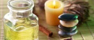
Recipes
Traditional medicine offers many options for using celandine. They help with both corns and calluses with a core. But in order for the recipes to be effective and not cause harm to the body, it is important to follow the recommendations for their use.
Removing skin growths with plant juice
Celandine juice can be used both fresh and canned as an alcoholic extract. They are used to precisely lubricate the problem area 1-2 times a day. The product should be applied daily for one to two weeks.
For best results, it is recommended to regularly take warm foot baths and remove dead tissue with pumice.
The juice has a bright color and is difficult to wash off from things. Therefore, you should use it in the evening before going to bed, putting thin cotton socks on top.
To get fresh juice, just cut or break the stem of the plant - drops of bright orange liquid will appear. It must be used immediately.
Laser excision of callus
Laser callus removal remains a priority treatment method.
Allows you to effectively eliminate the problem with minimal risk of recurrence.
The laser penetrates into the deep layers of the dermis, which makes it possible to remove the root completely, rather than partially.
The operation of the device is based on the reproduction of a laser beam with special light properties:
- Monochromatic: Has only one specific wavelength or color.
- Coherence: Vibrates in the same phase or synchronously.
- Divergence: the beam works in a given direction.
During the manipulation, the epidermal layer is “burned out”, one after another.
The manipulation requires the use of an anesthetic.
The average session duration is no more than 10-15 minutes.
Before using the laser, the callus is treated with antiseptic drugs.
After laser irradiation, a dense crust forms, which will disappear after 10-14 days.
Laser treatment to remove calluses leads to cell regeneration.
It has an anti-inflammatory effect and stimulates the local immune system.
Thus, it promotes rapid healing of wounds.
However, the main advantage of the laser is the complete removal of callus even with a deep root location.
Often, one visit to a dermatologist is enough to effectively eliminate the problem.
Radio wave removal of callus
Callus removal by radio wave is carried out using a special device “Surgitron”.
The Surgitron device uses advanced radio wave technology.
Ensures accuracy, versatility and safety of medical procedures.
The surgical electrode, which is a tungsten wire, emits 3.8 MHz radio waves and slides through the tissue without putting pressure on it.
The pathologically changed area is evaporated, which leads to the destruction of the callus.
An important advantage is that the electrode is self-sterilizing during use, which reduces the likelihood of infection after the procedure.
Other benefits of radio wave treatment:
- Excellent cosmetic results; the manipulation is not accompanied by the formation of scar tissue.
- Fast recovery for the patient, since the treatment process does not affect the healthy tissue surrounding the callus.
- Reduced postoperative pain; in addition, the manipulation itself is not accompanied by pain.
After radio wave removal, the callus becomes covered with a crust, which falls off on its own after 7-10 days.
After which a new, healthy layer of new skin appears, which over time blends with the natural color of the epidermis.
Features of corns on feet
In appearance, corns resemble dry calluses. It is located in areas of high pressure on the foot. Most often it is the heel, areas of skin on the metatarsus, and the pads of the toes. The way corns look does not depend on the patient’s age or the causes of the pathology.
On the heels, corns are often located in the center or border the edge. The skin is usually dry and rough to the touch. It differs in color from the surrounding tissues. Yellowish shades predominate, the edges of the corns are fuzzy, smoothly transitioning into healthy tissue.
With severe hyperkeratosis, the surface of the corn begins to peel off. It flakes off in the form of small scales or large layers of keratinized skin. Separation from the foot in the area of the corn occurs painlessly.
A characteristic symptom of corns is a decrease in skin sensitivity in the area of hyperkeratosis. Sensations from light touches and line touches are poorly experienced. But pressing on the lesion, especially in a vertical position, causes pain in the foot. In some cases, this leads to gait disturbances.
Corns with a rod
The thickness of the skin layer in the area of hyperkeratosis can vary greatly from patient to patient. But sometimes a dark dot appears in the center of the lesion. This is a corn with a stem, or a core callus. The reasons for the appearance of this form of pathology are precisely unknown. Presumably, the formation of the rod is associated with infection with certain types of human papillomavirus.
What does a corn with a stem look like: it is thickened white skin with a yellow tint. There is a dark brown dot in the center. When pressing on the center of the corn, a sharp pain appears. It is difficult to remove this type of corns on your own; this can lead to increased pain and the development of infectious complications.
Corns on fingers
Signs of hyperkeratosis appear less often on the toes than on the heel of the foot or metatarsal pads. The symptoms of corns on the fingers are not much different from those on other parts of the feet. More often they appear in areas experiencing increased loads. These are fingers 1 and 5, on the rest - less often.
Areas of hyperkeratosis may have a regular round or oval shape, but usually their contour corresponds to the area of increased impact. Corns can form not only on the plantar part, but also on the side surface; there can also be core forms on the fingers. This is observed with incorrectly selected shoes, which constantly rub in these areas.
Skin sensitivity in the area of corns is significantly reduced. But when you press on them with force or while walking, pain appears under the focus of hyperkeratosis. This forces you to select other shoe models and change the position of your foot when walking, which negatively affects your gait.
The addition of a fungal infection does not always make it possible to diagnose corns. The patient experiences itchy skin and peeling between the fingers. Hyperkeratosis can develop on the fingertip or tip. Over time, nails become involved in the pathological process. The following additional symptoms are characteristic of a fungal infection:
- thickening of the nail plate;
- cracks, splitting of the nail;
- discoloration, cloudiness or yellowing of the nail;
- increased fragility, longitudinal cracks.
Combined damage to the nail plate and foot requires treatment by a dermatologist.
Treatment of callus with medications
It is immediately worth noting that drug treatment of calluses has the least therapeutic effectiveness.
This phenomenon is associated with the inability of drugs to penetrate deep into tissues, eliminating the root of the formation.
The products used for core calluses often contain keratolytic components and can be effective at the beginning of callus development.
As a rule, products based on salicylic acid are used.
Salicylic acid is a keratoplastic and keratolytic agent.
In addition, it has an antibacterial and antifungal effect.
Salicylic acid works by softening keratin, a natural protein that is part of the skin's structure and produced by the body.
The drug has a softening effect on the stratum corneum of the dermis, which facilitates its removal.
Salicylic acid is often used in combination with other substances.
Allows for a higher therapeutic effect.
When using salicylic acid preparations, the simultaneous use of the following drugs is not recommended:
- Alcohol-containing medications.
- Any other local medications that contain dibenzoyl peroxide or retinoid.
- Soapy substances that have a drying effect.
- Cosmetics that have an exfoliating effect.
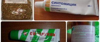
Traditional medicine in the treatment of calluses
Folk remedies cannot affect the development of callus and are not an effective treatment method.
The treatment and removal of callus must be carried out by a qualified specialist.
This will minimize negative consequences and complications.
It is necessary to begin treatment of callus in the early stages of its development.
When there is still a chance to get rid of the problem without resorting to surgical methods.
As it progresses, the root of the callus will penetrate into the deeper layers of the epidermis.
Then it may be necessary to use more radical and expensive methods of therapy.