Fungal infections are lesions of the nails, internal organs, skin and mucous membranes caused by pathogenic fungi. Fungal microorganisms are quite common in the environment, and some of them are constantly present in the human body (for example, the normal microflora of the intestines and oral cavity includes candida).
Some infections are caused by pathogenic fungi that live strictly in certain areas (for example, endemic mycoses). In such cases, the infection most often enters the human body in two main ways:
- aerogenic, when a person inhales spores of pathogenic fungi; the first sign of such an infection may be pneumonia;
- contact, when skin lesions develop through direct contact of the skin with soil that contains pathogenic fungi.
Opportunistic fungal infections can occur in immunodeficient states (for example, in HIV patients), as well as with a general decrease in the body's defenses. At the same time, fungi, which are constantly present in the human body, begin to grow and multiply rapidly.
The most common group of fungi are superficial mycoses of nails and skin. Their pathogens can be transmitted from person to person, and when pathogens spread from the environment to the skin. The disease does not develop in all cases, since different people vary in susceptibility to fungal infections.
Fungi in the body can actively multiply in humid and warm conditions. This is due to their frequent localization on nails, feet, skin of toes and skin folds.
There are many antifungal medications available to treat infections caused by fungi. These drugs can be used in the form of ointments, tablets, injections, creams, solutions for topical use: they must be selected depending on the severity of the disease and the type of infection.
What is eye fungus?
It is believed that this type of eye disease is quite rare, but the number of fungal organisms that provoke the disease is quite large.
Today, there are more than 50 types of fungus that can affect the eyes in this way .
Often the fungus is introduced into damaged areas of the eyeball: this happens with injuries of various types. Sometimes the fungus begins to affect the eye, moving from other diseased areas.
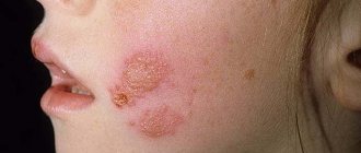
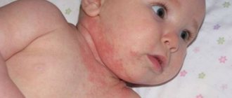
For example, with fungal skin diseases that are localized on the face, the disease can spread to the mucous membrane of the eye .
In children, such diseases are more common than in adults, and this disease is much more severe in childhood . Even the most harmless saprophyte fungi, which constantly exist around a person in large numbers, can provoke inflammatory and pathogenic processes in children.
Important! Conventional therapy, which is effective in treating bacterial and infectious eye diseases, is ineffective in such cases.
Antibiotics and topical corticosteroids are usually prescribed.
Causes of fungal infections of the cornea
Most often, fungal keratitis develops against the background of wearing contact lenses (42% of cases) and injuries accompanied by damage to the cornea (26%).
Currently, there is an increase in the incidence of fungal pathologies of the cornea throughout the world. This is explained by both new diagnostic capabilities and the growing influence of provoking factors - irrational use of antibiotics and steroids, immunodeficiency states.
Fungal keratitis can be caused by more than 70 species of fungi, with yeast and filamentous fungi recognized as the main pathogens. The development of inflammation is facilitated by problems with the natural protection of the eyes (epithelial defects, disorders of tear production and blinking). Often, fungi penetrate the corneal stroma during trauma, surgery, and various eye diseases. The ability of fungi to penetrate into the deep layers of the cornea through normal Descemet's membrane (an intermediate layer between the anterior epithelium and the corneal stroma) is known.
Immunosuppressants (eg, corticosteroids) increase the likelihood of fungal infections.
Types of eye fungus
The following types of fungal microorganisms can infect the mucous membrane of the eye::
- Aspergillosis . Molds that live on human skin and multiply on the mucous membrane, but in ordinary cases the organ of vision is not damaged in depth.
- Candidomycosis . Yeast-like fungi, which are found both in the environment and are present in the human body as opportunistic microflora.
- Sporotrichosis . A fungus that lives in the soil and is carried by animals, birds and people themselves.
- Actinomycosis . A form of fungus that is close in structure to anaerobic bacteria, which lives in large numbers in the human body, multiplying on the mucous membranes.
Most fungi are carried by animals and enter the human body if hygiene rules are not followed..
Granulomatous conjunctivitis of the eyes
This form of the disease often develops with coccidiosis, actinomycosis, sporotrichosis and other types of ophthalmomycosis. They are accompanied by distinctive symptoms: the eyes become red and swollen, and granulomatous growths appear on them. With conjunctivitis caused by the fungi Penicllum viridans, superficial ulcers of the mucous membrane with a greenish coating are observed.
With fungal conjunctivitis of the granulomatous form, purulent lymphadenitis can develop. Inflammation of the lymph nodes occurs, which begin to fester, and inside them there are micelles of fungi.
Symptoms of the disease
Regardless of the form and type of fungi, diseases caused by such microflora have the same characteristic symptoms:
- Burning and itching in the affected eyes.
- Redness , characteristic of all inflammatory processes.
- Purulent discharge of different colors (depending on the type of fungus).
- of a film on the mucous membrane .
- Copious and uncontrollable tearing .
- Painful sensations.
- Redness of the whites of the eyes.
- Whitish inclusions in the mucous membrane of the organs of vision.
- A feeling of blurred vision, the patient sees a veil and cloudiness before the eyes.
- Gradual decrease in visual acuity .
- and dense formations under the eyelid
- The appearance of ulcers and wounds on the eyelid.
Also, fungus in the eyes provokes the appearance of other ophthalmological diseases .
In this case, their treatment is carried out as an addition to the main course, but cannot be completely excluded, since against the background of a fungal disease, any other ailments can quickly become severe.
Exudative ophthalmomycosis
The causative agents of this type of eye pathology are Candida albicans and aspergillus. With the development of candidiasis, so-called pseudomembranes sometimes form on the conjunctiva - films of a gray or yellowish tint, which are easily removed. With aspergillosis, papillary growths occur, ulceration of the mucous membrane or cornea occurs - in this case, mycotic keratitis is diagnosed. Aspergillosis is also sometimes confused with a stye or chalazion.
Conjunctivitis of fungal etiology has minor clinical manifestations and scanty discharge from the eyes. However, they are difficult to treat with antibiotics - this is the main danger of ophthalmomycosis. A long course of fungal conjunctivitis of the eyes can lead to deformation of the edges of the eyelids, as well as their inversion. Complications often arise in the form of inflammation of the lacrimal canaliculus, and the corneal epithelium is affected.
Causes of eye fungus
With normal functioning of the immune system, a healthy person is completely protected from eye fungus. But in addition to immunity disorders, the development of diseases of this nature may be due to a number of other reasons :
- Injuries of various types (domestic, industrial, operating).
- Immunodeficiency (fungal diseases are often found in AIDS patients).
- The disease can appear with diabetes mellitus.
- Incorrect use of antibiotics in the treatment of other diseases.
- Failure to comply with the rules of hygiene and use of contact lenses (in particular, wearing such optical products for longer than the period specified by the manufacturer).
Important! The fungus is often carried with dust, so working in dusty industrial areas can also lead to this disease if special protective equipment is not used.
Prevention of fungal infection
Prevention of fungal infections is to comply with the following rules:
- Do not wear excessively narrow shoes;
- Make sure your underwear is fresh and change your socks daily;
- Avoid wearing wet clothing (sportswear and swimsuits);
- Dry your hands and feet after bathing;
- Follow the rules of personal hygiene, do not use other people's bed linen, towels, shoes and clothes;
- Don't go barefoot in swimming pools and gyms;
- Use antibiotics only if prescribed by your doctor.
Treatment
Correct and quick treatment is possible only by determining the cause of the disease and the type of fungus.
In the process of diagnosing the disease, the ophthalmologist is also interested in details regarding the patient’s intake of various medications during the appearance of fungal infections.
Information about all congenital and chronic diseases is important (such diseases can also become a factor in the appearance of fungus).
Treatment in most cases takes place on an outpatient basis, but in case of advanced disease, inpatient treatment under the supervision of doctors is possible.
Blepharitis
The focus of inflammation in this disease is localized along the edge of the eyelids. The causes of blepharitis are usually prolonged exposure to caustic substances, smoke, volatile liquids, the presence of a chronic infection in the body, or infection due to injury to the eyelids.
It is customary to distinguish 3 forms of the disease: simple, ulcerative and scaly.
Simple blepharitis is accompanied by redness of the edges of the eyelids with slight swelling, without spreading the process to the surrounding tissues. The process is accompanied by a sensation of a foreign body in the eye, which causes the patient to blink frequently. Sometimes foamy discharge is observed in the inner corners of the eye.
Scaly blepharitis is accompanied by severe redness of the eyelid margins and noticeable swelling. Its characteristic feature is grayish or pale yellow scales at the edges of the eyelids. Their mechanical removal leads to mild bleeding. The patient feels itching in the eyelids, sometimes there is a feeling of a foreign body, pain when blinking. Advanced cases occur with pain and photophobia. Visual acuity often decreases.
Ulcerative blepharitis is the most severe form of the disease. Its characteristic sign is dried pus at the roots of the eyelashes, which causes them to stick together. After the crusts are eliminated, slowly healing ulcers form. Eyelashes fall out and often then grow incorrectly. Often accompanied by conjunctivitis caused by the spread of an infectious agent.
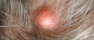
Fungus on the eyelids: photo
Under normal conditions, pathogenic and opportunistic fungal microorganisms do not pose a danger to humans. But even with the slightest disturbance in the functioning of the immune system, the risk of developing fungal eye diseases increases significantly .
Diagnosis of fungal conjunctivitis
The external symptoms of various eye inflammations are quite similar. When ophthalmomycosis is detected, additional diagnostics are also carried out to identify causative agents of conjunctivitis of other etiologies: allergic, bacterial, viral. The fungal origin of the disease may most likely be indicated by a long course, as well as the use of antibiotics or glucocorticosteroids before the development of eye pathology.
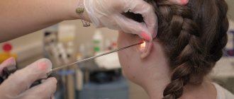
To conduct the study, a scraping is taken from the conjunctiva, a drop of purulent discharge, an imprint smear and then microscopic and cytological examination is carried out, as well as bacterial inoculation on nutrient media. If necessary, a consultation with a dermatologist is scheduled to examine scrapings for pathogenic fungi from smooth skin.
Useful video
Useful information about fungal eye infections can be found at the following links:
Fungal infections of eye tissue should be treated as quickly as possible. Properly organized therapy helps to get rid of dangerous complications.
Author's rating
Author of the article
Alexandrova O.M.
Articles written
2100
about the author
Was the article helpful?
Rate the material on a five-point scale!
( 6 ratings, average: 4.17 out of 5)
If you have any questions or want to share your opinion or experience, write a comment below.
Risk factors
The following factors may contribute to the development of blepharitis:
- traumatic eye injuries;
- chronic pathologies of the visual organs;
- failure to comply with hygiene rules;
- lack of vitamins and microelements in the body;
- diseases accompanied by a persistent decrease in immunity;
- diabetes mellitus, gastrointestinal pathologies, anemia and other chronic diseases;
- previous eye infections;
- exposure to unfavorable external conditions (dusty, dirty air).
Keratoconjunctivitis
A disease caused by an adenovirus that affects the conjunctiva and cornea.
Transmitted by contact.
From the moment of infection to the onset of the disease, 7-8 days pass. It begins with headache, chills, weakness and apathy. Then there is pain in the eyes and redness of the sclera, a feeling of a foreign body inside and profuse lacrimation with mucus discharge from the lacrimal canal.
The conjunctiva turns red, small bubbles with liquid inside appear on it - a characteristic manifestation of an adenoviral infection.
With timely treatment, after 5-7 days, signs of infection disappear, with the exception of photophobia. Cloudy lesions form in the cornea. Complete healing occurs after two months.
Dacryocystitis
Inflammation of the lacrimal sac of an infectious nature. Predisposing factors to the disease are the characteristics of the lacrimal canal and fluid stagnation in the lacrimal gland.
The disease is manifested by liquid purulent discharge and lacrimation. A tumor develops in the inner corner of the eye - a swollen lacrimal gland. When pressed, pus flows out of it. Sometimes hydrocele of the lacrimal gland may develop.
Dacryocystitis can be easily cured with timely treatment. Complications often include keratitis and conjunctivitis with a slight decrease in vision.
Keratitis
This is an inflammatory process localized in the tissues of the cornea. There are exogenous and endogenous forms of the disease and its specific varieties (creeping ulcer of the cornea).
Exogenous keratitis develops after a chemical burn and infection with viruses, microbes and fungi. The endogenous form occurs against the background of a creeping corneal ulcer or infectious diseases of a fungal, bacterial and viral nature (syphilis, herpes, influenza). Sometimes the cause may be some metabolic abnormalities or hereditary predisposition.
The disease begins with tissue infiltration, continues with ulceration, and ends with regeneration.
The infiltrate is a fuzzy yellowish spot. The area of damage varies from microscopic to global, over the entire area of the cornea. Its formation leads to decreased visual acuity, the development of photophobia, lacrimation and spasms of the eyelid muscles (corneal syndrome).
After some time, an ulcer forms. If left untreated, it spreads to the cornea and internal structures of the eye. Its healing is possible only with antibacterial therapy.
If the area of the ulcer is small, visual acuity does not decrease. With extensive damage, blindness can occur.
Choroiditis (posterior uveitis)
This is an inflammatory process behind the choroid. The reason for its occurrence is the introduction of microbes into the capillaries.
The disease is characterized by an initial absence of symptoms. As a rule, choroiditis is discovered incidentally during an ophthalmological examination.
If the focus of inflammation is localized in the center of the choroid, characteristic signs are sometimes observed: distortion of the contours of objects, flickering and light flashes before the eyes.
In the absence of timely antibacterial therapy, retinal edema develops with microscopic hemorrhages.
Structure and functions of the eyelids
The main task of the eyelids is to protect the eyes from foreign objects, adverse external factors, and also from drying out.
In this article
- Structure and functions of the eyelids
- How does blepharitis affect eyelid function?
- The most common causes of the disease
- Risk factors
- Main clinical forms of blepharitis
- What is fungal blepharitis?
- Fungal blepharitis - characteristic symptoms
- How is fungal blepharitis diagnosed?
- Treatment of fungal blepharitis
- Prevention of fungal blepharitis
In order for the eyelids to effectively cope with these functions, they have a special structure. Each eyelid consists of cartilaginous tissue, inside of which there are meibomian glands that produce sebaceous secretion (it forms the lipid layer of the tear film). On the inside of the eyelids there are glands that produce mucus, which ensures close contact of all layers of the tear film with the surface of the eye. The eyelids are connected to each other at two corners - the outer and inner. Near the internal angle there are lacrimal openings - the entrance holes into the lacrimal canaliculi. Along the edge of both eyelids, which are covered with skin on top, there are eyelashes.