Types of rashes
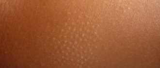
Spots are areas of a different shade from the surrounding skin. To the touch they can be smooth, protruding, or rough. Such manifestations on the surface of the skin are divided into 3 groups, which depend on the provoking factors and description.
- Vascular. Pinkish markings, available in red and purple.
Vascular spot - Pigment. The rash is white and brown in color. Appears against a background of deficiency or excess of melanin.
Pigment spots on the back - Artificial . Appear after the introduction of coloring substances under the skin - tattoo, permanent makeup.
In addition to this classification, there are separate types of marks related to certain pathologies. For example, ringworm. With it, papules disperse throughout the body. Depending on the type, they may also be on the head.
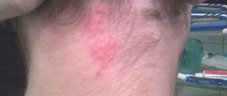
Vascular spots
| Name | Description |
| Hyperemic (blood) | Inflammatory and non-inflammatory. The first option arises against the background of current processes in the body. The size of the spots ranges from 2 cm (roseola) to 3-4 cm and above (erythema). The second option involves a rash that occurs as a result of an emotional outburst. Observed in the chest, neck, face |
| Hemorrhagic | Appear after a bruise or rough impact on the skin. They disappear on their own after 7-14 days. If bruises appear spontaneously and do not disappear for a long time, there is a risk of developing a disease that begins to affect blood vessels |
| Telangiectatic | These are spider veins that arise against the background of short-term dilation of blood vessels. Their occurrence is either congenital or acquired. There are many reasons for this - diet, bad habits, climate, temperature changes |
Dark spots
Markings of light and dark shades are observed on the skin if the amount of melanin has changed. The spots are:
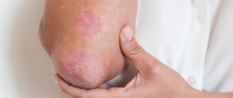
- Hyper. The reason is increased pigmentation in a certain area. Congenital and acquired. The first includes birthmarks, the second - freckles.
- Hypo. The reason is a decrease in the amount of melanin. They also arise from birth and are acquired throughout life. The latter occur with psoriasis, lichen, and vitiligo.
Spots on the back with vitiligo
Age spots may appear if you often go to the solarium. Also, the cause is prolonged lack of treatment in the presence of inflammation on the skin. For example, acne often leaves behind marks. This also includes burns, cauterization of pimples, and rough squeezing.
Pregnancy and hormone intake are common causes for the formation of spots. If a person is prone to freckles, age spots will not bypass him. Old age is also included in this list.
Before you start fighting pigmentation, you need to know what caused it. This is due to the fact that some stains go away on their own, without wasting money and time.
Video - Why do brown spots appear on the skin of the chest and back?
Clinical manifestations of coffee stain syndrome
Diagnosis of neurofibromatosis type I is based on the search for key criteria - we can talk about the disease if at least 2 of 7 signs are present:
- Six or more coffee spots or macules on the skin with a diameter of more than 5 mm in children (before puberty) and more than 15 mm in adolescents and adults (after puberty) ( Fig. 1 ).
- More than two spots in the groin or armpit areas.
- Two or more typical neurofibromas or one plexiform neurofibroma.
- Optic nerve glioma.
- Two or more hamartomas of the iris are usually detected by an ophthalmologist using a slit lamp.
- Dysplasia of the sphenoid bone of the skull or pathologies of the long bones of the skeleton (for example, pseudarthrosis).
- Family history - the presence of neurofibromatosis in close relatives (father or mother, brothers or sisters).
Many of these signs are not present until puberty, making diagnosis difficult in children. Moreover, if a patient at risk reaches 10 years of age without the appearance of at least one symptom (for example, coffee stains on the skin), in the future he will almost certainly not have neurofibromatosis type I.
Neurofibromatosis type I should be distinguished from type II, in which the skin manifestations are relatively poor, but patients have a tendency to develop meningiomas and bilateral acoustic neuromas.
Other syndromes in which coffee stains may appear on the skin (some of them were listed above):
- Silver-Russell syndrome - growth retardation and psychomotor development, triangular face, shortened fingers, syndactyly, characteristic light brown spots on the skin, etc.
- Bloom's syndrome - dwarfism, reduced immunity, increased photosensitivity of the skin, coffee stains, etc.
- Gorlin-Goltz syndrome is a hereditary basal cell carcinoma of the skin with erythematous lesions and skeletal abnormalities.
- Maffucci syndrome is a benign overgrowth of cartilage tissue with bone deformation and dark vascular formations on the skin.
These are not all syndromes in which characteristic café-au-lait hyperpigmentation may occur on the skin. However, they are united by two important factors - relative rarity and overall severity, i.e. the presence of other signs (except coffee stains) that have a much more significant effect on the body.
In general, the following patterns are noted:
- Multiple small coffee spots on the body indicate the presence of one of the above diseases (usually neurofibromatosis type I).
- Single large light brown spots, as a rule, are not associated with other pathologies and exist on their own.
Rice. 1. Coffee stains in the armpits and back (www.medscape.com)
Red spots on the skin
Every person has had strange spots on their skin at least once. Red spots appear for many reasons. However, they can be a slight irritation and indicate a deadly condition. If changes in the skin are observed along with fever, swelling, pain, itching, then it is important to seek help in time.
- Allergies to foods, sunlight, household chemicals, medications.
- Dermatitis.
- Lack of vitamins (seasonal).
- Low immunity.
- Infections.
- STD.
- Stress.
Rash due to illness
People, especially men, do not always pay attention to changes in skin tone in certain places. The reason for their appearance may be various factors. It is important to diagnose a provocateur. By eliminating it, the stains will also disappear. In the stronger half of humanity, red marks are more often associated with the following “pathogens”:
Infectious diseases
Measles, rubella, scarlet fever, chickenpox are infectious processes. Marks and blisters appear on the body. These diagnosed diseases imply that the patient is isolated from society.
Measles
It may manifest itself against the background of a cough or fever. The rash starts on the face and goes down to the heels. In appearance it looks like red bumps that can merge into one large spot. Even after the illness ends, marks may remain on the skin.
Rubella
This is a small rash accompanied by fever. The virus reaches humans through the air. At the initial stage, you feel unwell and weak. Body temperature does not exceed 380. A little later a runny nose is observed. Then a rash begins to appear in the face area, and further along the body. Spots – 6 mm. Complications from this pathology are more common in adults.
Table of contents
- Etiology and pathogenesis
- Clinical manifestations
- Principles of treatment
Coffee spots (café au lait spots, café au lait macules, CALMs) are hyperpigmented areas on the skin with uneven or smoothed edges, the color of which varies from light to dark brown. In foreign literature they are often called “café au lait spots”, which is a combination of the French “café au lait” (coffee with milk) and the English “spots” (spots).
In our company you can purchase the following equipment for removing coffee stains:
- M22 (Lumenis)
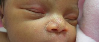
In newborns, coffee spots on the body occur with varying probability, depending on the skin phototype:
- 0.3% of children - phototypes I–III;
- 3% of children - IV phototype;
- 18% of children have V–VI phototypes.
Before puberty, such spots can appear with a probability of 13% in light-skinned people and 27% in dark-skinned people.
Diagnosis and treatment
The cause of the rash is determined by a specialized doctor. As a rule, a visual examination first follows, then tests and additional examination are prescribed. It is useless to describe the rash in words or in correspondence - it is impossible to make a diagnosis in this way, just like on your own. Therefore, if “unhealthy” spots appear on the skin, you need to make an appointment with a dermatologist. If there is a suspicion of a skin pathology, it may additionally be recommended to take a scraping from the affected area for further examination.
Treatment of rashes should be under the guidance of a physician. His task is to prescribe effective and appropriate therapy. In cases of chickenpox, measles and rubella, there is no need to take a course of antibiotics.
If we are talking about a rash due to the presence of scabies mites, then the treatment will be simple. Rashes of an allergic nature require taking samples and prescribing treatment to eliminate the pathogen. Non-infectious skin diseases do not go away on their own; proper treatment is required, taking into account all the characteristics of the body.
Any spot of unknown origin on the body should be shown to a dermatologist if there is cause for concern. Do not voluntarily take medications not prescribed by a doctor. If treated incorrectly, there is a risk that the disease will worsen and then more serious treatment will be required, including hospitalization.
Principles of treating coffee stains on the body
Before starting treatment for coffee stains on the skin, more serious pathologies should be excluded. For this patient, it is necessary to carefully interview, carefully examine, and prescribe laboratory and instrumental studies. It is impossible to do this in a cosmetology office, so it is important to maintain continuity and maintain contact with doctors of other specialties.
If coffee stains are associated with any diseases, maximum efforts should be directed toward treating the underlying pathology. If coffee stains exist on their own, or the underlying disease has already been treated, but areas of hyperpigmentation remain, you can use hardware methods.
One such technique is the Q-switched alexandrite laser . In a study of 48 people, it showed good to excellent effectiveness in more than half of the patients (51.4%) after an average of 3.2 sessions. Moreover, relapse was recorded in 10.4% of cases. Laser treatment for coffee spots carries a high risk of post-inflammatory hyperpigmentation - about 50%. If this occurs, therapy should be discontinued until hyperpigmentation resolves.
IPL therapy has also been successfully used to treat coffee stains . Intense pulsed light (IPL) selectively destroys melanin in areas of excess accumulation, resulting in lighter skin. Modern IPL devices, such as M22 (Lumenis), are equipped with light filters that allow the skin to be exposed to the required range of the radiation spectrum. Thanks to this, IPL can be safely used to treat coffee stains on dark skin - up to Fitzpatrick phototype V.
It has been noticed that if the coffee stain is completely removed, then the risk of its reappearance is minimal. If the stain is partially removed, the probability of recurrence is 50%.