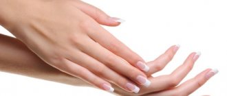
Often young women are perplexed: where did the strange lump appear on their hand?
?
Although painless, but quite large (from 0.5 to 6 cm in diameter), it cannot go unnoticed due to its location. So what is hygroma
- cancer or a purely aesthetic defect?
Although hygroma on the arm is a tumor-like formation, it, contrary to misconceptions, is not an oncological disease
, and also
never degenerates into cancer
. This disease is quite widespread (up to 50% of all neoplasms on the hands) and can be successfully treated. On the other hand, this subcutaneous formation can cause discomfort or even interfere with normal blood circulation in the hands.
Hygroma
(or, as it is also called,
ganglion
) is a capsule-bag that is dense to the touch, filled with a viscous and jelly-like protein liquid mixed with fibrin. The hygroma noticeably protrudes over the ligaments and tendons and practically does not move under the skin. Most often, synovial cysts are observed in women aged 20 to 30 years (⅔ of the total number of patients). Children are least likely to suffer from it.
In what cases should you worry about treating hygroma and can it be prevented?
Hygroma - formation in the form of a tumor
Types of hygroma
There are different types of hand hygromas:
Mucous cysts - hygromas occur against the background of deforming arthrosis of the joints. Most often they form at the distal interphalangeal joint - in this case, hygroma occurs in the area of the nail phalanx, at the base of the nail. Hygroma on the finger develops when osteophytes, present in deforming arthrosis, begin to irritate the skin, underlying tissues and capsular ligamentous apparatus. Because of this, a hygroma occurs - a formation that is hollow inside, in a transparent capsule, with transparent, jelly-like contents.
When a hygroma appears on the nail phalanx, it begins to put pressure on the growth zone of the nail and deform it.
Treatment of hand hygroma
Treatment boils down to excision of the cyst. Due to the fact that with mucosal cysts the skin over the lump on the hand becomes weak, the formation is excised along with the changed skin. After the operation, plastic surgery is performed - both with free skin grafts and with complex skin reconstructions.
A tendon ganglion is a cyst that is formed from the tendon sheaths and the walls of the tendon sheaths. Such hygroma of the hand can cause not only pain, but also limitation of motor functions.
Treatment of a tendon ganglion involves removing the formation - this does not pose any technical difficulty. After operations there are practically no relapses or side effects.
Synovial cysts in the area of the wrist joint (hand hygroma) are the most common type of hygroma.
Diagnosis of hygroma
To make an accurate diagnosis, you need to visit a doctor - an orthopedist, surgeon or rheumatologist. Diagnosis of hygroma requires a comprehensive study of the clinical picture, taking into account localization, external signs, and also, if necessary, the results of laboratory or instrumental studies. Typically, a patient examination begins with palpation of the neoplasm, examination and questioning of the patient, as well as checking joint mobility. In some cases, this is enough, and sometimes additional examination of the lump is required to exclude lipoma, fibroma, atheroma or traumatic cyst. If the hygroma is in the palm area, tumor formations in the bone or cartilage tissue should be excluded
To exclude a malignant origin of the formation, the doctor may prescribe a puncture (fluid sampling) from the cyst for histological and/or cytological examination, which is carried out using a syringe.
In the case of an atypical location of the lump, an X-ray examination or ultrasound is performed to make a diagnosis. Ultrasound examination of the cyst helps determine the density of its contents, the presence of veins and other vessels nearby that it can compress, as well as the uniformity of the lump or the presence of several nodules. In some cases, the patient may be recommended an MRI (to determine the structure of the walls, the presence of vessels in the hygroma, and the consistency and composition of the filling of the cyst).
It is important to diagnose hygroma in the early stages
Hygroma of the wrist joint
Most often, wrist hygroma occurs on the dorsum of the joint and looks like an ordinary lump on the hand - round and tumor-like. Hygroma is inactive, painless, dense, elastic consistency, with unchanged skin. According to statistics, almost 70% of hygromas are formed on the back of the wrist.
If the hygroma occurs on the dorsum of the hand, then it is clearly visible when the joint is flexed.
Much less commonly, wrist hygroma develops on the palmar surface of the wrist joint, in the area of the radial artery (in the place where the pulse is usually checked). This arrangement makes the surgical procedure very difficult.
Treatment of hygroma on the wrist
Treatment of such a hygroma is very difficult, since you have to very carefully isolate the cyst in the area of the radial artery, trying not to damage it. Carelessness during surgery can result in serious injury to the artery, which can disrupt the blood supply to the hand. Hygroma on the wrist requires the most professional and careful treatment, so only an experienced surgeon can perform a correct and effective operation.
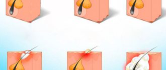
Causes of hygroma
The exact causes of hygroma on the hand have not yet been established. It is believed that joint injuries can lead to the formation of small cavities, which are filled with serous fluid and then merge into one large or several small cysts. Doctors identify several factors that contribute to the onset of the disease and the growth of education:
- tenosynovitis
(inflammation of the tendon sheath) or bursitis (inflammation of the joint capsule) with a subacute course; - hand injury
(bruise, sprain, fracture, ligament rupture); - chronic trauma to articular and periarticular tissues
(ligaments, tendons, cartilage lining), for example, due to repetitive professional movements; - previous hand surgeries
; - diseases of the musculoskeletal system localized in the hands
; - the need to write or draw a lot by hand
(especially if the position or selection of stationery is incorrect); - congenital weakness of the tissues that form the joint capsule
(in cases where the tendency to hygromas is inherited between family members).
It has been established that athletes (tennis players, golfers and others), musicians (pianists, violinists and others), as well as people whose work involves stress on their hands (painters, turners, loaders, typists, bakers, etc.) are most often susceptible to hand hygroma. hobby). Sewing, embroidery, beadwork and similar hobbies can also increase the risk of developing a hygroma.
The structure of the wrist joint
The wrist joint is very complex in its structure and this is largely due to its versatility. In order for us to make various movements of the wrist, various types of bones are “assembled” in the joint (radius, ulna, metacarpal bones, carpal bones, cartilaginous articular disc), connected by ligaments designed to ensure the strength of the joint. It is the ligaments that allow the wrist and hand to move, rotate and rotate in all directions. All these elements, merging in one place, form the capsule of the wrist joint, in the cavity of which there is synovial fluid.
Hygroma, “growing” in the wrist joint, gradually increases in volume, “pushes apart” the surrounding tissues and ligaments and, ultimately, protrudes from the back or palmar side of the wrist, resembling a moving ball in appearance.
Treatment of hygroma
Although sometimes a hygroma on the arm can be left without treatment, due to the risk of complications and aesthetic discomfort, doctors usually recommend its removal. Sometimes a synovial cyst can resolve spontaneously if the factor that provokes it (for example, occupational stress) is eliminated in a timely manner - then the synovial fluid will simply stop flowing into the capsule.
The indication for treatment of hygroma is its multi-chamber nature, rapid growth of formation, development of the inflammatory process, pain syndrome, and limited mobility in the joint.
The preferred treatment for hygroma is surgical removal.
, since puncture or aspiration of the tumor is usually ineffective or poses a risk of complications.
Thus, conservative treatment of wrist hygroma is characterized by recurrence of the disease in approximately 80-90% of cases
, while for surgical treatment the same figure is only
8-20%
.
Surgery
Doctors do not recommend delaying surgical treatment of hygroma, since a large, albeit benign, formation soon begins to displace blood vessels, muscles, ligaments and nerves. Their non-physiological position significantly complicates the operation and may have consequences for the patient
.
Removal of a hygroma on the arm is usually carried out under local anesthesia with a tourniquet and lasts no more than half an hour. The operation is performed using a scalpel or laser equipment. In advanced cases, large or unusual formations may require general anesthesia. When carrying out the procedure, it is important to remove all degenerative cells without exception (otherwise the altered tissue may again develop into a cyst), wash the capsule, and also bandage the so-called. the mouth of the hygroma, which is associated with the joint and feeds the lump with synovial fluid. The drainage tube for fluid drainage is removed 1-2 days after the procedure, and postoperative sutures after complete healing of the incision are removed on days 7-10. After this, it is recommended to immobilize the operated area with a splint and a pressure bandage for several weeks and avoid serious stress on it for 30-60 days.
.
Recently, surgeons have given preference to endoscopic removal of wrist hygroma, in which the capsule and its contents are removed through a small incision a couple of centimeters in size. Rehabilitation after such an intervention occurs much faster.
Conservative treatment of hygroma
As a conservative treatment for hygroma of the hand, crushing it (without penetration and formation of a wound) is occasionally used, followed by the application of a compressive bandage. This method should never be used on infected synovial cysts due to the risk of sepsis, but it can be used to treat sterile lumps. The main disadvantage of this approach is the rapid recurrence of the disease: as soon as the burst edges of the capsule grow together, it will again begin to fill with serous fluid.
A somewhat more successful technique is aspiration puncture of the formation, in which its contents are sucked out through a small puncture, and glucocorticoid solutions and sclerotization preparations are injected in its place. This allows the capsule to be filled with connective tissue instead of liquid. This method is also indicated for administering antibiotics and treating infected hygroma.
Conservative techniques do not eliminate degenerative cells
, which provoke the formation of hand hygroma and are therefore considered ineffective.
There are several methods for treating hygroma
Physiotherapy
A small cyst that does not grow, does not become inflamed and does not cause pain can often be treated conservatively using physiotherapeutic techniques. They help reduce the volume of formation and ensure the outflow of its contents, slow down cell growth. Although physiotherapy cannot completely cure hygroma, in combination with chondroprotectors
it helps keep the tumor under control without surgery.
Sometimes physiotherapy is the optimal solution for non-standard location of the lump
, in which excision can be a rather traumatic procedure.
This type of treatment helps relieve compression of nerve endings, eliminate pain and inflammation, improve tissue regeneration, and relax the muscles around the tumor.
In modern physiotherapy for hygroma of the wrist and other joints, the following is used:
- electrophoresis
; - ultra high frequency therapy
; - phonophoresis with hydrocortisone
; - magnetotherapy
; - paraffin applications
; - mud baths and wraps
; - UV therapy
.
During the entire course of physiotherapy, and also, as prescribed by the doctor, after it, a tight, compressive bandage is applied to the area affected by hygroma, which prevents further accumulation of fluid in the capsule.
If the tumor is too large or continues to grow, there are signs of inflammation, the attending physician, together with the patient, usually decides on surgical intervention.
Drug treatment
For the medicinal treatment of hygroma, a wide range of anti-inflammatory drugs are used, which are applied to the surface of the cone or injected directly into its cavity.
However, anti-inflammatory therapy provides only a temporary and symptomatic effect
,
and therefore used in combination with other techniques
.
In case of a purulent process inside the cyst, and also as a preventive measure before surgery to remove it, the attending physician selects a specialized antibiotic for the patient - usually in the form of injections.
But the most effective medicine for the treatment of tendon ganglion are chondroprotectors
.
They are helping:
- establish metabolic processes in the tissues adjacent to the hygroma, strengthen muscles and ligaments, prevent the destruction of synovial cartilage;
- improve the quality of synovial fluid - as a result, the joint begins to produce it in smaller volumes, and the nutrition of the hygroma is reduced;
- slow down further growth of tumor formation;
- relieve symptoms of inflammation in the area of hygroma;
- significantly reduce pain (many patients note a reduction in pain by 50-80%);
- cope with discomfort from the tendon ganglion, which cannot be operated on due to its small size.
Chondroprotectors do not allow the joint on which the hygroma occurred to “starve” due to poor supply of nutrients. They also relieve swelling and restore normal mobility in the fingers, hands and wrist joint. In addition, they have a systemic effect on the body, improving the condition of the musculoskeletal system and preventing the appearance of new hygromas, as well as the recurrence of old ones. Due to the almost complete absence of side effects, chondroprotectors can be taken to prevent synovial cysts even before they appear
,
during treatment of a neoplasm
, as well as
after surgery
.
Chondroprotectors stimulate tissue repair after surgery and prevent the re-formation of modified cells. The standard course lasts from 3 to 6 months per year. Compared to other drugs in this series, Artracam
, the course of which lasts 1.5 months, and then can be repeated after 2 months if necessary. It is made exclusively from natural ingredients - seafood.
Drug Artracam
is one of the most effective chondroprotectors for hygroma of the wrist and hand.
Causes of hygroma of the wrist joint
Hygroma forms when a “weak spot” appears in the capsule, which occurs when it becomes thinner due to damage or changes in it.
The main reasons that can lead to this condition:
- Joint injury;
- Consequences of surgery;
- Persistent minor injuries to the hand (and joint) that occur as a result of repetitive activities, such as playing tennis;
- Inadequate physical activity on the arms and especially the hand.
Possible complications of hygroma
Although hygroma is a disease with a positive prognosis and a relatively low risk of complications, it can lead to pathologies that require prompt surgical intervention
. First of all, this concerns painful or rapidly growing formations that can compress blood vessels and soft tissues and impair their trophism. This condition directly damages the bone and cartilage tissue in the hands, leading to starvation and degenerative changes in the wrist and interphalangeal joints, deterioration of muscle and ligament function, as well as fine motor skills of the fingers.
With an “unfortunate” location, the hygroma of the hand can compress veins and nerve bundles
, disrupting the conduction of nerve impulses in the hands, causing aches and severe pain after exercise. There is also a risk of traumatic crushing of the tumor.
In the worst case scenario, a large synovial cyst can cause:
- thrombophlebitis (inflammation of the venous wall with the formation of a blood clot, which leads to blockage of the vessel), venous stagnation (accompanied by swelling and cyanosis of the tissues around the hygroma);
- rupture of the hygroma’s own capsule, as well as blood vessels with hemorrhage;
- pinched nerves and disruption of the normal innervation of the hand, which is accompanied by numbness in the fingertips and tingling;
- bursitis and even purulent tendovaginitis;
- suppuration and sepsis if the wall of the hygroma bursts;
- contracture (persistent loss of mobility in the hand, finger or other affected parts of the body);
- re-development of benign formations (single or, more often, multiple) after injury or removal of hygroma.
If you encounter mechanical crushing of a hygroma on your hand, do not try to remove its contents yourself.
!
Treat the affected area with an antiseptic and apply a sterile bandage, then contact your surgeon within a few hours
.
Due to damage to the skin over the formation, infection of the tendon ganglion
(hygromas). The development of complications and the onset of the inflammatory process is indicated by:
- increase in general body temperature (up to 38-40°C);
- impaired mobility in the joint;
- changes in the structure of the skin over the hygroma (loss of elasticity, redness, roughness, peeling);
- soreness.
Treatment of hygroma in this case should be carried out by an orthopedist-traumatologist or surgeon. It is not worth leaving a crushed hygroma without treatment: after the capsule wall heals, it will fill with liquid again, and new formations may appear around it.
Hygroma of the finger
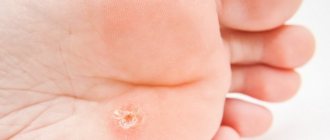
The appearance of a finger hygroma is a tumor-like formation (similar to a lump) localized on the finger, in the area of one of the joints (in some cases, multiple). Often the formation reaches 2-3 centimeters in a few days.
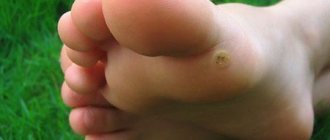
Often, hygroma of the finger looks like an ordinary wart, and only a doctor can accurately determine the pathology during examination.
A hygroma on a finger (a photo of which can be found on the Internet) can spontaneously open and, at first glance, disappear from the finger. But, as a rule, after some time the tumor appears again, since the capsule of the formation itself does not disappear anywhere, but continues to produce fluid and be filled with it. In addition, this can provoke the appearance of one more or even several hygromas on the finger (photo).
Of course, hygroma is not life-threatening, but painful and unpleasant sensations can affect the quality of life for the worse.
Hygroma symptoms
In approximately 35% of cases, a synovial cyst may be asymptomatic
, except for the compaction under the skin.
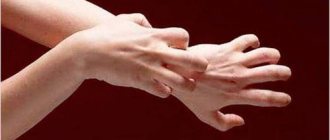
Therefore, the main symptom of hygroma is considered to be the appearance of a lump
(one or several)
in the area of the wrist joint
,
fingers or hand
;
extremely rarely - on other parts of the body. Typically, such a lump reaches 1, 2 or 3 centimeters in size, but an advanced synovial cyst can be up to 6 cm in diameter. In the scientific literature there are references to even larger seals
! But there is usually no need to worry about its size, if it does not cause severe inconvenience, because the hygroma grows very slowly and does not form a truly large tumor-like body.
Synovial cyst
has a characteristic location: on the back of the wrist or ankle joint, as well as the hand or foot.
In addition to the elastic dense lump, patients may experience:
- difficulty moving the affected area;
- dull, usually aching, pain in the hand;
- deterioration of sensitivity in the hand;
- weakness of muscles and ligaments;
- discomfort that increases with physical activity on the hands (especially the supporting one);
- crawling sensation, tingling or burning sensation, as well as other symptoms of compression of nerve endings or blood stagnation;
- increased sensitivity of the skin in the area of the lump.
Special mention should be made of hygroma, in which the growing lump is located under the ligament
, and therefore
does not form a distinct protrusion above the surface of the skin
. In this case, patients complain of:
- constant or periodic pain (pressing or dull), which can radiate to the hand, fingers, forearms;
- pain when pressing;
- roughening, peeling of the skin over the formation;
Unlike other similar diseases, hygroma on the arm, as a rule, does not cause pain on palpation (sometimes it can only be slightly painful). It is characteristic that the skin over the lump does not undergo any changes - it remains elastic and retains its normal color, and does not “grow” onto the tumor. The formation, as a rule, can be moved from side to side, but it does not roll under the skin. One of the main differences between a hygroma and a real tumor is that it can shrink and increase in size. As a rule, hygroma loses volume during the rest period and swells after exercise.
Symptoms of hygroma may appear or intensify in women after the birth of a child.
How to treat
Conservative ways of solving the problem have been practiced for a long time. The formation was kneaded, crushed, the liquid was removed from it through punctures, and sclerosing drugs were injected. They used therapeutic mud, ointments with an anti-inflammatory effect, and physiotherapy. However, only in 10% of cases was it possible to achieve a positive effect: in the rest, relapses occurred.
After scientists discovered the ability of degenerated hygroma cells to restart the process of tumor formation, the conservative methods were done away with. The only treatment today is surgical excision, after which not a single fragment of tissue remains. Therefore, if in the case of other formations it is possible to remove atheroma with a laser or radio wave method, then with hygroma you will have to trust the surgeon and go for surgery, although some doctors practice endoscopic techniques.
Intervention is simply necessary if the tumor progresses seriously, causes severe pain and limits movement in the joints. You shouldn’t turn a blind eye to the aesthetic side of the issue: a lump on the wrist is always a cosmetic defect. After its removal, performance is restored and a normal lifestyle is established, and if endoscopic intervention is chosen as an intervention, recovery time is significantly reduced.
Prevention of hygroma and its complications
For standard prevention of hygroma of the wrist and other joints, you should:
- avoid injury and hypothermia;
- promptly treat arthritis, arthrosis and other connective tissue diseases;
- minimize the load on the joint, incl. monotonous (from time to time take a break from work or monotonous activities to warm up);
- pay attention to hereditary and metabolic diseases that can negatively affect the health of the musculoskeletal system (gout, diabetes);
- follow safety precautions and exercise rules when playing sports;
- Avoid self-medication for injuries and other lesions in the hands and wrists;
- Monitor your body weight and maintain a low-calorie diet rich in vitamins, minerals and amino acids.
If in your family or for your occupation there is a high predisposition to hygromas and other diseases of the musculoskeletal system, you should think about additional measures. Rheumatologists recommend an annual course of prophylaxis with chondroprotective drugs. They help strengthen connective tissue and improve its metabolism, preventing the development of degenerative cells. Artracam is ideal for this purpose.
- due to the high bioavailability of glucosamine sulfate, pleasant taste and convenient sachet form.
Take care of yourself!
Images designed by Freepik
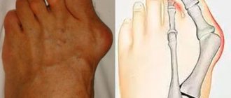
Symptoms
Symptoms include:
- bumps grow on the joints of the fingers;
- difficulty bending the finger;
- Difficulty in straightening the finger;
- pain when moving;
- increase in leukocytes in the blood;
- redness and swelling in the area of the affected joint.
In the case when the results of a general blood test show low lymphocytes, this fact may indicate the presence of an inflammatory process in the body, including one that causes the formation of tubercles on the fingers. Lymphocytes are cells of the human immune system. They are responsible for creating antibodies to viruses, bacteria and other substances that are foreign to the body.