Causes of lichen
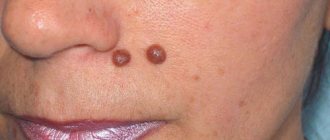
The diagnosis of “lichen” is made on the basis of the clinical picture - small rashes that rise slightly above the skin level and look like a localized area of rash with clear contours (Fig. 1).
Figure 1. Plaques of pityriasis rosea usually have a clear outline. Source: PHIL CDC
Since we are talking about various diseases, the causes of lichen may be different. Most variants of pathology occur due to exposure to harmful microorganisms (viruses, fungi, bacteria), which are especially active against the background of weakened immunity. In some cases, contact with sick animals plays a decisive role. For certain types of deprivation, the question of the reasons for development still remains open - there are hypotheses that require further study.
Predisposing factors for the development or exacerbation of the disease are:
- stressful influences,
- hypothermia,
- previous infections,
- failure to comply with hygiene rules.
The risk group includes both children and adults who have a decrease in immune defense, hormonal imbalances, and neurological disorders. The hereditary factor is considered only for some types of lichen with an unknown infectious cause of development.
All types of lichen that occur due to exposure to microorganisms on the body are transmitted by contact and are therefore considered quite contagious. For other variants of the disease, there is no convincing data on the danger to others.
Types of lichen
Depending on the cause of development and characteristics of manifestation, several variants of lichen are distinguished:
- Pityriasis rosea (Giber's disease). The disease is characterized by a viral nature - the cause of pityriasis rosea is considered to be herpes viruses types 6 and 7.
- Shingles (herpes zoster). Origin - the herpes virus (Herpes Zoster), which causes chickenpox in childhood. Often appears after hypothermia, the rash is usually localized in the direction of the nerves.
- Pityriasis versicolor (variegated, solar) lichen. Occurs due to the action of a fungus of the Malassezia species. The peak of the disease is the summer season; infection often occurs on the beach, in shared showers. Without proper treatment, it can become chronic.
- Ringworm. Most often diagnosed in children. The cause of the disease is fungi of the Microsporum and Trichophyton species, which can be transmitted from sick people or animals.
- Lichen planus. It occurs quite rarely, its appearance is not associated with infection. The cause is considered to be autoimmune reactions of the body.
- Scaly lichen (psoriasis). It also does not apply to infectious and “contagious” lichen; the reasons for its development are still unknown. One of the theories discussed is an autoimmune variant of the disease - systemic damage to connective tissue. Characterized by seasonality, deterioration after stress. Increases the risk of developing arthritis and other systemic manifestations of autoimmune disease.
Lichens of infectious origin with contact transmission also differ in the degree of “infectiousness” (Table 1).
Table 1. The degree of infectivity of various types of lichen.
| Type of lichen | Contagiousness degree |
| Shearer | The tallest |
| Pink | Low (there is an opinion that lichen is not contagious) |
| girdling | High, especially in the phase of active rashes |
| Pityriasis | Low (there is an opinion that lichen is not contagious) |
Can humans or pets become infected?
Every dog owner should know what type of lichen is transmitted to humans and other animals. Regarding ringworm, the answer is clear - this disease is highly contagious and very contagious. When diagnosing it, the animal must be isolated from other pets and household members (especially children).
Ringworm does not pose a danger to others, as it is eczema of allergic origin. Regular hygiene measures when caring for a sick pet are quite sufficient.
The possibility of transmission of infection with pityriasis rosea is currently questionable. The opinions of experts of different specializations contradict each other: some believe that this disease is contagious, others say the opposite. In general, it is generally accepted that if a person has a strong immune system, then pityriasis rosea is not dangerous for him. If the immune system fails (due to age, illness and other reasons), then it is best to stay away from the animal.
Pityriasis versicolor also does not pose a danger to humans and animals, since the fungus is also present on the surface of their bodies. There is a theoretical probability that household members will develop the disease if their immunity is greatly reduced, so if your apartment contains a dog with pityriasis versicolor, it is better to play it safe and isolate it from children, the elderly or weakened people.
Symptoms and manifestations
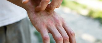
The rash of lichen is accompanied by a change in skin color (redness), moderate or severe itching and peeling. The appearance of scales can be isolated, or may be associated with constant scratching of itchy areas. If the outbreak is localized on the scalp, hair loss occurs in this area. It is important that the elements of the rash do not transform into others, but retain their appearance until the lichen is cured.
Although skin symptoms for different types of lichen are similar, they often differ in location and additional manifestations:
- Pityriasis rosea. Spots on the skin up to 10 cm in diameter acquire a pink tint, and the rash is accompanied by peeling. There is usually no itching or pain. Typical localization is limbs, groin area. On average, the disease goes away in 3–8 weeks; with the right approach to treatment, complications are unlikely.
- Shingles. Rashes most often appear along the intercostal nerves, accompanied by itching and pain - neuralgia. With proper treatment, symptoms disappear within 2–4 weeks; in the absence of antiherpetic therapy, the disease can be complicated by severe damage to the nervous system.
- Pityriasis versicolor. The rash is characterized by pinkish and yellow-brown spots without itching. Typical localization is the skin of the chest, back and shoulders. If you follow medical recommendations, the rashes disappear within 2–3 weeks, leaving no serious consequences.
- Ringworm. Localization of the rash is the scalp, areas of smooth skin, and nails. The hair at the site of the lesion takes on the appearance of being cut off, and a white coating appears on the skin. The incubation period of the disease lasts up to 1.5 months, and the duration of treatment can be up to 6 weeks. Typically, ringworm goes away without complications.
- Lichen planus. It is distinguished by a rash in the form of dense elevations with a characteristic depression in the center, the color of the rash is red with a purple tint. The acute form of the disease disappears in a few weeks, the subacute form lasts up to 6 months and can turn into a chronic process. Possible complications of the pathology are associated with the addition of an infection, a mental reaction to constant itching and the risk of developing squamous cell skin cancer in the absence of proper treatment.
- Squamous lichen. Psoriasis is a chronic disease that manifests itself differently depending on the extent of the skin damage. The main element of the rash is voluminous spots with white scales without itching, cracks due to a violation of the integrity of the skin. The skin of patients takes on a repulsive appearance, and due to the disease, the risk of arthritis, pathologies of the cardiovascular and nervous systems, and the gastrointestinal tract increases significantly.
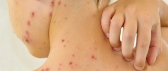
In addition to characteristic changes on the skin (Fig. 2), lichen often manifests itself as weakness, deterioration of health, and even fever.
Figure 2. Some types of lichen. Source: ResearchGate, PHIL CDC
Microsporia is a fungal disease caused by fungi of the genus Microsporum. It affects the skin and hair, in some cases – nails, causes damage to the skin similar to damage caused by trichophytosis, and is also called “ringworm”.Methods of infection with microsporia:
Microsporia is transmitted to humans from other people and from animals. The main carriers of microsporia are cats and dogs or through things that have skin flakes infected with fungi. In addition, fungal spores can remain viable in the soil for 1-3 months. The situation is aggravated by the fact that very often in pets the disease proceeds unnoticed, without any special external symptoms.
In our country, microsporia is caused by two types of fungi:
- Microsporum canis is a microsporia transmitted from animals, the so-called zoophilic microsporia;
- Microsporum ferruginium is a disease transmitted from person to person, anthropophilic microsporia.
Microsporia is the most contagious disease of all cutaneous mycoses: this means that a person who encounters the fungus has a high risk of becoming infected with it, and the disease spreads easily. In external manifestations and by its nature, this disease has much in common with trichophytosis. With microsporia, skin rashes develop faster and tend to spread across the skin and merge. In the foci of rashes, figures of double and triple rings are formed, which is excluded with trichophytosis.
An increase in the incidence of microsporia is observed at the end of summer, since in summer children’s contacts with animals are more frequent than in the cold season. The peak incidence occurs at the end of autumn.
Incubation period:
— with zoophilic microsporia lasts 5-7 days.
- with anthroponotic microsporia lasts 4 - 6 weeks.
Types of microsporia:
The two main types of microsporia are microsporia of smooth skin and microsporia of the scalp. They differ in external manifestations, the nature of the disease, and the method of treatment.
With microsporia of smooth skin, the focus of the disease looks like a ring. The manifestation of the disease begins with a red, slightly hyperemic spot. After a short time in the middle of the affected area, signs of inflammation disappear. The skin peels off, blisters and crusts may appear on it. There is almost no discomfort; sometimes the affected skin may itch slightly. Typically, microsporia of smooth skin is characterized by a small number of lesions, their size is also small, from 1 to 3 centimeters. They can be located on the skin of the face, neck, forearms, and shoulders.
Microsporia of the scalp usually affects children under 12-13 years of age. With microsporia of the scalp, the lesions have a diameter of 2 to 5 centimeters, with reddened skin, sometimes small ones with a diameter of half a centimeter can be located around the main spot. The location is mainly the crown of the head, the temporal regions. The hair begins to break off already on the 6th – 7th day, leaving “stumps” 5-6 mm long. With improper treatment, a suppurative form of microsporia develops, the surface of the skin becomes covered with abscesses, and when pressed on, pus appears on the outside.
Diagnostics:
Methods used to diagnose microsporia are luminescent, microscopic and cultural studies.
The luminescent method is based on the fact that hair affected by the microsporum fungus exhibits an emerald green glow when exposed to a Wood lamp (a lamp emitting in the “soft” ultraviolet range). Examination under a Wood's lamp makes it possible to assess the extent of fungal damage and subsequently serves to adjust therapy, determining its effectiveness.
For microscopic examination, hair scales are taken from the lesions, and in case of microsporia of the scalp, hair fragments are taken. Under a microscope, filaments of mycelium, spores on the surface of the hair, and disturbances in its structure are visible.
A cultural study consists of inoculating the fungus in a nutrient medium and serves to accurately determine the pathogen. Hair scales or fragments are also used as material for sowing. Results usually appear on the third day after sowing.
Prevention measures:
1. Children with microsporia should not attend schools and kindergartens until they have fully recovered.
2. It is necessary to observe sanitary and hygienic rules, that is, use only individual hats, clothing, have a separate bed, towel, comb, washcloth and other personal items.
3. Children should not be allowed to communicate with stray animals. Kittens or puppies adopted into the home should be shown to a veterinarian. Don't let children take pets to bed.
4. In case of contact with a sick animal, you must wash your hands with soap, lubricate scratches and abrasions with 5% iodine, change clothes, boil the removed items or iron them with a hot iron.
5. Wash domestic cats and dogs with soap and water as needed, keep them free of fleas, and be sure to show them to a veterinarian at least once a year.
6. If flaky spots or lesions appear on the skin or head, you should immediately consult a dermatologist.
REMEMBER!
Failure to consult a dermatologist in a timely manner or self-medication complicates diagnosis, leads to the spread of rashes and the disease becoming chronic, which can ultimately lead to irreversible hair loss and the spread of infection in the environment!
Diagnostics
Sometimes, to determine the type of lichen, an external examination and collection of anamnesis is enough - a history of the development of symptoms of the disease and possible connections with risk factors (hypothermia, contact with pets). But in some cases, the diagnostic search turns out to be more difficult.
To correctly determine the type of lichen, the dermatologist carries out:
- visual inspection;
- examination using a Wood's lamp (“black lamp”);
- examination of scraping damaged skin under a microscope.
Also, to assess the general state of health, doctors often prescribe blood and urine tests, and a study of the immunological status.
How to prevent your dog from licking ointment and scratching
Foci of lichen cause severe itching in the pet. When he scratches the affected area, the infection spreads to adjacent healthy tissue, which significantly delays recovery. The same thing happens if the animal licks the applied ointment.
If the lesions are single, you can seal them with a plaster or bandage them, after applying a gauze swab with ointment to the affected area. It is possible to prevent scratching of large areas using a special collar. If the dog resists such a device and removes it, you should put a jumpsuit on it.
Treatment of lichen in adults and children
Glucocorticosteroid creams are effective against shingles caused by herpesvirus infection.
Photo: AGLPhotoproduction/Depositphotos Therapy for each of the variants of this group of diseases depends primarily on the cause of the lichen, as well as on the severity of the changes. In adults and children, treatment regimens are almost the same, only in childhood the dosages of drugs may change, and some “adult” drugs are prohibited for use at an early age.
Treatment of the main variants of the disease:
- Therapy for pathologies caused by the herpes virus (pink and herpes zoster) is reduced to the prescription of antiherpetic drugs (acyclovir), anti-inflammatory and antiallergic drugs (antihistamine tablets), as well as the use of local steroidal anti-inflammatory ointments.
- Fungal types of pathology (pityriasis versicolor) are treated with antifungal, antimycotic ointments.
- To quickly treat ringworm, you need to get rid of the hair and then use antifungal ointments. Between applications of medicinal compositions, it is necessary to lubricate the stains with iodine.
For types of disease with unknown causes, specific treatment regimens have not been developed; therapy is selected individually. There is information about the incurability of scaly lichen - only an asymptomatic course of the disease is possible.
What can you do to speed up your recovery?
How to cure a dog from lichen without causing complications? The main condition is strict adherence to the recommendations of the veterinarian. The animal must be given medications in strict accordance with the prescribed course and dosage, without skipping doses. Unauthorized replacement of one medicine with another, as well as termination of therapy prematurely, is not allowed. The dog owner should be prepared for the fact that in some cases treatment may take a month or more.
Treatment with special shampoos that contain antifungal components will help speed up the recovery of your four-legged friend and prevent the spread of infection. Such hair care products are gentle on the surface of the skin, but using them for a long time is not recommended. To avoid excessive dryness of the epidermis, follow the instructions.
Self-medication: consequences and dangers
Scratching the rash can cause scars to form on the skin.
Photo: LaCameraChiara / Depositphotos Treatment of lichen without the help of doctors most often turns out to be ineffective - it is almost impossible to independently determine the exact cause of the disease. Self-medication can also be dangerous - for example, erroneous use of antibiotics for herpes zoster not only affects the manifestation of a viral disease, but also causes a toxic effect on the body and the development of antibiotic resistance in the future. A common complication of herpes zoster is postherpetic neuralgia. If you do not consult a doctor in a timely manner, any of the variants of the disease can progress, become more difficult to treat, become a chronic pathology and cause the formation of irreversible changes on the skin - scars.
How to apply ointment correctly
The better prepared the affected area of a dog's skin for lichen, the greater the effect that can be achieved from the medicinal ointment, so every owner should know the algorithm of actions when treating an infectious source.
- Prepare the following items in advance: medical rubber gloves, blunt-tipped scissors, a metal container and matches, soap solution, antiseptic (furacilin, chlorhexidine, hydrogen peroxide), tweezers, gauze wipes, medicinal ointment.
- Before proceeding directly to the procedure, put on gloves.
- The affected area of skin is treated with an antiseptic solution.
- If there is hair along the periphery of the lesion, the hair is carefully cut at a distance of approximately 5 mm from the affected epidermis. The cut hair is immediately burned.
- If there are scabs, soak them in a soap solution until completely softened.
- The scabs are removed with tweezers, and the area is again treated with an antiseptic.
- Use a napkin to remove any remaining moisture.
- Apply ointment.
Upon completion of treatment of all affected areas, the clipped wool and napkins should be burned.