The withering sidelong glances of neighbors, and simply those around you, mercilessly pierce the soul and chop the heart into pieces.
They say, what kind of parents are they who allowed such disgrace? And, probably, not everything is in order with the sanitary culture in the family, others pick up, savoring the topic, sprinkling salt on the wounds. At the same time, children are prohibited from communicating, giving the suffering child a categorical label - contagious.
Agree, it is a painful and extremely unpleasant condition. And you won’t explain to everyone that this happened completely by accident - you can’t put a scarf on every mouth.
Unfortunately, here we have to talk not only about medical obscurity and an ordinary lack of understanding of the cause-and-effect relationships of the occurrence of the disease, but also about the banal callousness of others.
What is the disease?
Somehow it became a tradition that any peeling on the skin, especially if it is outlined in a pinkish circle and has an inflamed appearance, is usually called lichen. Here, rather, the principle of excessive vigilance comes into play, although there is no such thing as too much vigilance. And this is probably correct, but we’ll look into this a little later.
Medicine uses a general term to describe and state a group of skin diseases that have different etiologies (causes), but are united by one (priority) risk factor - a reduced immune structure.
In addition, there are other causal circumstances motivating the disease trigger:
- Predisposition along the line of inheritance.
- Consequences of infection: cutaneous candidiasis, colitis, dysbacteriosis.
- Exorbitant, exhausting physical activity.
- Stress, depression, anorexia.
- Autoimmune skin disease.
- Allergic predisposition.
- Pathology of the endocrine system.
- Poor personal hygiene.
- Excess solar exposure.
The reader, of course, noticed that these reasons apply to everyone - both children and adults.
But the answer to the question - what causes lichen appears - is not this, these are only provoking factors.
In the recent past, a conditional generalization in the name of the disease allowed dermatologists to include here all small plaques, inflamed spots or itchy spots that did not transform over time into other formations, such as oozing, erosion or blisters.
Today's medical instruments make it possible to identify the cause of this phenomenon. Various fungi and viruses can act as provocateurs. In fairness, it should be noted that the latter are so insidious and subject to mutation that it is not always possible to identify their origin and verify it.
Looking ahead, we emphasize that depending on the cause and, of course, the pathogen, therapy involves exclusively individual drug rehabilitation.
Video from Dr. Komarovsky:
Bibliography:
- Zhdanov B.M. and Gaidamovich S.Ya. Virology, p. 433, M., 1966, bibliogr.;
- Luria G. and Darnell D. General virology, trans. from English, M., 1971;
- Rozentul M. A. General therapy of skin diseases, p. 135, M., 1970;
- Screen to in Yu. K. and Sharapova G. Ya. Skin and venereal diseases, p. 210, M., 1972;
- Modern issues of clinic, pathogenesis and therapy of dermatoses, ed. V. A. Rakhmanova, p. 85, M., 1968, bibliogr.; Shubladze A.K. and Maevskaya T.M. Herpes, M., 1971, bibliogr.;
- Shcherbakov I. M. Application of interferons and interferonogens in dermatology, Vestn, derm, i ven., No. 3, p. 8, 1973, bibliogr.;
- Nasemann T. Die Infektionen durch Herpes simplex - Virus, Jena, 1965, Bibliogr.;
- Partridge JBA Millis RR Systemic herpes simplex, infection in newborn treated with intravenous idoxuridine, Arch. Dis. Childh., v. 43, p. 377, 1968.
Varieties
The types of diseases are striking in their diversity. Undoubtedly, for parents, especially young and inexperienced ones, the question is relevant: what does lichen look like in children? And of course - what are the symptoms and how to treat lichen?
It should be noted that most cases of the disease occur in children. And this is understandable. The reason is the restlessness and curiosity of the little ones, as well as the significant location of their communication. Everything is interesting to them. I want to try everything, cuddle and caress.
And as soon as we talk about a new cat or dog that has appeared in the yard, then expect a skin “epidemic”. They will immediately catch this infection and will certainly “reward” each other with it.
It is not for nothing that it is included in the TOP 10 anti-rating among all dermal diseases.
Types of lichen with names, contagiousness and ways of communication:
| Variety | Transmission path | Degree of aggression |
| Pink | Personal contact | Minimal. Some dermatologists are inclined to believe that it is not contagious at all. |
| Flat red | Not fully studied | Minor. There is a risk of transformation into squamous cell malignancy. |
| Colored (pityriasis) | Personal contact | Low and low risk. |
| girdling | Personal contact | High. Especially during the rash stage. |
| Shearer | Personal contact | Extremely high. |
Types of lichen in children in the photo:
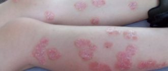
Flat red
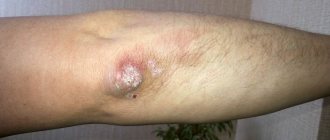
girdling
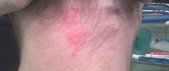
Pityriasis
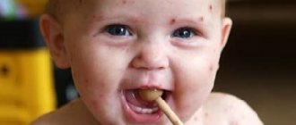
Shearer
Shearer
If the doctor writes microsporia in the diagnosis of a skin disease, then you can rest assured that your child “won” the jackpot and became the owner of ringworm. We understand that this is not entirely appropriate sarcasm. Although it is not a fact that it was the Microsporum fungus that provoked the pathology.
The fungus Trichophyton tonsuran has no less vile properties. Such dirty tricks can be expected from him. Remember, at the beginning of the article we talked about causal diversity.
There can be several ways of infection:
- Tactile contact (through the senses of touch) is the simplest and most common channel, because with the help of it your child learns about the world.
- Using other people's personal items: comb, towel, bedding in kindergarten, soft toys, etc.
- On the street, in the yard, on a walk in the park, through contact with grass, leaves, hay, where the fur of sick animals can become embedded. In the same way, pathogenic fungi can end up in a sandbox or soil.
Ringworm in children in the photo:
Anthropophilic trichophyton may not begin to manifest its sinister essence immediately, but only two or more weeks after contact with an infected object. This makes it much more difficult to identify the location and subject (source) of danger. Although no more than 7 days can pass from the moment of infection directly from the animal.
It is extremely important to see and recognize symptoms at an early stage and prevent the transformation from acute to chronic form:
- A distinctive visual sign of a problem is an inconspicuous round spot. This is how a maternal plaque is formed. How and where on the body it will choose a convenient place for further development is difficult to say. This can be anywhere on the child's head, neck or face. Carefully examine the entire baby - the number of spots will increase very quickly. When mature, the plaque gives some swelling on the body.
- Further, peeling and disturbing, obsessive itching will appear around the affected area.
- A clear sign of trichophytosis are characteristic spots on the hair. This is how the fungus “decorates” the scalp.
- Parents should not ignore enlarged lymph nodes and fever - these are also symptoms of an early stage.
There can be not only a different immune response of the body to the causative agent of the disease. The forms of flow are also radically different from each other.
| View | Description | Affected areas |
| Surface | The disease is mild. There is subtle damage to the dermis. With a progressive development trend, the spots increase in size. | Neck, face, areas of the head covered with hair. Another location of the body is rarely affected. |
| Chronic. | Dark spots appear on the head. Their diameter does not exceed 10 mm. The surrounding scars peel off. The hair color takes on an unnatural color: from bluish to pinkish. The affected area expands its boundaries, the contours become blurred. Itching and an unaesthetic appearance of the skin appear. | Lichen in a child is localized on the temporal and occipital parts, elbows, head, and less often in other places. |
| Infiltrative-suppurative (deep) | Only infected animals are capable of causing this problem. Signs:
| Almost always this is the scalp. |
It is important to know that the disease develops successfully in the presence of the following favorable conditions:
- A painful condition characterized by a chronic lack of vitamins is hypovitaminosis.
- Diseases in the active phase.
- Chronic infection.
- Reduced immune resistance.
- Skin injury.
Causes and treatment of microsporia in a video from Dr. Malysheva:
Pink
This phenomenon has another name – Zhiber’s disease and refers to an infectious-allergic pathology of the dermis. The idea that it is contagious is wrong. The disease can develop only in conditions that are comfortable for it - a cold and problems with the immune shield.
That is why the peak of diseases occurs in early spring and late autumn, when the vitamin warehouse is empty, immunity decreases and the likelihood of problems associated with colds increases.
This circumstance objectively indicates the seasonality of the disease.
The definitive causative agent is not known for certain. Although most dermatologists say that the culprit of the problems is the herpes virus type 7. It is he who is characterized by such deceit. But in the other camp this opinion is not accepted unambiguously.
Causes? They were briefly discussed above.
But there are other negative accompanying factors:
- Skin damage.
- Recent infections and colds.
- Stress, although this applies more to adults, of course.
- Pathology of metabolic processes.
- Allergic reaction to vaccinations and medications.
- Frostbite or sunburn.
- Poisonous insect bites.
In general, the background of the course of the pink “disgrace” is characterized as favorable, but it will be useful for parents to know some of the “individual” features of this disease:
- At the initial stage, you should be alert not only to the deterioration of the child’s general condition: lethargy, drowsiness, pain in the joints and head. The main thing that should immediately catch your eye is the appearance on the forehead, legs, neck, elbows, and back of a huge pink formation rising above the skin. This is the decisive sign - maternal plaque.
- It remains on the body for up to ten days, showing amazing “fertility.” The result of her work is small daughter spots.
- After a week they will turn yellow, begin to wrinkle and peel.
- Once peeling is complete, the spots change color. They can darken or become lighter.
- After two months, the spots will disappear without leaving a trace.
Pityriasis rosea in a child in the photo:
Parents should know:
- The disease occurs extremely rarely in children. And then, this is favored by an infection that has penetrated the intestines and, not surprisingly, by routine vaccination.
- Gibert's disease can occur in an atypical form - without the formation of a maternal plaque. On a baby's bottom, lichen may appear as a result of physical abrasion from a washcloth or clothing, or excessive sunbathing.
Video from an expert:
girdling
Herpes zoster, also known as herpes zoster, is definitely a viral disease that covers almost all areas of the skin with a painful rash.
In 90% of cases, the abdomen, chest and pelvis are affected. In addition, it is he who causes chickenpox, in other words, chickenpox.
Having penetrated the body in early childhood, the virus continues to stay for a long time, until favorable conditions arise, which are characterized by the following circumstances:
- Infectious diseases.
- Excessive ultraviolet exposure (summer tanning).
- Abnormal hypothermia or overheating of the body.
- Unbalanced diet and lack of vitamins.
Therefore, you need to understand that chickenpox and shingles are two troubles of the same nature.
Speaking about the “ferocity” of the Herpes Zoster virus, it is necessary to significantly emphasize its contagiousness and virulence, i.e., almost 100% probability of damage when it enters the body.
For a baby to “catch” the virus, banal everyday contact with a sick person is enough, because the airborne communication channel is the most favorable for this infection.
However, we can talk about more dramatic cases. And this applies to expectant mothers.
If in the third trimester she, God forbid, gets sick with herpes, then there is an extremely high probability that the newborn will develop herpes after birth. Although it is possible that it will make itself felt on the baby’s body a few weeks after birth.
Symptoms that should prompt you to see a doctor immediately:
- A debilitating malaise.
- Weakness turning into chills.
- An exorbitant increase in temperature, crossing the 39° mark.
- Swollen lymph nodes.
- Severe headaches and body aches.
- Nausea progressing to vomiting.
The following stages are distinguished:
- After entering the body, viruses from the nerve node endings rush to the surface of the dermis.
- After two days, spots that closely resemble swelling form in the areas most affected by the invisible enemy.
- Already on the fourth day, the swelling transforms into extremely painful transparent blisters.
- At the same time, the lymph nodes become enlarged.
- After seven days, the blisters begin to dry out and form a crust.
- The end of all periods of the disease: the first symptoms, the formation of bubbles, their drying out and falling off occurs in about a month.
The above clinic is typical for 90% of cases.
But there are also atypical forms, which are characterized by:
- Muscle weakness.
- Formation of blood fluid in blisters.
- Formation of a single large bubble, instead of a scattering of small ones.
- There are cases when preliminary rash is completely absent.
- Extensive damage to the skin around the eyes, with temporary loss of vision (ocular form).
- The same is true for the auricular form, where the concha is affected and hearing is lost.
Important! Parents need to be prepared and not panic when a little patient may exhibit pseudosymptoms of encephalitis, keratitis, stomatitis, iridocyclitis or conjunctivitis. Only a doctor can make a final diagnosis!
Video from Dr. Malysheva:
Flat red
Experts do not have a uniform interpretation of the reasons for this phenomenon.
The majority is inclined to the autoimmune nature of the motivating factors, naming the likely “culprit” - this is therapy, during which the uncontrolled use of medications occurs, such as:
- Anti-inflammatory.
- Pain relievers: Diclofenac, Aspirin, Ibuprofen and similar medications.
- ACE inhibitors, reducing renal and heart failure.
- Antimalarials.
Please understand correctly – this is not anti-advertising. Just be extremely careful when giving your baby medications. Especially strangers or without doctor’s recommendations.
Speaking about other motivating triggers, part of the medical fraternity is inclined to reduce the problem to gastrointestinal pathology, as well as a treated (untreated) infection in the mouth.
Numerous clinical symptoms of this skin disease sometimes make diagnosis difficult in children, but parents need to be aware of them.
There are several forms, the most common:
| Clinical form | Symptoms and description |
| Typical | The first signs of lichen planus in a child or adult are the appearance on the body of a monoform rash, identical in size and shape. These are red flat plaques up to 5 mm in size. During further transformation, they are able to connect and create a large location of the lesion. In every fourth case, the affected area is the oral or vaginal mucosa in girls. |
| Warty | Hyperkeratosis (pathological thickening) forms on the body. Its color is usually brown or purple. This formation vaguely resembles warts. The front of the shin is a favorite area. |
| Atrophic | The affected area experiences a process of atrophy with significant thinning of the skin. If the disease affects the scalp, it is often accompanied by baldness. Dangerous consequences are the scars and scars that may remain after recovery. |
| Pemphigoid or vesicular | Its characteristic symptomatology is the appearance of vesicular formations filled with bloody intercellular fluid. Vesicles can develop to large sizes - up to 10 mm in diameter. Most often, bullae (transformed vesicles) “sit down” on the feet or legs. |
| Moniliform | This is a very specific form with an exclusive rash. It attacks in a group at once and decorates the neck, elbows and outer surface of the hands with a necklace-shaped rash, sparing the rest of the skin. |
| Pigmented | This form is characterized by dry hyperpigmentation of the skin. |
| Pointed | After infection, papules form on the body, and a thickening is called hyperkeratosis in the form of a spike. The neck, shoulder blades and legs are most often affected. |
| Ring-shaped | The geometry of the rash resembles a circle. The spread occurs from the center to the periphery. |
| Erosive-ulcerative | It is distinguished by a generalized attack on the oral cavity - erosion and painful ulcers. |
Lichen ruber in a child in the photo:
Colored (pityriasis)
This type of skin pathology “appropriated” such cheerful names for itself - colored, solar fungus, multi-colored, but this did not add joy to either children or parents. To say the least, the disease puts pressure morally, causing psychological trauma.
The disease is of fungal etiology with a seasonality bias. During the period of active sun, the spots are the brightest; in winter, the “beauty” fades away.
The disease does not pose a danger to health, but we repeat - internal discomfort is obvious.
Children whose skin suffers from ringworm are most often at risk.
An amazing fact - the majority of the planet's inhabitants are carriers of an opportunistic fungus from the line of yeast-like mycotic organisms. But why do only a select few become the object of attention from the disease?
There are a number of reasons for this:
- Endocrine imbalance.
- Deficiency of immune resistance.
- Pathology of the digestive structure.
- Functional skin problems.
- Excessive sweat production and its exclusive chemical composition.
The disease manifests itself in various forms:
| Form of the disease | Symptoms |
| Spotty-flaky | Pinkish-yellow spots appear at the base of the hair follicle (bulb). Formations with an unusual color can be located on the neck, chest and abdomen. As they develop, they merge into a single affected field. Some time later, their color changes to red, brown and brown. A little later, peeling appears, the so-called “chips” effect. There is no itching with this pathology. After recovery, the previously affected areas do not tan and look like painful white islands. |
| Follicular | Papules appear on the surface of the skin in the area of the back, arms, legs and chest. Their size does not exceed 5 mm. But the distinguishing feature of this form is painful itching. |
| Inverted | Skin folds are a favorite location. In these places, redness and peeling of the dermis occurs. Itching is possible. |
Pityriasis versicolor in a child in the photo:
In terms of symptoms, white is similar to pityriasis. Although it is less common, it has the same fungal etiology. And after itself it leaves the same unpleasant white spotty surface.
Lichen alba in a child in the photo:
But it is distinguished by several exclusive features:
- painful itching at the site of the white spot;
- pain;
- getting the affected surface wet;
- in rare cases, the course is accompanied by inflammation of the affected areas.
After reviewing a significant part of the types of disease, the reader may be slightly confused and a logical question will arise: and yet, how to distinguish lichen from dermatitis or allergies and not confuse a spot that looks like lichen with a real fungal disease?
The table below will help you figure this out:
| Symptoms | Lichen | Allergy |
| Rash on the body | Skin spots have a strictly defined location. At first they are single and located at some distance from each other. Affected areas are diagnosed in specific areas: stomach, back, scalp, feet, neck or genital area. | The location of the rash cannot identify the system - it randomly covers the body. The spots vary in size. They always have the appearance of swelling, merging with each other, forming a single area, but can be located separately. |
| Itching | Only when infected with ringworm or lichen planus does painful itching occur. | Allergies are always accompanied by itching. Treated exclusively with symptomatic therapy. |
| Heat | Almost all types of similar skin phenomena are accompanied by a temperature abnormality. | For older patients, hyperthermic syndrome is extremely rare, which cannot be said about children - it is rather a common occurrence. |
| Enlarged lymph nodes | This symptom is characteristic of all types of fungal skin diseases. | As a rule, allergies do not affect biological filters. |
Pathohistology or the structure of pathologically altered tissues in herpes simplex (lichen blisters)
Pathohistology : so-called ballooning degeneration of the Unna epithelium is observed, expressed by focal changes in the cells of the spinous layer, which are rounded, increase in size and, taking on the appearance of balls, are separated from one another; at the same time, as a result of amitotic division, multinucleated large cells are formed. The serous exudate formed in the epidermis separates the altered cells, a vesicle cavity is formed, filled with exudate with epithelial cells suspended in it; Acidophilic inclusions are noted in the nuclei of degenerated cells. In the dermis there is swelling of the papillary layer, a significant dilation of blood and lymphatic vessels and an infiltrate consisting mainly of lymphocytes and neutrophils, a small amount of which penetrates into the cavity of the vesicle.
In tissues affected by the herpes virus, characteristic giant multinucleated cells with intranuclear inclusions are revealed; the latter in the early stage are diffuse and basophilic, later they become dense and eosinophilic. There is marked degeneration of the nuclei and cytoplasm of cells. Viral particles are found in the cells of affected tissues. Using the method of detecting fluorescent antibodies, a herpetic antigen was detected in the cells. Data from histochemical studies indicate a distortion of the metabolism of cells affected by the virus, and their loss of the ability to utilize, in particular, glycogen.
How to treat?
Question: how to treat lichen in children is a question for the average layman who requires a universal ointment or tablet.
It is immediately necessary to emphasize that treatment tactics, as well as rehabilitation time, depend solely on the severity and type of illness.
In addition, it is possible that temporary isolation of a sick baby will be required in order to localize the area where the infection is spreading.
The regimen, dose and procedure for taking medications is the exclusive prerogative of the attending physician.
All means are good for treating your child, be it traditional therapy or “grandmother’s” recipes that have come down to us from the depths of the gray Lukomorye, but have not lost their magical power.
| Medicines | Folk remedies |
| Shearer | |
| Antimycotics – antifungal medications (oral): Itraconazole, Fluconazole, Ketoconazole, Orungal, Griseofulvin. Ointments: Salicylic. Sulfuric. Sulfur-tar. Mikospor. Wilkinson's ointment. Lamisil. Prescription ointments (manufactured in pharmacies): Vidal's milk, Lassora paste. | Grated boiled beets, mixed in equal parts with honey. Rub into the affected area. Buckwheat decoction. One glass of buckwheat and two water. Boil until tender, pass the broth through a sieve. Cool and lubricate the sore spot. Cabbage leaf pulp . Grind in a blender. Mix 1:1 with sour cream - the life-giving potion is ready. Chamomile infusion . Pour a small spoon of chamomile petals with 200 grams of boiling water. Let cool. Wipe the affected area with a swab three times a day. Do not wash and let dry on your own. |
| Pink | |
| Erythromycin is an antibiotic. Acyclovir is an antiviral. Tavegil is an antihistamine. Hydrocortisone – hormonal, anti-inflammatory effect. Tsindol - dries the body and blocks inflammatory processes on the skin and the proliferation of microorganisms. Activated carbon – removes toxins. | Calendula ointment. Grind 10 g of dry calendula, add and mix with 50 grams of Vaseline. Treat the sore spot three times a day. Chamomile decoction and cabbage paste. The recipe is described above. Tar ointment . Birch tar and butter are mixed one to one. At night, lubricate the napkin and cover the sore spot. Yeast dough : a faceted glass of milk, 30 grams of raw yeast, 800 grams of flour, one egg, 2 large spoons of honey and 4 of the same spoons of vegetable oil. Apply the finished dough for two hours three times a day. Paper ash . Burn a blank white sheet of paper to ashes. Add 5 grams of alcohol. Rub the resulting composition into the affected area three times a day. |
| girdling | |
| Antiviral drugs: Acyclovir, Famvir, Valacyclovir, Valtrex. Antiviral ointments and gels: Zovirax. Acyclovir, Viru-merz. Hormonal agents: Prednisolone, Hydrocortisone. Strengthening the immune system: Neovir, Arbidol, Rimatodin. | Wormwood infusion. Pour a small spoon of chopped herbs with 100 grams of alcohol or a glass of vodka. Let it brew for a week. Shake regularly. Apply a cloth soaked in the solution to the affected area for 20 minutes. At the end of the procedure, lubricate the area with castor oil. Burdock decoction. Grind the burdock leaves. Pour 100 grams of boiling water over a large spoon of the starting material and steam for 5 minutes. Remove and cool. At night, moisten a napkin and wrap the inflamed area. Porridge from marsh cinquefoil . Pass the fresh herbs of the plant through a meat grinder. Apply the paste to the wound overnight. Herbal decoction . Take, mix and chop in equal parts: willow bark, blackberry leaves, tea rose petals, calendula flower, horsetail. Pour 50 grams of the mixture into three glasses of water. Bring to a boil, turn off the heat and simmer for another 15 minutes. Cool the resulting broth and filter. Before going to bed, moisten a napkin and cover the affected area. |
| Flat red | |
| Corticosteroids: Prednisolone, Metipred. Synthetic interferons: Neovir, Ridostin, Interferon-alpha 2b. Antihistamines: Parlazin, Zyrtec, Clemastine, Tavegil, Suprastin, Loratadine, Diazolin. Antibiotics: Metacycline, Tetracycline, Azithromycin, Erythromycin, Sumamed. Hormonal ointments: Hydrocortisone, Betamethasone, Flumethasone, Cloveit. Antiallergic ointments: Gistan, Fenistil. | Calendula oil. Pour calendula flower (50 g) into a clean bowl (200 g), pour in olive oil. Leave for 1.5 months, remembering to stir. Once ready, lubricate the sore spot regularly. Calendula ointment. Steam 200 g of calendula oil and 20 g of beeswax. Cool and refrigerate. During the day, treat the rash every four hours. Herbal infusion . Grind nettle leaves, elderberry flowers, juniper berries, dandelion roots and St. John's wort herbs - equally. Take two large spoons of the mixture and mix with boiled water (half a liter). Insist for an hour. Filter and refrigerate. Drink one hundred grams before eating. |
| Colored (pityriasis) | |
| Antifungal drugs (tablets): Ketoconazole, Oranozol, Orunit, Mycozoral, Fungavis, Itraconazole, Canditral, Itrazol. Creams, ointments: Bifonazole, Mycospor, Bifosin, salicylic lotion with chamomile, Clotrimazole, Terbinafine. | Tar soap. At any time of the day, soap the affected area once. Boric acid. Lubricate during the day after 4 hours. Vinegar solution . We dilute one to one 3% vinegar and iodine. We wipe the stains once a day. Wipe the affected area with 3% vinegar twice a day. A decoction of string or celandine . A large spoon of string and a glass of boiling water - brew. We insist for one hour. Filtering. We wet a napkin and cover the affected area for an hour. |
Video:
Etiology or causes and conditions of herpes simplex (vesicular herpes)
The disease is caused by the herpes simplex virus , which belongs to the group of DNA viruses and is widespread. For the first time, the infectious nature of herpes simplex was established by Grüter (W. Griiter, 1912), who caused this disease in a rabbit by introducing pathological material from a sick person; in tissues affected by the virus, Lipschutz (B. Lipschutz, 1921) discovered acidophilic inclusions, and Cowdry (E. Cowdry, 1934) identified them as inclusion bodies of the herpes virus.
Most researchers divide the herpes simplex virus into two types. Strains of the first type are assigned the role of causative agent of nongenital lesions; strains of the second type, in their opinion, cause predominantly genital herpes.
Prevention - how to protect a child?
Some readers will find this section simply unnecessary. Because it will list measures that are simple to the point of naivety, which self-respecting people must, but also must, observe and demand the same from their children.
But don’t consider this as intrusiveness - we will repeat the lesson with you again:
- When returning after a walk, immediately wash your hands with soap. During the summer season, we strongly recommend using disinfectants. Don’t be lazy - check the thoroughness of the hygiene procedure performed by your baby.
- Strictly exclude children from contact with stray animals.
- Do not allow children to use other people's personal hygiene items: combs, washcloths, toothbrushes, towels.
- Do not allow yourself and forbid your children to try on other people’s clothes, shoes and hats.
- Regularly strengthen your child's immune system. To do this, use every opportunity: walks in the air, hardening, dousing with cold water, taking natural vitamins, a healthy and balanced diet.
- During the seasonal risk period, “get hooked” on a hypoallergenic diet. These are vegetables, meat, rice, potatoes, dairy products. They will become a natural shield for you during a viral attack.
We hope that after reading this educational material you have a real tool in the fight against this skin infection. And the folk wisdom that it is better to prevent trouble than to fight it will come in handy when it comes to skin disease.
Why is lichen dangerous in children?
The greatest danger for children are the following consequences of deprivation:
- addition of a bacterial infection;
- spread of pathology to internal organs (bones, brain, etc.);
- development of immunodeficiency.
However, the degree and nature of complications after the disease depend on the type of lichen. Thus, pityriasis rosea occurs without side effects and does not cause deterioration in the child’s health. Trichophytosis leads to the formation of bald patches, and pityriasis versicolor, ringworm and red lichen cause a secondary infection, which significantly complicates treatment.
Herpes zoster can cause postherpetic neuralgia, as well as:
- impaired motor activity of the arms and legs;
- reduction and partial loss of sensitivity of the limbs;
- inflammatory process in the structures of the eye (eyeball, cornea, etc.);
- inflammation of the tissues of internal organs (liver, membranes of the spinal cord and brain, etc.).
Treatment of herpes
Treatment of this pathology, especially recurrent and chronic forms, currently presents significant difficulties. This is explained by the persistence of this virus in the human body, as well as specific immunodeficiencies that form in the body of patients. It must be taken into account that frequent exacerbations, untimely consultation with a doctor and the low effectiveness of many treatment methods used by patients independently cause a significant deterioration in the physical and mental condition of patients.
The required amount of treatment for a patient with herpes simplex is determined by the clinical form, stage and severity of the disease. To prescribe the most optimal treatment methods, patients have to undergo clinical and laboratory examination. Today, with significant development in the pharmacological field, quite strong drugs have appeared to treat this viral infection. Therefore, timely treatment can prevent further development of the disease and significantly improve the patient’s physical and mental condition; it is worth remembering that in case of complications of the disease, the treatment process is long; at the slightest source of infection, consult a doctor.
Diagnostics
Diagnosis of white lichen, first of all, consists of taking into account the external manifestations of the disease. If a person has very fair skin, then the white spots on the body may be almost invisible. In this situation, the patient's skin is examined using a Wood's lamp .
A biopsy of the lichen spot can reveal the absence of pigment cells. But it is also worth noting that their presence cannot rule out the diagnosis. Lichen alba must be differentiated from secondary hypochromic changes, which can occur in the presence of certain skin diseases.
When to see a doctor
Since the early period of the disease is asymptomatic, they do not seek medical help at this stage. At the time of the progressive stage, when children become restless, whiny, and there are scratches on the head and rashes, parents take their children to the doctor.
It should be remembered that psoriasis (scaly lichen) cannot be completely cured, but does not affect the quality of life. If it is detected, you should consult a dermatologist about symptomatic treatment of the child.
For ringworm and pityriasis versicolor, lighting under a Wood's lamp will give a positive result. Accordingly, the dermatologist will prescribe antifungal and supportive therapy. For pityriasis versicolor, the method with iodine is effective - the staining of the affected area will turn brown.
In case of lichen planus and rosea, the polymerase chain reaction of the biomaterial will have a positive result, which indicates a viral etiology, when the study of scales and illumination will not give any result. Additional blood tests (clinical and biochemical) will help establish the inflammatory component (increased erythrocyte sedimentation rate, leukocytes).
For a quick cure and to prevent possible complications, you should immediately seek medical help at the first stage of development of lichen, without self-medicating.