Pyoderma, what is it? Symptoms, causes and treatment
Pyoderma is a dermatological disease that occurs under the influence of pyogenic (pyogenic) bacteria. With various forms of pyoderma, the skin becomes covered with ulcers of various sizes.
Rashes, inflammation of the epidermis, redness, itching are the result of the activity of pathogenic microorganisms. Increased proliferation of streptococci, staphylococci, and fungi leads to extensive damage to the skin.
About staphylococci
Of the 27 strains of staphylococci, only 3 strains pose the greatest danger to humans:
- Staphylococcus aureus, which is the cause of more than 100 diseases. Represents the greatest danger to humans.
- Staphylococcus epidermidis (Staphylococcus epidermidis) is always present on human skin and causes virtually no harm. The development of infection mainly occurs in weakened people and pregnant women. Bacteria enter the body during catheterization, prosthetics and drainage.
- Saprophytic staphylococcus (Staphylococcus saprophylicus) lives in the urinary system of women and often causes inflammation of the bladder, urethra and kidneys in women.
In appearance, staphylococci resemble balls (cocci) up to 1.5 microns in diameter.
Clusters of bacteria resemble a bunch of grapes (Staphylе - bunch of grapes).
Bacteria are extremely resistant in the external environment: to direct sunlight, drying, 100% ethyl alcohol, hydrogen peroxide, phenol solution and a number of antibiotics. At a temperature of 150° C, microbes die only after 10 minutes. Staphylococci persist for a long time in food products, dust and household items.
Pathogenic staphylococci synthesize and secrete many substances that allow this type of microorganism to survive in the human body, damaging its organs and tissues.
The pathogenicity of staphylococci is determined by enterotoxins, exotoxins, enzymes and allergenic components. The main ones are:
- The ability of bacteria to coagulate plasma leads to early blockade of the lymphatic system, which helps limit the spread of infection. Clinically, this condition is manifested by the development of infiltrative-necrotic and suppurative processes.
- Staphylococci secrete a number of enzymes that have multidirectional effects (hyaluronidase, fibrinolysin, DNase, flocculent factor, etc. They facilitate the adhesion of microbes to human tissues and the penetration of the pathogen deep into the tissues, damaging them; they destroy the sebaceous plugs of the hair follicles, which facilitates the penetration of infection deep into the tissues; cause the coagulation of areas of blood plasma around microbes, which, like a cocoon, envelops staphylococcus, protecting it; protect the microbial population from the action of antibiotics.
Epidemiology
The infection is spread by patients and carriers of pathogenic strains of staphylococcus. Open purulent wounds, purulent inflammation of the eyes, mouth and pharynx, pneumonia and intestinal disorders are the source of staphylococcal infection. Food, contact and airborne droplets are the main ways of spreading infection. Surgical interventions, intramuscular and intravenous injections, various implants are also sources of infection. The infection can be transmitted to the fetus in utero, during childbirth and after the birth of the child.
Healthy carriers working in medical institutions, maternity hospitals and catering units are the most dangerous spreaders of infection.
Risk factors for developing staphylococcal infection
The following factors (risk factors) contribute to the development of staphylococcal infection:
- The use of catheters in hospital settings, the use of ventilation, infection can be transmitted during surgical procedures through surgical instruments.
- Immune suppression before transplantation or implantation.
- Carrying out hemodialysis.
- Intravenous nutrition of premature infants.
- Diseases accompanied by decreased immunity (AIDS, diabetes, cancer, some lung diseases, skin and heart diseases).
- Intravenous drug administration.
- Piercing, tattooing.
Rice. 3. In the photo there is staphyloderma - deep folliculitis.
Causes
The leading cause of pyoderma is considered to be the penetration of coccal microbes into the tissue of hair follicles, sweat and sebaceous glands with ducts, and damage. However, the causes of secondary forms of pyoderma, including ulcerative and gangrenous types, are still being studied, since in such cases the contamination of skin areas with pyogenic agents is secondary, that is, it occurs after the development of a certain pathology.
Key provoking factors have been identified:
- heredity;
- shifts in the functioning of endocrine organs (thyroid, hypothalamus, pituitary gland, adrenal glands, gonads), hormonal imbalances;
- skin damage (wounds, injections, abrasions, bites, scratches, burns);
- acute or long-term decrease in general and local immunity;
- skin pathologies, including allergy-related dermatitis, tick-borne infections;
- diabetes;
- introduction of pyogenic flora during surgical procedures;
- increased sensitivity to allergens and reaction to pyococci;
- increased humidity, absorption capacity, alkaline skin reaction;
- intolerance to certain medications;
- frequent hypothermia or overheating;
- disorders of a neurological nature and the thermoregulatory system
- lack of personal hygiene;
- periodic injury to the skin in certain areas;
- long-term worries and severe physical fatigue;
- exhaustion, any long-term illness;
- obesity, fat and carbohydrate metabolism disorder;
- contamination of the epidermis with paints, kerosene, solvents, oils, varnish, coal dust, gasoline, cement;
- vascular diseases, varicose veins, thrombophlebitis, hematopoietic disorders, gastrointestinal diseases;
- focal infections with an inflammatory process in a specific organ or tissue, including the stomach, intestines, nasopharynx and ear area, reproductive organs.
Classification of staphyloderma
Pyoderma is divided into staphylococcal and streptococcal, superficial and deep. Often pyoderma is of a mixed nature.
Superficial staphylococcal pyoderma
- Staphylococcal impetigo (ostiofolliculitis).
- Superficial folliculitis.
- Sycosis vulgar.
Staphylococcal pyoderma deep
- Folliculitis is deep.
- Furuncle.
- Carbuncle.
- Hidradenitis.
Staphyloderma by location
- Superficial folliculitis includes staphylococcal impetigo.
- Deep folliculitis includes sycosis vulgaris and deep folliculitis.
- Perifulliculitis: furuncle and carbuncle.
- Purulent lesions of the sweat glands: hidradenitis and pseudofurunculosis.
- Staphyloderma of smooth skin: epidemic pemphigus of newborns.
Staphyloderma in children
- Miliaria and vesiculopustulosis.
- Multiple abscesses or Finger's pseudofurunculosis.
- Epidemic pemphigus of newborns.
- Ritter's exfoliative (leaf-shaped) dermatitis of newborns.
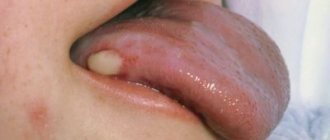
Rice. 4. In the photo, staphylococcal pyoderma is a stye on a child’s eye.
General characteristics of staphyloderma
Staphylococcal pyoderma is usually associated with skin appendages - hair follicles, apocrine and eccrine sweat glands, where they predominantly live. Staphyloderma is characterized by the purulent nature of inflammation. The main morphological element of staphylococcal impetigo in adults is the follicular pustule. Pustules have a hemispherical or conical shape and a dense, tense tire, in the center of which a cavity filled with pus is formed. Purulent exudate has a yellow-green color and a thick consistency. Along the periphery of the follicular pustule there is an erythematous-edematous zone (corolla) with pronounced infiltration. The purulent-inflammatory process mainly spreads in depth. The rashes do not tend to merge and grow peripherally.
In newborns, due to an underdeveloped follicular apparatus, blisters (bullas) appear during staphylococcal infection. The cause of bullous staphyloderma is staphylococci of the 2nd phage group. They produce an exfoliative toxin that damages the desmoids of the spinous layer of the epidermis, causing its detachment with the subsequent formation of blisters and crevices.
Simultaneous infection with staphylococci and mycoplasma causes more severe damage than monoinfection.
Folliculitis is a purulent inflammation of the hair follicle. If the inflammatory process affects only the mouth of the follicle, then ostiofolliculitis (staphylococcal impetigo) develops. When inflammation penetrates 2/3 of the hair follicle, superficial folliculitis develops. When the entire follicle is involved in the inflammatory process, deep folliculitis develops. Inflammation of the hair follicles in the area of the mustache, beard, and less commonly the pubis in men is called sycosis vulgar. Purulent-necrotic inflammation of the hair follicle and surrounding tissues is called a boil. When several follicles are involved in the inflammatory process and a deep inflammatory infiltrate is formed with the formation of several purulent-necrotic rods, they speak of a carbuncle.
Rice. 5. In the photo, the boil is in the stage of suppuration. A cone-shaped pustule is formed in its thickness with the formation of a necrotic rod in the center.
Therapeutic cosmetics La-Cri for children with staphyloderma
Children's skin is especially vulnerable because its protective functions have not yet developed like those of an adult. During and after staphyloderma, the epidermis suffers even more, so during this period it is especially important to switch to hypoallergenic care products.
La-Cri natural cosmetics enjoy an excellent reputation among pediatricians and parents. The series of products for the care of children's and sensitive skin includes creams, gels and emulsions.
La Cree soft gel for face and body is ideal for cleansing vulnerable skin. It is designed specifically for skin prone to redness, itching and irritation and is suitable from the first days of life. This gel gently cleanses and restores the skin thanks to the presence of medicinal plant extracts, avocado and olive oils. There are no parabens, hormones or other harmful chemicals in La Cree cosmetics.
After washing, it is advisable to apply cream for sensitive skin. It effectively relieves itching, fights inflammation and flaking. The parsley extract included in the composition helps reduce rashes, while violet extract and bisabolol soothe the skin. In addition, the cream contains panthenol, which accelerates skin regeneration. Walnut extract improves wound healing and fights germs.
Staphylococcal impetigo
Staphylococcal impetigo (ostiofolliculitis, Bockhart's impetigo) belongs to the group of superficial staphyloderma. With the disease, purulent inflammation of the mouth of the sebaceous-hair follicle structures develops. The disease is more often registered in adult men suffering from uncleanliness, excessive sweating and/or as a result of constant skin irritation during shaving. Ostiofolliculitis in some cases is combined with other itchy dermatoses; it is rarely recorded in children.
Staphyloderma is caused by Staphylococcus aureus or Staphylococcus epidermidis. Both exogenous (mechanical irritation, maceration, minor injuries, overheating, pollution and dustiness of the skin) and endogenous factors (decreased immunity, acute infections, disorders of all types of metabolism, reduced nutrition, diseases of the gastrointestinal tract, etc.) contribute to the development of osteophylliculitis. .
The main places where ostiofolliculitis appears are the mustache and beard areas, scalp, chest and extensor surfaces of the limbs.
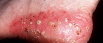
On the affected parts of the skin, pustules appear, permeated with hair, the size of a pinhead or slightly larger, with a dense covering and an inflammatory halo along the periphery. Pustules are filled with gray-white pus, thick consistency, not prone to fusion and peripheral growth. They are single and multiple. After 2–5 days, the pustules dry out to form a yellowish-brown crust. In their place, after the crust falls off, pigmented spots and slight peeling remain, which subsequently disappear without a trace. In some cases, the inflammatory process spreads deeper with the formation of deep infiltrates.
Staphylococcal impetigo should be distinguished primarily from folliculitis and streptococcal impetigo.
Rice. 5.1. In the photo there is folliculitis on the face (chin area).
Rice. 10 and 11. The photo shows superficial folliculitis.
Rice. 12 and 13. The photo shows superficial folliculitis (ostiofolliculitis).
Rice. 14 and 15. The photo shows superficial folliculitis (ostiofolliculitis).
Rice. 16 and 17. In the photo there is staphyloderma - staphyloccal impetigo.
Prevention
Compliance with basic rules of personal hygiene is the main preventive measure. In addition, it is recommended:
- Healthy food;
- sunbathing;
- avoid stress and overwork;
- regularly carry out vitamin therapy;
- maintain a daily routine;
- provide the child with adequate sleep;
- cut your nails regularly;
- strengthen immunity;
- promptly treat any damage to children's skin;
- fight excessive sweating.
All preventive measures that help avoid infection of children with pyoderma are familiar to parents. These are their direct parental responsibilities. If a child is instilled with a healthy lifestyle from childhood, he will not be afraid of any cocci. The only exceptions are unforeseen external circumstances that cannot be predicted (accidental microtrauma of the skin, contact with a pathogen carrier, etc.).
Folliculitis deep
Deep folliculitis is a purulent inflammation of the entire follicle, including the hair papilla. The disease develops when infection penetrates from ostiofolliculitis. The morphological element is a follicular pustule surrounded by a raised inflammatory ridge. Unlike a boil, there is no necrotic core. The disease is accompanied by severe pain. Patients with anemia, diabetes mellitus and immunodeficiencies of various etiologies are at risk.
Deep folliculitis is most often localized on the back, but can be recorded on any area of the skin where follicles are located. With the disease, deep follicular pustules of a conical shape form on the skin, accompanied by severe pain. Initially, the inflammatory infiltrate (nodules) has a bright red color, which after 3 days turns into a conical pustule. The pustules reach a diameter of 0.5 - 1 cm in diameter; an inflammatory corolla is located along the periphery. After opening the pustule, the crust dries out or an erosive surface is exposed. Healing occurs with the formation of temporary pigmentation or a small scar, and the hair follicle dies. The duration of deep folliculitis is up to 5 - 10 days. Deep folliculitis should be distinguished from acne, perioral and seborrheic dermatitis, herpes, epidermomycosis, etc.
Rice. 18 and 19. In the photo there is deep folliculitis.
Rice. 20. In the photo there is deep folliculitis.
Rice. 21 and 22. In the photo, deep widespread folliculitis.
Clinical researches
Conducted clinical studies prove the high efficiency, safety and tolerability of products for daily skin care of children with mild and moderate forms of atopic dermatitis and during remission, accompanied by a decrease in the quality of life of patients.
In the course of research conducted by the Union of Pediatricians of Russia, it was found that La-Cree cream for sensitive skin:
- reduces itching and irritation;
- relieves skin redness;
- moisturizes and gently cares for the skin.
Sources:
- Molochkova Yulia Vladimirovna, Dermatology. Brief reference book, GEOTAR-Media, 2022.
- Kildiyarova Rita Rafgatovna, Pediatrician for every day. Guide for doctors, GEOTAR-Media, 2022.
- Cohen Bernard A. Pediatric dermatology, MEDpress-inform, 2015.
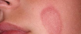
Sycosis vulgar
Sycosis vulgar (non-parasitic) is multiple folliculitis with a recurrent course and ostiofolliculitis with a pronounced inflammatory reaction. Inflammatory elements are localized in the area of beard growth, mustache, pubis, armpits (growth area of long hair) and eyebrows (bristly hair).
The cause of staphyloderma is Staphylococcus aureus. Shaving, a runny nose and other types of pollution, as well as an imbalance of sex hormones (damage to seborrheic zones) contribute to infection.
Clinically, sycosis is manifested by the development of superficial and deep folliculitis. The follicles penetrated by the hair become inflamed. A dense, painful infiltrate of bluish-red or bluish-brown color forms around them. When neighboring follicles are involved in the inflammatory process, pustular-inflammatory infiltrates are formed with a long course. The pustules gradually open up, the purulent infiltrate shrinks into crusts. When pulled out, a gelatinous glassy coupling soaked in pus is visible on the roots of the hair.
The course of the disease is long-term, relapsing – many months and even years.
Resolution of vulgar sycosis occurs without scar formation. Sometimes staphyloderma is complicated by eczematization.
Sycosis should be distinguished from the infiltrative form of trichophytosis, complicated scabies and herpes.
Rice. 7 and 8. The photo shows vulgar (ordinary) sycosis.
Rice. 23 and 24. The photo shows vulgar sycosis.
Rice. 25 and 26. In the photo, widespread vulgar sycosis.
Rice. 26.1 and 26.2. In the photo, common vulgar sycosis.
Treatment
It is advisable to begin therapy for pyoderma with a study to determine the type of pathogen and the sensitivity of patients to antibiotics, as well as determining whether this form of the disease is contagious.
In accordance with this, drugs for antimicrobial treatment are selected and a regimen for their administration is prescribed:
- the course of antibiotic therapy can be up to two weeks. Antibiotics such as Tetracycline, Rifampicin, Metacycline and others have proven themselves well.
- To strengthen skin blood vessels, angioprotectors are used: Trental, Actovegin, Sodium Nicotinate.
- In order to enhance regeneration processes, treatment of pyoderma in adults is accompanied by vitamin therapy, taking microelements and immunomodulating drugs.
- chronic deep forms of this pathology require the use of hormonal and cytostatic agents (Prednisolone, Methotrexate).
- in some cases, treatment with vaccines (staphylococcal medicinal, staphylococcal acellular) is prescribed.
In diabetes mellitus, treatment of pyoderma is based on strict adherence to dietary recommendations, rational rest and avoidance of stressful situations.
Local treatment of pathological formations includes: removal of crusts and treatment with aseptic preparations, the use of stretching liniments and ointments, and the application of sterile dressings.
In adults
The peculiarities of the course of the disease in mature people lie in the state of their immune system. Typically, it is its weakening that contributes to the development of pyoderma in adults.
Of the superficial forms, they are most often diagnosed with boils, carbuncles and various types of impetigo.
Deep types of disease develop against the background of serious damage to the central nervous system, internal organs and hormonal system. Therefore, young and mature people can develop: ulcerative and gangrenous pyoderma, Hoffmann's undermining folliculitis, atrophic dermatitis of the lower extremities.
In children
Since many systems are just being formed and improved in infants and toddlers in early childhood, even simple pyoderma is more difficult for them. Its superficial types are usually observed in the area of the umbilical ring, in the folds of the skin, on the face and limbs.
More often in children, symptoms of pyoderma are associated with increased body temperature, nervous excitability and anxiety, and enlarged lymph nodes.
For successful treatment of the disease, it is necessary to identify the causative agent and identify the form of the disease in time. In childhood they are usually diagnosed with:
- periporitis associated with excessive sweating of the baby; small ulcers appear on his skin in the area of the body, face and head, they burst and form crusts, become wet and itchy;
- diaper dermatitis, characterized by severe redness, pastiness and the formation of boils in the perineum or buttocks; without treatment, these formations ulcerate;
- panaritium, forms near the inflamed nail plate and represents small foci with suppuration.
In childhood, close attention should be paid to such processes and immediately consult a doctor; timely treatment will help avoid complications (abscesses, phlegmon and infection of other organs and systems).
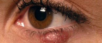
Barley
Barley is an acute purulent inflammation of the hair follicle or sebaceous gland of Zeiss. Barley can be external or internal. With external barley, the Zeiss or Mohl gland becomes inflamed, with internal barley, the meibomian gland. The infection gets into the eyes with dirty hands and when using shared towels. The disease can be unilateral or bilateral. Most common in children. The causative agent is Staphylococcus aureus.
Signs and symptoms of external stye. The disease begins with the appearance of hyperemia, edema and painful infiltration on the eyelid, in the center of which a purulent pustule appears after 2 - 3 days. On the 3rd - 4th day, the abscess opens and the purulent contents come out. In some cases, several styes may appear.
When abscess formation occurs, headaches appear, body temperature rises, and regional lymph nodes become enlarged.
Healing often occurs spontaneously and quickly. Sometimes antibiotic therapy is required, rarely surgical treatment.
Signs and symptoms of internal stye. Internal barley (meibomitis) is an acute inflammation of a bacterial nature (usually Staphylococcus aureus) of the meibomian gland (modified sebaceous glands located at the edges of the eyelids). The purulent infiltrate is visible through the conjunctiva of the eyelid, and the breakthrough of purulent masses occurs into the conjunctival cavity. Treatment sometimes requires surgery.
With barley, spontaneous self-healing is often recorded, but in some cases pyoderma takes a chronic, relapsing course.
Rice. 27. In the photo, styes are external (left) and internal (right) of the lower eyelid.
Rice. 28 and 29. In the photo there is a stye on the eye of children.
Rice. 30 and 31. The photo shows phlegmon of the child’s orbit. The disease develops as a result of the spread of microbes from skin lesions into the fiber and connective tissue of the orbit.
Rice. 32 and 33. In the photo there are multiple styes.
Rice. 34 and 35. Stye on the eye.
Rice. 36 and 37. Stye on the eye.
Rice. 38 and 39. Stye on the eye.
Rice. 40 and 41. Stye on the eye of the upper eyelid.
Rice. 42 and 43. In the photo there is stye on the lower eyelid.
Rice. 45 and 46. Stye on the eye can cause the development of an abscess and phlegmon of the eyelid.
How to treat pyoderma?
Treatment of pyoderma is carried out under the supervision of qualified specialists. As a rule, the doctor prescribes medications for external and internal use, including restoration of immune strength.
General uses:
- diagnosis and treatment of concomitant diseases (hormonal imbalance, diabetes, immunodeficiency);
- elimination of adverse effects on the skin (damage, pollution, exposure to high or low temperature);
- nutrition with a limitation of refined carbohydrates, a predominance of proteins, plant fiber, and fermented milk products;
- prohibition of washing (shower, bath), you can only wash local unaffected areas of the skin with great care so as not to spread the infection;
- trimming hair in the affected area;
- treating the skin around the ulcers twice a day with a solution of salicylic alcohol.
A special low-carbohydrate diet must be followed. The following medications are used to treat various types of pyoderma:
- It is recommended to carry out antibiotic therapy using semisynthetic macrolides, Penicillin, Tetracycline, aminoglycosides, and latest generation cephalosporins);
- in case of severe development of the disease, glucocorticosteroid drugs are used (Hydrocortisone, Metipred, etc.);
- for pyoderma, hepatoprotectors are prescribed (Essentiale Forte, Silibor, etc.);
- the use of angioprotectors (Actovegin, Trental) is recommended;
- cytostatics (Methotrexate) are prescribed.
To antisepticize erosive ulcerations, it is necessary to use ointments with a bactericidal effect. Most often used in the treatment of pyoderma:
- zinc ointment or salicylic-zinc paste;
- Levomekol;
- tetracycline ointment;
- lincomycin ointment;
- erythromycin ointment;
- hyoxysone ointment, etc.
In addition, there are drugs for complex treatment that have antibacterial, anti-inflammatory and antifungal effects. The most popular ointments are Thymogen and Triderm.
When pyodermatitis is accompanied by the appearance of ulcers, the inflammatory foci should be washed with aseptics after removing the scab (Tannin, Furacilin, boric acid, Dioxidine, Chlorhexidine, etc.).
For carbuncles, boils, hidradenitis, you can apply a sterile bandage with Ichthyol + Dimexide, Chymotrypsin and Trypsin to the affected area. In addition, a Tomicide bandage is often applied to the affected parts of the body.
Furuncle
Furuncle is an acute purulent-necrotic inflammation of the hair follicle, sebaceous gland and surrounding subcutaneous fat. Boils can be localized on any area of the skin where follicles are present. Most often they appear on the face, back of the neck, thighs and buttocks - places where subcutaneous fatty tissue is most developed. Localization in the area of the nose and upper lip is dangerous, due to the possible penetration of staphylococci into the venous network of the brain.
A boil can develop from folliculitis or an inflammatory nodule in the dermis.
In the first case, the disease begins acutely with deep folliculitis with a powerful perifollicular infiltrate, in the center of which necrosis quickly develops, or gradually with superficial folliculitis. Around the inflamed follicle, a purple-red, painful infiltrate (nodule) forms over the course of several days, which gradually increases in size. A cone-shaped pustule forms in its thickness with the formation of a necrotic core in the center. After 1–2 days, the infiltrate opens. The resulting ulcerative defect is filled with granulation tissue and scarred. Healing lasts 1 – 2 weeks.
Boils reach large sizes when localized in places where subcutaneous fatty tissue is well developed - the face, thighs and buttocks. Particularly painful boils are localized in places where soft tissues are minimally developed and tendons and nerves pass through - the external auditory canal, the scalp, the anterior surface of the legs, the dorsum of the fingers.
Localization in the area of the nose and upper lip (nasolabial triangle) is dangerous, due to the possible penetration of staphylococci into the venous network of the brain, with the subsequent development of thrombophlebitis of the cavernous sinus, facial veins, meningoencephalitis and sepsis.
The disease may be limited to the appearance of one boil, or multiple ones, and then they speak of furunculosis. With furunculosis, the presence of boils is noted, which are at different stages of development. There are local and general furunculosis. Both options can be acute or chronic. The disease lasts a long time - months and even years. Furunculosis often affects people with reduced immunity and those suffering from diabetes.
Furuncle should be distinguished from pseudotuberculosis, hidradenitis, carbuncle, erythema nodosum and deep trichophytosis.
Rice. 47 and 48. The photo shows a boil in the stage of suppuration (left) and cleansing - rejection of purulent masses (right).
Rice. 49 and 50. In the photo there is a boil on the thigh and chin in the stage of suppuration.
Rice. 51 and 52. The photo shows a boil on the face.
Rice. 53 and 54. The photo shows boils on the nose.
Rice. 55 and 56. The photo shows a boil in the area of the auricle.
Rice. 57 and 58. The photo shows a boil of the ear canal (left) and the auricle (right).
Rice. 59 and 60. The photo shows a boil on the buttock and finger.
Rice. 61 and 62. The photo shows a boil in the healing stage.
Rice. 63 and 64. The photo shows furunculosis.
Complications
Severe consequences of pustular diseases are observed with:
- untimely access to a medical institution;
- weak immunity;
- undergoing an incomplete course of therapy;
- use of questionable treatment methods;
- poor hygiene;
- preservation of provoking factors.
Complications:
- inflammation of the lymph nodes;
- abscesses;
- scars in areas of self-removal of pustules; infection of bone tissue;
- blood poisoning;
- thrombosis of cerebral vessels;
- meningitis;
- inflammation of internal organs.
Carbuncle
Carbuncle is an acute purulent-necrotic inflammation of a group of hair follicles united by a common infiltrate. Of all staphyloderma, it is the most severe inflammation. The disease is registered mainly in adults. The areas of the body most susceptible to contamination are most often affected - the back of the neck, back and lumbar region. Carbuncles often occur in people with reduced immunity, suffering from diabetes mellitus and other diseases debilitating the body, as well as with massive contamination of the skin and non-compliance with the hygienic regime.
The carbuncle is a purple or dark red nodule up to 5–10 cm in diameter, not clearly demarcated from the surrounding tissues, with several pustules on the surface. Opening the focus of suppuration is accompanied by the release of a purulent-bloody mass and rejection of necrotic rods. After opening the carbuncle, an extensive ulcer with undermined edges forms on the skin. Its bottom is covered with a coating of mucopurulent consistency and bleeds easily. Gradually, the ulcerative defect is cleared of plaque and filled with granulations. Healing occurs within 2–4 weeks with the formation of a deeply retracted scar. The disease occurs with pronounced symptoms of intoxication.
Carbuncles are most often single. Of the complications, the most dangerous are phlebitis, thrombosis of the cerebral sinuses and sepsis.
Rice. 65 and 66. The photo shows a carbuncle (left) and a boil (right).
Rice. 67 and 68. The photo shows a single carbuncle of the interscapular region (left) and the lateral surface of the body (right).
Rice. 68.1. The photo shows multiple carbuncles of the shoulder region.
Rice. 69 and 70. The photo shows a huge carbuncle in the back of the neck. Numerous pustules are visible, from which pus is released, like from a sieve.
Rice. 71 and 72. The photo shows a carbuncle on the face.
Rice. 73 and 74. The photo shows a carbuncle on the neck.
Rice. 75 and 76. The photo shows a carbuncle on the back.
Rice. 77 and 78. The photo shows a carbuncle on the neck (left) and multiple carbuncles (right).
Rice. 79 and 80. The photo shows a carbuncle on the back and finger.
Rice. 81 and 82. In the photo there is a carbuncle. The opening of the abscess is made in the form of a cross-shaped incision.
Rice. 83. In the photo there is a carbuncle on the chin. The opening of the abscess is made in the form of a cross-shaped incision.
Rice. 84, 85 and 86. The photo shows a carbuncle on the back. The stages of cleansing the ulcer are shown.
Rice. 87 and 88. The photo shows a carbuncle on the neck. Wound healing in the process of treating a source of suppuration.
Rice. 89 and 90. In the photo there is a carbuncle. The ulcerative defect has been cleared.
Diagnostics
The main diagnostic criteria are the characteristic elements of rashes on the body (pustules, conflicts).
To establish the exact type of disease and the causative agent that caused it, a microscopic method is used to examine the discharge of purulent elements. For deep tissue damage, a biopsy may be used. In case of severe diseases, it is recommended to draw blood to determine glucose levels (the goal is to exclude diabetes mellitus). When performing a general blood test, an increase in leukocyte counts and ESR is often noted.
Differential diagnosis is carried out with skin manifestations of tuberculosis, syphilis, parasitic and fungal infections of the epidermis, candidiasis, microbial eczema.
Hidradenitis
Hidradenitis is a purulent inflammation of the apocrine sweat glands. The disease is especially often registered in women from puberty; in old age it practically does not occur due to the extinction of the activity of the sweat glands. The causative agent of hidradenitis in most cases is Staphylococcus aureus.
Apocrine glands are located in the armpits, around the navel, areola of the mammary glands, and the anogenital area. Most often, hidradenitis is localized in the armpits, less often in the perianal and groin areas.
Clinically, the disease is manifested by the appearance of a painful node in the deep layers of the skin the size of a pea, which within a few days increases to the size of a hazelnut and even a walnut. The skin over the infiltrate becomes purplish-red. Multiple suppurations in the armpit have a conical shape (popularly called “bitch udder”). After 4–5 days, the foci of suppuration open with the release of purulent masses mixed with blood. After drainage, inflammatory phenomena quickly subside. Healing occurs with the formation of cord-shaped scars. Hidradenitis often occurs with severe symptoms of intoxication. The inflammatory process lasts about 2 weeks and often has a recurrent course. The disease is especially severe in obese people.
Rice. 91 and 92. The photo shows hidradenitis. Multiple suppurations in the armpit have a conical shape (popularly called “bitch udder”).
Rice. 92.1 and 92.2. The photo shows hidradenitis. The most common location is the armpits.
Rice. 93 and 94. The photo shows hidradenitis in women.
Rice. 95. The photo shows the consequences of hidradenitis in a woman (rare localization).
Rice. 97 and 98. The photo shows hidradenitis. Localization of inflammatory infiltrates in the perineal area and around the anus occurs more often in men.
Rice. 96. In the photo, hidradenitis in a man, the consequences of the disease.
Rice. 99 and 100. In the photo, hidradenitis in the armpit is multiple scars formed after healing of the fistula tracts.
Rice. 101 and 102. The photo shows the consequences of hidradenitis - multiple hypertrophic and keloid scars formed after healing of the fistula tracts.
Rice. 103 and 104. In the photo, the consequences of hidradenitis are ugly, retracted scars.
Rice. 105 and 106. The photo shows hidradenitis, the condition after healing.
Rice. 107 and 108. The photo shows hidradenitis, the condition after plastic surgery.
Staphyloderma in infants
Staphylococcal infection occupies a leading position among all diseases in young children due to the anatomical features of the structure of their skin.
Vesiculopustulosis
Vesiculopustulosis or periporitis is a purulent inflammation of the mouths of the eccrine sweat glands. The causative agent of the disease is Staphylococcus aureus. When the excretory ducts are blocked, sweat accumulates in the ducts, stretching them. Blockage occurs at different levels, which determines the clinical picture of the disease.
With crystalline prickly heat, the ducts are blocked at the level of the stratum corneum - close to the surface of the skin. The disease is characterized by the appearance on the skin of painless vesicles (bubbles), filled with clear liquid (sweat), with a diameter of up to 1 - 2 mm. The bubbles look like droplets of water, sometimes they are draining in nature.
With prickly heat, blockage occurs at the level of the middle and terminal layers of the epidermis. Red itchy nodules (papules and papulovesicles) appear on the skin, surrounded by a red inflammatory ridge. The nodules reach 2 mm in diameter and are surrounded along the periphery by an inflammatory halo. The rash is accompanied by itching and burning. The itching can be quite severe and intensifies with sweating and increased ambient temperature. Miliaria rubra always requires medical treatment. The duration of the disease is about 2 weeks.
When the mouths of the eccrine sweat glands become infected, vesiculopustulosis develops against the background of existing miliaria. The disease is characterized by the appearance on the skin of superficial pustules-vesicles, the size of a millet grain, filled with milky-white contents (miliaria alba), surrounded by a hyperemic corolla. After the pustules are opened and the pus is rejected, the surface becomes covered with serous-purulent crusts, after the rejection of which scars or pigment spots remain. The duration of the disease ranges from 2 to 10 days. When inflammation spreads deeper, as is observed in premature babies, multiple abscesses develop.
Rice. 109 and 110. In the photo, vesiculopustulosis is one of the symptoms of staphylococcal infection in infants.
Rice. 111 and 112. The photo shows vesiculopustulosis.
Rice. 113 and 114. The photo shows vesiculopustulosis.
Rice. 114.1. The photo shows vesiculopustulosis.
Multiple abscesses in children (Finger's pseudofurunculosis)
The disease may occur primarily or be a continuation of vesiculopustulosis. The disease is characterized by the involvement of the excretory ducts of the sweat glands and even the entire glomerulus of glands in the infectious process. At risk are children suffering from rickets, malnutrition, excessive sweating, hypovitaminosis and anemia. Finger's pseudotuberculosis occurs with a violation of the general condition of the child. Nodules and larger (1-2 cm) nodules appear on the skin of the scalp, buttocks, back or inner thighs, the skin over which becomes bluish-red and thins. After opening the infiltrates, thick pus is released. Healing occurs with a scar.
Rice. 115 and 116. The photo shows staphyloderma in newborns - multiple abscesses (Finger's pseudofurunculosis).
Epidemic pemphigus of newborns
Epidemic pemphigus of newborns (pyococcal pemphigoid) occurs in the first week of a child’s life and is a common superficial purulent, highly contagious lesion of the skin.
The source of pyogenic microbes (staphylococci and streptococci) is the birth canal and purulent infection in the mother, infection in staff and children, care items, diapers and the surrounding air. The disease progresses in waves, with new rashes occurring every 7-10 days, which is accompanied by dyspepsia and vomiting in the child.
The disease goes through several stages of development. First, red spots appear, in place of which bubbles filled with clear liquid soon form. Then the liquid becomes purulent and the blisters burst. Crusts form in place of the bubbles. Healing occurs without scarring. The larger the affected area, the more severe the disease. In severe cases of pemphigus, blisters appear on the mucous membranes of the nose and mouth.
Rice. 117 and 118. In the photo In the photo there is staphyloderma - pemphigus of the newborn.
Exfoliative dermatitis (Ritter's disease)
Ritter's exfoliative dermatitis or acute epidermolysis of the newborn is a malignant form of pemphigus, the most severe form of staphylococcal pyoderma. Premature babies are at risk. The disease develops in children in the first days of life, is severe and long-lasting, accompanied by high fever and symptoms of intoxication. Clinically, the disease is manifested by redness of the skin, the appearance of cracks, desquamation of the epidermis around the mouth or in the navel area, serous impregnation of the skin, epidermolysis, the appearance of flabby blisters, and the symptom of “scalded skin.” In some cases, the mucous membranes and visceral organs are affected. Resolution occurs without scarring. The illness lasts about 15 days. A complicated course is often observed with the development of pneumonia, pyelonephritis, otitis, purulent conjunctivitis, abscess, phlegmon, and sepsis.
Rice. 119. Ritter’s photoexfoliative dermatitis in a newborn.
What it is?
Pyoderma is a pustular skin disease caused by pyogenic bacteria, the main of which are staphylococci and streptococci, and a little less frequently - Proteus vulgaris and Pseudomonas aeruginosa. Pyoderma is more common in childhood and among workers in certain types of industry and agriculture.
An increase in incidence is observed in the autumn-winter period - the cold and damp season. The humid climate of hot countries is the reason for a large number of patients with mycoses and pustular skin diseases.