Neurodermatitis (or atopic dermatitis) is a common skin disease accompanied by itching and inflammation of the skin. Among all allergic skin diseases, neurodermatitis occurs in 25-30% of cases. Atopic dermatitis occurs more often in women, and residents of large cities with unfavorable environmental conditions are also more susceptible to the disease. Poor nutrition and environmental pollution lead to an increase in the incidence of allergies throughout the world. Treatment of neurodermatitis usually requires time and the complex use of medicinal and non-medicinal methods.
Causes
Atopic dermatitis is a disease with a hereditary predisposition. The main factors in the development of the disease:
- Atopic constitutional abnormality (the body's predisposition to an excessive response when in contact with allergens).
- Features of the skin condition, weakening of its barrier function.
- Unfavorable course of pregnancy, childbirth (a factor in the development of the disease in the child).
- Irrational feeding in the first year of life (including early cessation of breastfeeding).
- Disorders of the digestive system: dysbiosis, biliary dyskinesia.
- Dysregulation of the central nervous system.
- Stress, traumatic situations.
- Overwork.
- Bad habits (smoking, alcohol abuse)1.
The risk of developing neurodermatitis in a child whose both parents suffer from allergic diseases (bronchial asthma, allergic rhinitis, urticaria) can reach 75%, so it is very important to monitor the condition of his skin from the first days of life. Various allergens provoke exacerbations of neurodermatitis:
- pet hair;
- antibacterial drugs;
- flavorings;
- plant pollen;
- house dust mites.
Atopic dermatitis is also exacerbated by upper respiratory tract infections during seasonal outbreaks of ARVI.
Complications
In general, neurodermatitis is not cause for concern and is not associated with serious health problems, but in the acute phase it can significantly affect the quality of life. Excessive dry skin, rashes, sores and sores increase the risk of bacterial and fungal skin infections. It is not always possible to notice them in time, for example, if foci of dermatitis involve the scalp. In this case, it may be difficult to properly care for irritated skin, which also contributes to the development of inflammation.
The second common complication of neurodermatitis is the formation of scars at the site of the rash. The main reason for their appearance is uncontrolled scratching of inflammation.
Symptoms
Neurodermatitis can occur acutely, subacutely or chronically with exacerbations3. There are several criteria to recognize the disease:
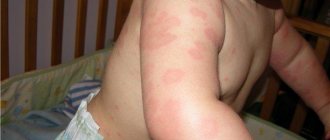
- A variety of skin rashes. There is redness of the skin, the appearance of spots of various shapes, scales, crusts, areas of thickening of the skin, increased skin pattern, roughness, and pigmentation disorders. The severity and type of elements of the rash depend on the form and severity of the disease3.
- Symmetrical rashes. Most often, rashes occur on the skin of the face, head, neck, shoulders, legs, elbows and popliteal fossae. The rash may be widespread or localized.
- Long-term course with periodic exacerbations. Onset usually occurs in childhood or early adolescence. The disease occurs with periods of remissions and exacerbations.
- Changes in the nature of the rash with age. As you get older, the rashes change their location. Inflammatory elements give way to areas of dryness and thickening of the skin.
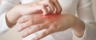
- Severe itching. The itching is debilitating, worsens at night, and leads to sleep disturbances.
- The addition of a secondary infection of the skin occurs due to scratching of the rash.
- Paleness, dry skin, dull hair, flaking on the eyelids (the so-called “atopic face”).
- The presence of other diseases of allergic origin (asthma, seasonal rhinitis).
The manifestations of neurodermatitis change with age. During infancy, the course of the disease is acute, there is redness of the skin, swelling, exudation with subsequent formation of crusts. From the third year of life, neurodermatitis in children occurs in a chronic form with symptoms of dryness and thickening of the skin2. Characterized by redness of the skin, the presence of itchy papules, areas of peeling, and pigmentation disorders. In severe cases, it may be accompanied by a bacterial infection with the appearance of pustules, increased body temperature and general intoxication of the body. Atopic cataracts and visual impairment are a rare complication of severe atopic dermatitis, occurring in approximately 1% of patients.
Atopic dermatitis, or neurodermatitis
To characterize skin lesions in the form of papular-vesicular rashes, hyperemia, infiltration, dryness, peeling, lichenification, the terms “atopic dermatitis” and “neurodermatitis” are more often used. According to the clinical course, atopic dermatitis is divided into limited and diffuse. For limited atopic dermatitis, the typical localization area is the back of the head, back of the neck, elbow and popliteal folds, forearms, and inner thighs. With limited atopic dermatitis, there are one or two, less often several, lesions. The disease is accompanied by dry rashes; weeping is not typical.
Diffuse atopic dermatitis differs from limited atopic dermatitis in a more widespread process. Sometimes the process takes on a generalized character. Along with vaguely limited brownish-red areas of infiltrated and lichenified skin, which can cover large surfaces, individual flat shiny nodules and multiple excoriations are noted. The skin of patients with diffuse atopic dermatitis is dry. Some patients experience vesiculation and oozing. At the same time, the clinical picture of the disease is dominated by, along with infiltration and lichenification, swelling of the skin, papules, vesicles, erosions and erythema of an acute inflammatory nature. Patients are bothered by incessant itching. Temporary hyperpigmentation of the skin remains in areas of resolved rashes. In diffuse atopic dermatitis, a chronically relapsing course is observed with frequent and prolonged exacerbations. Very often, patients with atopic dermatitis experience respiratory manifestations of atopy: bronchial asthma, rhinoconjunctivitis.
| Along with atopic dermatitis, or neurodermatitis, which is well known to dermatologists, there is severe, generalized atopic dermatitis, which occurs with constant symptoms of eczematization and pyoderma. It is characterized by a combination with atopic diseases of the respiratory tract in the form of allergic rhinosinusitis, conjunctivitis and atopic bronchial asthma. The severe course of the disease often leads to disability. Treatment of these patients seems extremely difficult, due to common drug intolerance |
One of the features of the course of atopic dermatitis is a purulent skin infection, manifested in the form of pyoderma, which is characterized by small pustular rashes that are superficial. Typical for these patients is herpes simplex, which can be localized on the skin of the face, arms, buttocks, and mucous membranes and is capable of migrating to new areas. The predisposition of patients with atopic dermatitis to staphyloderma and herpes infection is considered a clinical sign of immune deficiency. Conjunctivitis in atopic dermatitis is sometimes seasonal and is a manifestation of hay fever, although in most cases it occurs year-round. When cultured from the conjunctiva, Staphylococcus aureus is often identified.
Thus, we can distinguish a syndrome that is characterized by a combination of severe generalized atopic dermatitis with alternating widespread eczematization and pyoderma and respiratory manifestations of atopy. In this case, there is an extremely high level of IgE, sensitization to almost all atopic allergens. The combination of allergic IgE-mediated reactions and immunodeficiency, clinically manifested in recurrent pyoderma, herpes infection and confirmed by the results of an immunological study, is one of the pathogenetic mechanisms for the development of this syndrome. The particular severity of the disease, the young age of the patients, and the ineffectiveness of traditional methods of therapy made it possible to isolate this disease and call it severe atopic syndrome (Yu. A. Poroshina, E. S. Fedenko, V. D. Prokopenko, 1985).
Unfortunately, traditional methods of therapy are not effective enough in the treatment of severe atopic syndrome. Glucocorticosteroids, which have a pronounced anti-inflammatory and antiallergic effect, often lead to steroid dependence and cause a number of severe complications in the form of exacerbations of foci of infection and pyoderma. Exacerbation of pyoderma causes the need for antibacterial therapy, which, as a rule, aggravates atopic dermatitis or is complicated due to the patient's drug intolerance. Due to the ineffectiveness of traditional therapy, the Institute of Immunology of the Ministry of Health of the Russian Federation has developed and carries out methods of extracorporeal immunopharmacotherapy (EIPT, plasmacytopheresis with diuciferon, prednisone and other drugs).
For the period 1995–1997 In the Clinic of Neurosis, 2686 patients were examined who consulted an allergist regarding various itchy skin rashes. Of these, 612 were diagnosed with atopic dermatitis (205 men and 405 women aged 16 to 35 years, average age 22–25 years).
Limited atopic dermatitis was observed in 319 patients, widespread atopic dermatitis in 205, and severe atopic syndrome was detected in 88. The diagnosis was made on the basis of clinical, allergological and immunological examination.
All patients underwent general clinical examination, including functional, laboratory instrumental examination, consultations with specialists (ENT, gastroenterologist, ophthalmologist, etc.).
In addition to a carefully collected allergological, pharmacological and food history according to a specially developed scheme, an allergological examination was carried out with standard domestic sets of household, pollen, epidermal and food allergens and included skin tests - prick or scarification (in patients with severe skin lesions in the forearms, tests were performed on back), provocative nasal and conjunctival tests, as well as the test of inhibition of natural emigration of leukocytes (TTEEL) in vivo according to Ado A.D. with medications.
The immunological examination included the determination of general and specific IgE in the blood serum and the study of indicators of primary immune status: the number of leukocytes, lymphocytes, T- and B-lymphocytes, T-helpers and T-suppressors, immunoglobulins of class A, M, G, as well as neutrophil phagocytosis .
Clinically, skin manifestations were characterized by the presence of papular-vesicular rashes, in some places with hemorrhagic crusts on the surface, severe dryness, widespread erythema, and lichenification. Foci of bright hyperemia, swelling and exudation were noted in the elbow bends and popliteal areas. Subjectively, I was bothered by a painful itch. In 28 out of 88 patients with severe atopic syndrome, pyoderma was observed - superficial pustules with purulent discharge. Pyoderma was widespread in 9 patients, limited in 19, and was combined with furunculosis in 5 patients. 82 people suffered from recurrent herpes simplex.
Table 1. Respiratory manifestations of atopy in 365 patients with atopic dermatitis
| Disease | Number of patients | |
| (in abs). | % | |
| Dust allergic rhinoconjunctivitis | 83 | 23,8 |
| Dust bronchial asthma | 96 | 26 |
| Hay fever (pollen bronchial asthma and rhinoconjunctivitis) | 152 | 42 |
| Combination of dust rhinoconjunctivitis, bronchial asthma and hay fever | 34 | 9 |
| Total | 365 | 100 |
In addition to manifestations of atopic dermatitis and pyoderma, many patients (365) had respiratory manifestations of atopy, which are presented in Table 1.
The majority of patients (427) suffered from concomitant diseases of the gastrointestinal tract (chronic gastritis, peptic ulcer of the stomach and duodenum, dyskinesia of the biliary tract and colon, intestinal dysbiosis, etc.).
In 362 patients, pathology of the ENT organs was identified (chronic sinusitis, polypous ethmoiditis, chronic tonsillitis, chronic subatrophic pharyngitis).
As can be seen from Table 1, a significant number of patients (41%) with atopic dermatitis were diagnosed with hay fever. Half of the patients (49.1%) had respiratory manifestations of household allergies.
During an allergological examination of 612 people suffering from atopic dermatitis, sensitization to non-bacterial allergens was detected in 428 patients. Table 2 presents the results of an allergological examination of these patients according to skin (prick and prick), nasal and conjunctival tests, as well as the determination of specific IgE in the blood serum.
As can be seen from Table 2, the largest number of positive tests were obtained with household and pollen allergens, which, as mentioned above, is due to the presence of not only skin, but also respiratory manifestations of atopy.
Table 2. Results of allergological examination of 428 patients with atopic dermatitis
| Allergens | Number of patients with positive tests | |
| (in abs). | % | |
| Household allergens (house dust, feather pillows, library dust) | 179 | 42 |
| Grass pollen | 63 | 14 |
| Tree pollen | 37 | 8 |
| Pollen of Asteraceae and Chenopodiaceae | 52 | 12 |
| Epidermal allergens (cat, dog, rabbit, sheep, horse dander, human hair) | 42 | 10 |
| Food allergens (milk, fish, egg) | 9 | 2 |
| Combination of different groups of allergens | 46 | 11 |
| Total | 428 | 100 |
105 patients with a history of drug intolerance underwent TTEEL with drugs. In 38 people it was positive.
The clinical course of widespread atopic dermatitis and severe atopic syndrome with relapses of pyoderma and herpes in the presence of chronic foci of infection (chronic maxillary ethmoiditis, chronic tonsillitis, intestinal dysbiosis, etc.) suggested that this category of patients has a combination of allergies and immunodeficiency. An immunological examination revealed the following changes in humoral and cellular immunity: a decrease in the number of peripheral blood lymphocytes due to a decrease in T-cell populations, a decrease in neutrophil phagocytosis. There was an increase in the production of immunoglobulins of class A, M, and especially IgE. In all examined patients, a significant increase in the level of total IgE was recorded, sometimes 10–30 times compared to the level in healthy people.
Along with the term atopic dermatitis, which reflects the allergic nature of this disease, the older definition of neurodermatitis is still widely used (especially by dermatologists). This name was not given without reason, since in almost all patients the exacerbation of the skin process is caused by emotional stress factors and a traumatic situation. In turn, constant itching, which bothers the patient, causes irritability, short temper, and leads to insomnia.
Example
Patient Ch., born in 1972, was in the clinic for treatment from 02/18/93 to 04/29/93 for neurotic depression, severe asthenic-depressive syndrome.
Concomitant diagnosis: atopic dermatitis, a common form in the acute stage (remission at discharge). Hay fever. Allergic rhinoconjunctivitis. Sensitization to tree pollen. Food allergies to nuts, apples, carrots. Chronic gastritis with reduced secretory function. Dyskinesia of the colon.
Complaints of constant itching of the skin, rashes, irritability, short temper, tearfulness, sleep disturbance, headaches, increased fatigue, decreased mood.
From the anamnesis: my grandmother suffered from eczema. My aunt has bronchial asthma. She was born as her first child, on time. According to the mother, pregnancy and childbirth proceeded without complications. From the age of five months she was bottle-fed and almost immediately developed infantile eczema. At the age of five, neurodermatitis was diagnosed. Was observed by dermatologists. There were long periods of relative well-being, when limited dermatitis remained (in the area of the elbow joints, hands). There is always improvement in summer, worsening in September-October. Since 1992, symptoms of rhinitis appeared in May. There was an exacerbation of dermatitis after eating nuts, chocolate, eggs, and citrus fruits. In August 1993, angioedema occurred after eating apples; after that, she did not eat apples. Since childhood, a sharp exacerbation of dermatitis due to stressful situations has been noted. The last deterioration was about a year against the background of a traumatic situation in the family (divorce from my husband). Skin rashes have spread, constant itching bothers me, and sleep has been disturbed because of this. I lost 7 kg in a year. I took suprastin and diphenhydramine with little effect. Local treatment prescribed by a dermatologist (the patient does not know the names of the ointments) gave only short-term improvement.
On admission: general condition was satisfactory. The skin is dry, against the background of areas of hyperemia, multiple scratches, small papular, vesicular scattered rashes, crusts. There is pronounced lichenification in the neck, elbow joints, and wrists. There is no swelling. There are no symptoms of rhinitis or conjunctivitis. The mucous membrane of the oral cavity and pharynx is not changed. There is vesicular breathing in the lungs, no wheezing. Heart sounds are clear, rhythmic, blood pressure is 110/70. Pulse 88 per minute, satisfactory properties. The abdomen is soft, painless in all parts. The liver is not enlarged. There is no dysuria. Stool is not regular, tendency to constipation. Asthenized. The mood is low. Fixed on her condition and on the traumatic situation in the family.
Results of clinical and laboratory examination: class. an. blood – HB-124, L-5.0; p.1, p.46, e.12, l.36, m.5, soe-8 mm/hour. Biochem. an. blood: total protein 80.0; urea 4.4; creatinine 89.1, cholesterol 6.1; beta lipoproteins 6.1; bilirubin total 14.8; ALT–0.25; AST-0.34; glucose 4.7; seromucoid 0.10. SRB - negative, Wasserman district negative.
An. urine – without features.
ECG: without pathology.
X-ray of the PPN - the transparency of the sinuses is not impaired.
EGDS: gastritis, duodeno-gastric reflux.
Cons. ENT: healthy.
Cons. gynecologist: healthy
Cons. gastroenterologist: chronic gastritis with reduced secretory function. Dyskinesia of the colon.
Cons. neurologist: vegetative-vascular dysfunction of the hypotonic type.
Immune status: L:5.0; Lf-36%, - 1.9; phagocytosis 74%, Tlf-61%-1.1; Vlf 5% -0.09; IgA 230, IgM 110, IgG 1400, IgG-tot. more than 1000.
Allergological examination: scratch tests are positive with allergens from birch pollen +++, alder +++, hazel +++, oak ++, ash ++, poplar +, with epidermal and household allergens – negative. Certification tests with food passive allergens - nuts (walnuts, hazelnuts, almonds, peanuts), carrots, apples (Starkin, Golden) - positive. Testing was carried out on the back.
Treatment: individual hypoallergenic diet, IV drip of Dexon 8 mg per person. r-re, two days; IV drip Dexona 4 mg. On physical solution – 3 days, total 28 mg; IV drip Ascorbic acid 5.0 per physical. solution 5 days; zaditen 1 t. x 2 times, festal 1 t. x 3 times during meals, intal 2 drops. x 3 times (dissolved in water) in 20 minutes. before meals, activated carbon 1 t. x 3 times 1.5 hours after meals; IV drip tavegil 2.0 for physical solution - 2 days, tavegil 2.0 IM at night - 5 days, retabolil 1 t. at night, local skin treatment with celestoderm ointment and Unna ointment, physiotherapy (electrosleep, ultraviolet irradiation), acupuncture, complex psychotherapy, therapeutic relaxation gymnastics, group and individual sessions with a psychologist. HBOT – 5 sessions.
As a result of the treatment, pronounced positive dynamics were noted: the skin rashes completely disappeared, lichenification remains in the area of the elbows and wrists, and there is almost no itching. Sleep returned to normal. I became calmer, more cheerful, more active. My mood has improved.
Recommendations given upon discharge:
- Observation of an allergist at the place of residence.
- Following a hypoallergenic diet with the exception of apples, nuts, honey, carrots, stone fruits, and histamine-liberating products.
- Continue taking zaditen 1 t. x 2 times, topically Unna cream.
- In May, it is advisable to travel to a different climate zone.
- Herbal medicine, penicillin drugs, nicotinic acid, B vitamins, and radiocontrast agents are not indicated.
- Avoid contact with dust and household chemicals.
- Frequent change of underwear and bed linen.
- Before surgery, premedication: IV or IM Dexon 8 mg, IM Tavegil 2.0; honey. vaccinations only for health reasons and with the same premedication.
Treatment
Since neurodermatitis is a chronic disease, it requires long-term complex treatment. Main directions of therapy:
- Limiting contact with allergens. It is important to follow a diet that excludes food allergens. These most often include citrus fruits, coffee, eggs, smoked foods, products containing dyes and flavors. In residential areas, you need to get rid of fungal mold and dust mites.
- Correction of underlying diseases. Since the development of neurodermatitis is influenced by the state of the central nervous system and gastrointestinal tract, it is necessary to ensure their normal functioning. You should avoid stress, follow a sleep and rest schedule, and maintain a balanced diet. If necessary, use enzyme preparations and prokinetics.
- Drug treatment. Use sedatives and antihistamines, emollients, vitamins, glucocorticosteroids (for sudden, severe exacerbations under the supervision of a physician). For local treatment of inflamed skin, moisturizing, antipruritic ointments and gels are used. They often contain components such as menthol, anesthesin and tar. For bacterial skin infections, antibiotic ointments are prescribed1.
Physiotherapeutic procedures – phototherapy, laser therapy, plasmapheresis – can be used in complex treatment.
Diagnostics
Diagnosing neurodermatitis with a dermatologist is not difficult. This is due to the clear clinical picture. Initially, anamnesis is collected. During a conversation with a patient, the doctor is primarily interested in the presence of atopic diseases in relatives. He will also be interested in other questions in particular:
- Seasonality of exacerbations of pathology.
- Negative reactions to vaccination.
- Presence of allergic reactions.
A general blood test is important for making a diagnosis. First of all, the level of eosinophils and leukocytes, as well as ESR indicators, are assessed. Additionally, skin tests and cultures are performed. An immunogram is also performed, which allows you to evaluate the main indicators of the immune system. During diagnosis, other studies may be prescribed to rule out scabies, psoriasis, eczema and other diseases with similar symptoms.
To diagnose the presence of concomitant chronic diseases, consultations with highly specialized specialists are required. Based on their recommendations, the required laboratory and instrumental studies are carried out. Patients must undergo a psychological examination. This allows you to confirm or exclude the presence of malfunctions in the autonomic nervous system.
Cetrin for neurodermatitis
Cetrin is a second generation antihistamine used for acute and chronic allergic diseases. The active substance, cetirizine hydrochloride, blocks H1-histamine receptors4. This prevents the development of allergic reactions, which result in redness, swelling of the skin, and severe itching.
Advantages of the drug:
- Practically does not cause drowsiness in a therapeutic dosage.
- Take 1 tablet (10 mg) 1 time per day, regardless of food intake.
- Begins to act 20 minutes after administration.
- The effect lasts up to 3 days after the last dose of the drug.
- Suitable for use in children over 6 years of age4.
Cetrin is suitable for the treatment of exacerbations of neurodermatitis in adults and children. It relieves itching, the severity of skin hyperemia, and improves the quality of life of people suffering from atopic dermatitis1.
What it is
Neurodermatitis is an inflammatory skin disease that occurs due to its increased reactivity to allergens or some nonspecific irritants.
Many people know it under another name: atopic dermatitis. Neurodermatitis is not as rare as we would like. Up to 12% of people suffer from its manifestations, with women more often than men².
Most often, the disease is observed in infants and young children, but sometimes symptoms remain with a person for life. In this case, coping with dermatitis can be difficult, but with the help of modern medications, patients with a chronic form of the disease can achieve stable remission and live a full life.
It should be noted that despite its frightening appearance (the disease manifests itself as a rash, dryness and wounds on the skin), neurodermatitis is not contagious and does not pose a threat to life, although it can cause a lot of trouble, mainly due to itching.
Compound
| Ointment for external use | 1 g |
| active substances: | |
| betamethasone dipropionate | 0.643 mg |
| (equivalent to 0.5 mg betamethasone) | |
| clotrimazole | 10 mg |
| gentamicin (as gentamicin sulfate) | 1 mg (1000 IU) |
| excipients: liquid paraffin - 50 mg; soft white paraffin – qs up to 1 g |
| Cream for external use | 1 g |
| active substance: | |
| betamethasone dipropionate | 0.643 mg |
| (equivalent to 0.5 mg betamethasone) | |
| clotrimazole | 10 mg |
| gentamicin (as gentamicin sulfate) | 1 mg (1000 IU) |
| excipients: petroleum jelly - 150 mg; propylene glycol - 100 mg; cetostearyl alcohol - 72 mg; liquid paraffin - 60 mg; macrogol cetostearate - 22.5 mg; benzyl alcohol - 10 mg; sodium dihydrogen phosphate dihydrate - 2.995 mg; phosphoric acid - 0.03 mg; phosphoric acid or sodium hydroxide - until pH is established; purified water - qs up to 1 g |
special instructions
Cannot be used in ophthalmic practice.
Prolonged topical use of antibiotics can sometimes lead to the growth of resistant microflora. In this case, as well as if irritation, sensitization or superinfection develops while using the drug Triderm®, treatment should be interrupted and appropriate therapy should be prescribed. Cross-allergic reactions with aminoglycoside antibiotics have been observed.
Any side effects that occur with the use of systemic corticosteroids, including suppression of the function of the adrenal cortex, can also be observed with local use of corticosteroids, especially in children.
Systemic absorption of GCS or gentamicin when applied topically will be higher if treatment is carried out over large surfaces of the body or when using occlusive dressings. Avoid applying gentamicin to open wounds or broken skin. Appropriate precautions should be taken in such cases, especially when treating children.
It is recommended to discontinue the drug after long-term use gradually.
Use in pediatrics
Children from 2 years of age are prescribed only according to strict indications and under medical supervision, because Systemic side effects associated with betamethasone may develop. When using the drug Triderm®, like many other topical corticosteroids on large surfaces and/or with an occlusive dressing, the function of the hypothalamic-pituitary-adrenal system may be suppressed, a decrease in the excretion of growth hormone, as well as an increase in ICP may be observed.
Impact on the ability to drive vehicles and operate machinery. No effect of Triderm® on the ability to drive vehicles or operate machinery has been identified.
Side effects
Very rarely, when using the drug Triderm®, the following are observed: burning sensation, erythema, exudation, pigmentation disorders and itching.
Caused by local corticosteroids (especially when using occlusive dressings): burning sensation, itching, dry skin, folliculitis, hypertrichosis, acne, hypopigmentation, perioral dermatitis, allergic contact dermatitis, skin maceration, development of secondary infection, skin atrophy, stretch marks, prickly heat.
Caused by clotrimazole: erythema, tingling sensation, blistering, peeling, local swelling, itching, urticaria, skin irritation.
Caused by gentamicin: transient skin irritation (erythema, itching), usually not requiring discontinuation of treatment.
Overdose
Symptoms: with long-term use of local corticosteroids in high doses, suppression of adrenal function with the development of secondary adrenal insufficiency and symptoms of hypercortisolism, including Cushing's syndrome, is possible. An overdose of clotrimazole when applied topically does not lead to the appearance of any symptoms. With a single overdose of gentamicin, the appearance of any symptoms is also not expected. Long-term treatment with gentamicin in high doses can lead to the growth of insensitive flora.
Treatment: symptomatic. Acute symptoms of hypercortisolism are usually reversible. If necessary, electrolyte imbalance is corrected. In case of chronic GCS toxicity, gradual withdrawal of GCS is recommended.