Today you rarely meet a person with perfect skin without a single tumor. According to statistics, 70% of the population is infected to varying degrees with the human papillomavirus (HPV). Any growths on the skin cause unpleasant emotions, painful sensations or simply discomfort, both psychological and physical, which interferes with leading a full life.
Cost of services in our clinic
| Appointment with a dermatologist, candidate of medical sciences | 1500 rub. |
| Consultation with a dermatologist (KMS) when removing 2 tumors | 0 rub. |
| Removal of a neoplasm (wart, mole) using the radio wave method | 500 rub. |
| Make an appointment by phone: 8-800-707-15-60 (toll-free) |
| *The clinic is licensed to remove tumors |
Warts and papillomas are the most common manifestations of the human papillomavirus. Due to their common nature, they have many similarities, but there are also many differences between them. Initially, warts and papillomas appear on the body as benign formations, but some of them, under certain conditions, can degenerate into a malignant form. This necessitates their removal not only for aesthetic reasons.
What are warts
The content of the article
Warts are rounded elevations of the skin in the form of a papilla or nodule with clear contours and a dense structure, arising due to the active growth of the upper layers of the epithelium. They can appear in absolutely every person, at any age and on any part of the skin, from the head to the soles of the feet. The most common “targets” are exposed areas of the body that are often subject to injury: arms, elbows, knees, head. Warts can reach sizes from 1 mm to 15 mm: this directly depends on its type and location of formation. They are located on the body singly or in small groups, and their distribution depends on the functioning of the immune system. There is also a rapid increase in the number of growths when the integrity of the wart is damaged. Sometimes several warts merge, forming a large tumor with a wide base in the form of a cone or hemisphere. When they appear, the growths are flesh-colored, and over time they can acquire a brown or even black tint, sometimes gray or yellowish. Their surface can be smooth, slightly rough or pimply. Outwardly, they resemble acne or calluses, so they are quite easy to confuse. A feature of warts is the high probability of spontaneous remission, especially if they arose in childhood.
Malignant growths:
Lymphomas
Tumors formed due to diseases of the lymphatic tissue. Characterized by enlarged lymph nodes. On the skin they appear in the form of mycosis fungoides, Sezary syndrome, primary anaplastic form, lymphomatoid papulosis.
Myelomas
Formations arising from blood plasma cells. Initially formed in the ribs, spine and bones of turtles. Secondary myeloma may appear on the skin.
Carcinomas
Cancers of epithelial tissue. Carcinomas grow rapidly, metastasizing to cells and organs. A type of carcinoma develops on the skin - basilioma . The tumor appears on the neck or face, forming ulcerating nodules. Basilioma most often does not metastasize, but during development it destroys surrounding tissue. Another type, squamous cell carcinoma , forms nodules, cracks, ulcers, plaques on the skin and quickly grows, metastasizing. A common cause of carcinoma is excess ultraviolet radiation.
What are papillomas
Papillomas are also benign formations. They come in a variety of shapes - leaf-shaped, spherical, filamentous or lobed, often resembling cauliflower in appearance. The growths have a soft, loose structure and are located on a thin stalk. Initially, their size is about 2 mm, but gradually they are able to grow up to 1-2 cm. The color of papillomas also varies from white to dark brown. The favorite location is areas of the body with thin and delicate skin, folds and mucous membranes, i.e. these are the neck, eyelids, nasolabial fold, armpits, area under the mammary glands, genitals, groin area, perianal fold, around the anus, in the rectum, urethra, bladder, etc. Quite rarely, papillomas can form in the oral cavity and on the vocal cords. The growth can be either single or multiple. Basically, there are 5-20 papillomas in one area. Their prevalence largely depends on the immune system and its ability to resist the virus. The main feature of this type of neoplasm is the increased threat of transition to a malignant form. This is especially true for papillomas that form on the mucous membranes of the female genital organs. If left untreated, they cause cancer.
Diagnosis of moles and papilloma
If a neoplasm is detected on the skin or mucous membrane, you should not wait for it to resolve on its own.
This will not happen.
To dispel all doubts regarding the oncogenic activity of the growth, you should immediately go to a specialist, preferably an oncologist-dermatologist.
The doctor will examine the tumor and decide what tests need to be taken.
What is the difference in tests and histology for papillomas and moles?
It all depends on what kind of neoplasm it is.
- 1. If it is a nevus that does not show signs of degeneration, then it will be removed, and the biomaterial will be sent to the laboratory to determine the nature of the neoplasm. If malignancy is present, histological analysis will show a positive result.
- 2. If HPV is suspected, the patient will be examined for the presence of papillomavirus and its type. After removing the growth, a histoanalysis will be performed to determine the presence of cancer cells.
There is no difference in performing dermatoscopy and the initial examination of moles and papillomas.
The use of a dermatoscope is necessary so that the doctor can evaluate the structure of the skin formation and identify possible changes.
Causes of warts and papillomas
The appearance of warts and papillomas is a consequence of the human papillomavirus (HPV) entering the body. And the well-known theory that warts appear from touching a frog is just a misconception. You can become infected with papillomavirus in several ways:
- sexually;
- during childbirth;
- contact and household.
- Speaking in more detail about the household contact method, we can highlight the following ways of infection entering the body:
- direct skin contact (handshake, kiss);
- through objects of common use (in the gym, in transport);
- when using personal items (razor, toothbrush);
- wearing someone else's shoes, walking barefoot in public places (baths, swimming pools, etc.);
- when carrying out cosmetic procedures with instruments that have not been disinfected (epilation, manicure).
The virus penetrates through microcracks, cuts or abrasions on the skin. After penetration, the pathogen goes into standby mode and is activated under certain conditions. That is why there is no exact definition of the incubation period of HPV. In people with a strong immune system, neoplasms do not form; the pathogen is destroyed and eliminated from the body within a year. The following factors can “activate” the virus and thereby cause the growth of warts and papillomas:
- infectious diseases that weaken the immune system;
- stressful situations and nervous overstrain;
- pregnancy;
- obesity;
- decreased immunity;
- undergoing chemotherapy;
- avitaminosis;
- diabetes;
- hormonal imbalance;
- heavy and prolonged physical activity;
- lack of hygiene;
- bad habits;
- diseases of the gastrointestinal tract;
- long-term use of antibiotics that suppress the immune system.
Older people and children are most likely to become infected with the virus. This is due to the condition of the skin in this age category. Genetic predisposition also influences HPV susceptibility to some extent.
Prevention
To avoid possible risks, you need to be careful about your skin. First of all, this applies to people who have fair skin, blue eyes and a lot of freckles on their body. It is this category that is more susceptible to cancer processes.
To avoid the growth of moles and the formation of papillomas on them, it is not recommended to spend a lot of time under the sun. Equally important is proper nutrition and healthy lifestyle.
Moles and papilloma individually are not dangerous to human health. However, if a papilloma growth is formed from the nevus itself, then in this case you need to be extremely careful, and it is better to consult a doctor, since such a condition may signal a possible process of malignancy.
Differences and similarities between warts and papillomas
It is often difficult to distinguish a wart from a papilloma, since they are very similar to each other. In some cases, it is difficult even for a dermatologist to do this during a visual examination. Warts and papillomas have the following common features:
- formation on the surface of the epithelium;
- the nature of the neoplasms is viral: they develop due to the entry of the human papillomavirus into the body;
- the initial character is benign;
- under certain conditions they can become malignant;
- may cause itching, irritation, swelling of nearby tissues;
- have blood vessels, so they bleed when injured;
- education and development are affected by the state of the immune system;
- After removal there is a risk of relapse.
Why do papillomas appear?
The main reason for the formation of papillomas is infection of the body with HPV, which most often occurs through casual sexual contact.
Factors that can trigger the growth of warts include:
- decreased immune defense of the body
- alcohol abuse
- presence of chronic, regularly recurring infections
- pathology of the digestive system
- stress conditions and depression
- long-term antibiotic treatment
- visiting the swimming pool, public bath, sauna
The distinctive features of papillomas are:
- rapid appearance of growth
- spread of papillomas due to rapid growth
- increased risk of cancer
- high contagiousness of papillomas
- merging of warts into one large growth
When multiple neoplasms appear on the surface of the skin, this should be the reason for a full diagnosis for HPV infection and urgent treatment.
Despite many similarities, neoplasms have an impressive list of differences
| Index | Warts | Papillomas |
| Form | Mostly round with clear contours | Mostly leaf-shaped or lobed with fuzzy, ragged contours, looks like cauliflower |
| Color | From flesh to light brown, sometimes pink, can even acquire a black tint due to the accumulation of dirt | White to dark brown |
| Localization | On open areas of the body: arms, legs, scalp, etc. | In folds and mucous membranes: armpits, genitals, neck, face, etc. |
| Structure | Dense | Soft |
| Fastening | Wide base | "Leg" |
| Size | Capable of shrinking and growing | Only increasing |
| Peculiarity | Capable of self-destruction without treatment | Do not disappear on their own, must be removed |
| Treatment | Removal, possibly drug treatment | Delete only |
| Tendency to malignancy | Almost never reborn | High probability of transition |
Treatment of papilloma
Papillomas are removed with an electrocoagulator or radio wave scalpel (Surgitron), or excised with suturing and sent for histological examination. Removing papilloma is a quick, painless procedure; thanks to the use of modern technologies, there are practically no scars left.
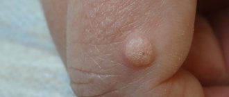
One of the varieties of papilloma - a plantar wart (shown by an arrow in the photo) - is an intradermal formation with pronounced keratinization and a “rod” in the center. Usually located on the plantar surface of the feet.
Plantar warts are characterized by fairly severe pain, especially when walking or applying pressure.
Types of warts
Today there are over 100 types of HPV, each of which gives rise to a certain type of warts and papillomas. The incubation period is about 2-6 months, but it largely depends on the human immune system: a strong and healthy body easily fights the occurrence and proliferation of growths. The following types of warts are distinguished:
- ordinary (simple, vulgar);
- plantar (pinuses);
- flat (juvenile);
- senile (age-related keratomas).
Ordinary (simple, vulgar) - warts in the form of small dense dry nodules with an uneven and rough surface, variable sizes and a rounded shape. They do not cause itching, pain or discomfort. Simple warts reach from 3 to 10 mm in diameter. They are usually flesh-colored, sometimes pink, yellowish, or light brown. Due to the accumulation of contaminants in the porous surface, they can turn dirty gray. The favorite localization of simple warts is the back of the hands and fingers. Sometimes they are observed on the knees and elbows, rarely on the face and feet. After some time (it’s different for everyone), the tumor increases in size, and the skin in its place begins to peel off. The progression of the pathology is manifested by the formation of a large “mother” wart with a scattering of small ones around. When removed, small ones usually self-destruct over time. Plantar warts (spikes) are warts in the form of several fused nodules with a roll of keratinized skin around them. Inside the growths, many small black dots are visible - thrombosed capillaries. Plantar warts are round in shape, rise above the skin by 1-2 mm and can reach up to 2 cm in diameter. However, 75% of the tumor is located in the deep layers of the epithelium, so their main feature is inward growth. Outwardly, these warts resemble calluses, which is why they are often confused with each other. It's important to remember that, unlike a callus, a ring of dead skin forms around a wart. Spines are localized on the feet, rarely on the palms. Usually colored yellow or dark brown. This type of wart causes significant discomfort, itching and pain, aggravated by walking, and may even bleed. In 50% of cases it self-destructs without treatment, but this may take from 8 to 18 months. As the pathology progresses, the number and size of warts will increase, which may lead to the inability to walk due to unbearable pain. Flat (juvenile) - warts in the form of small flat papules with a smooth (sometimes scaly) surface, slightly raised above the surface of the skin. Traditionally it affects people aged 10 to 25 years. They are formed in multiple clusters or singly, which is quite rare. Usually the growths are flesh-colored, sometimes white, brown, yellowish or pink. They appear on the face, neck, knees, elbows, back, legs and arms (especially on the fingers and back of the hands), sometimes on the head of the penis. As a rule, they are painless and do not cause discomfort unless they are subjected to mechanical pressure or damage. Flat warts can suddenly disappear on their own just as they appeared, especially in childhood. But sometimes they are quite difficult to treat. Senile (seborrheic keratoses) are warts in the form of dark, flat, round or oval plaques that appear in old age. They reach a diameter of 0.2-3 cm, sometimes 4-6 cm. The pathogenesis is not clear, but their appearance is definitely not associated with HPV. Senile warts can affect any part of the body except the palms, soles of the feet, and mucous membranes. As a rule, they appear in multiple clusters (about 20 foci). Initially, they appear as small light brown spots or papules with clear contours. Over time, keratoses may retain their appearance and resemble freckles, or they may harden, forming a warty surface covered with easily removable thin crusts. In most cases, senile warts take on a mushroom shape and are dark brown or black in color. They develop very slowly, over decades, never degenerating into a malignant form. Typically, seborrheic keratoses are removed for aesthetic reasons or because they become itchy and irritating. If the growths, especially large and warty ones, are injured (rubbed against clothing, touched by something), they may bleed or become inflamed.
What is the difference
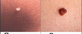
The difference between a mole and a wart can be identified by external characteristics (color, shape, size, surface), depending on the reasons for its appearance and location.
| Distinctive feature | Wart | Nevus |
| Form | Fuses with the skin, rarely have smooth edges and regular shape. | Smoothly defined edges, representing a spot on the skin. |
| Color | Brown, yellow, flesh. | The predominant color is brown, less often pink or red. |
| Surface | Rough, the stratum corneum is thickened. | Soft. |
| Number of formations | As a result of infection of healthy skin, new warts appear. | There is no infection of the skin, multiple nevi are rarely formed. |
| The main reason for the appearance | Manifestation of HPV in weakened immunity. | Increased production of melanocytes. |
| Transmission path |
| Not getting through. |
| Localization | Hands, feet, face, neck, genitals. | Whole body. |
| Presence of hairs | Never. | Indicates that the nevus is benign. |
| Is it necessary to treat | Papilloma is a symptom of a viral disease; therapy should be based on its treatment. We need to strengthen our immunity. | If the nevus does not bother you in any way and does not change, then no treatment is required. |
The table is provided for reference only. The diagnosis is carried out by a doctor.
Types of papillomas
- The following types of papillomas are distinguished:
- filamentous (acrochords);
- anogenital.
Anogenital (genital condylomas) - papillomas in the form of flesh-colored or pale pink growths of an acute shape, prone to fusion. When rubbed, they become crimson and begin to bleed. Such growths reach 1-1.5 cm and in appearance they resemble cauliflower or cockscomb due to their lobed structure. They are usually transmitted sexually and have a specific localization: on the genitals, in the perineum, on the walls of the vagina, on the foreskin, scrotum and around the anus. Although with less frequency, even in the absence of anal sexual intercourse, they can even grow in the anus and urethra, causing bleeding and discomfort during bowel movements. If the virus enters the body through oral sex, then warts appear on the mucous membrane of the oral cavity, in the pharynx, on the vocal cords or trachea. Sometimes they can form in the armpits, under the mammary glands, and in children - in the nasolabial folds. Anogenital papillomas are quite dangerous for humans for many reasons, the main one of which is the high probability of degeneration into a malignant form. Being inside a woman's vagina, they cause itching and burning, pain in the lower abdomen, pain and discomfort during sexual intercourse, and after it - spotting. Therefore, regardless of position, shape and size, genital warts should always be removed. Filiform - neoplasms in the form of thin, long, racemose shoots. They are also called facial because they are usually localized on the face, especially the eyelids and nasolabial folds, and neck. Rarely they can occur on the legs, in the groin folds, under the mammary glands and in the armpits. Those. they appear in areas of delicate and thin skin, in folds characterized by high humidity. It mainly affects older people. The process of formation of a growth begins with the appearance of a small nodule; over time, the formation acquires an elongated oval shape or remains round on a thin stalk. Traditionally, thread-like growths are flesh-colored, which is why they often go unnoticed for a long time, but sometimes they become yellow, brown or pink. They grow about 0.5-1 cm, exceeding that is extremely rare. The risk of acrochords degenerating into a malignant form is minimal, but if they are injured, an inflammatory process may develop. This type of neoplasm does not self-destruct and is prone to spreading to other parts of the body and increasing in size and number. For most people, this causes psychological discomfort, emotional stress and decreased self-esteem. Therefore, a cosmetic defect is the main reason for removing filamentous growths.
Symptoms
Papilloma developing from a mole is quite difficult to notice immediately. First of all, an alarming signal is a change in the shape of a mole, as well as its growth.
What causes papillomas?
Experts identify a wide variety of causes for the appearance of papillomas.
In addition, the following signs will indicate transformation:
- blurred boundaries;
- pain during touch;
- change in shade;
- bleeding;
- characteristic discharge from a mole.
It is worth noting that papillomatosis is distinguished by its rapid development, despite the fact that the first symptoms of manifestation may go unnoticed.
Why are neoplasms dangerous?
The main danger that warts and papillomas pose is the threat of degeneration into a malignant form. The degree of their danger depends on the type of papillomavirus:
- high (oncogenic) – 16, 18, 31, 33, 35, 39, 45, 52, 58, 59, 68;
- low (non-oncogenic) – 6, 11, 26, 41, 42, 50, 61, 73, 82, etc.;
- absent – 1, 2, 3, 4, 5.
There is a high probability of malignancy if warts and papillomas are constantly injured (rubbed against clothing, touched by something, etc.). In this case, infection with harmful bacteria may occur. To prevent the degeneration of a benign tumor into a malignant one, it is necessary to regularly visit a dermatologist and not hesitate to detect suspicious symptoms. Particularly dangerous are genital warts that affect the internal genital organs of a woman, since they can cause serious diseases: pseudo-erosion of the cervix, ectopia of the columnar epithelium, and cervical cancer. In addition, their presence is manifested by a number of unpleasant symptoms: burning and itching in the vagina, unpleasant odor, pain in the lower abdomen, pain and discharge of ichor during sexual intercourse. In men, the lack of treatment for papillomas leads to decreased libido, erectile dysfunction, and infertility. It can also provoke dangerous diseases: adenocarcinoma, anal cancer, diseases of the penis, oral cavity or larynx.
Many varieties are not dangerous, but ignoring the treatment of warts and papillomas will provoke an increase in their number. And the constant attack of the body by viruses will ultimately lead to a deterioration in the overall health of a person.
Treatment of moles
Mole removal. It makes sense to remove moles only if they are constantly injured (for example, by clothing) or create severe cosmetic discomfort.
Typically, moles are excised within healthy tissue and sent for histological examination to exclude melanoma.
In rare cases, a mole can transform into a malignant melanoma. Risk factors are light skin, an abundance of moles, a family history of melanoma, etc. Degeneration can be facilitated by constant trauma to the mole.
Diagnostics
It is extremely important to correctly determine the type of tumor, taking into account oncogenicity and external similarity. Initially, the doctor conducts a clinical examination of the growth, if located on the genital organs - gynecological in women, proctological and urological in men. This is not enough to make an accurate diagnosis. To establish a reliable clinical picture, it is recommended to conduct research at the cellular level using special diagnostic methods, namely:
- cytological examination of cell morphology - to identify DNA mutations;
- colposcopy - examination of the vulva, vagina and cervix using a special microscope - colposcope;
- biopsy - sampling of cells and tissues for the purpose of diagnosing the pathogen;
- histology - a type of biopsy - to determine a precancerous condition;
- polymerase chain reaction (PCR) techniques - to identify all types of virus;
- Digene test. It is the most reliable and informative diagnostic method: it accurately determines the presence and type of HPV, the level of oncogenicity, and the degree of concentration of the pathogen in the body.
Most often, the diagnosis is made based on the results of using several methods, since individually each of them is erroneous to a certain extent.
Doctors to contact regarding this issue
Denezhkina Natalya Nikolaevna
Reshanova Lyudmila Mikhailovna
Varfolomeeva Oksana Igorevna
Chaplitsky Evgeniy Aleksandrovich, urologist-andrologist, specialist
Appearance mechanism
Need advice from an experienced doctor?
Get a doctor's consultation online. Ask your question right now.
Ask a free question
The mechanism of occurrence of moles and warts is different. In the first case, the vegetation is congenital, in the second case it is acquired as a result of infection. Warts must be removed; nevi should not be touched if there are no unpleasant symptoms.
Warts
Formations occur in patients with weakened immunity in the presence of HPV in the body. They are papillary or round growths of a yellowish-gray color. Inside the seal you can find black inclusions and roots. The surface is hard and may cause pain when pressed.
If HPV is left untreated, the growths become multiple.
Methods for removing tumors
Treatment of warts and papillomas should only be performed under the guidance of a qualified specialist. There should be no self-medication, since the pathology will not only not be eliminated, but will also significantly worsen, and serious complications may arise. Under no circumstances should you resort to your grandmother’s methods of tying the growth with thread or sealing it with tape in order to deprive it of blood flow. This can only injure the neoplasm, which will inevitably lead to the appearance of new growths and increase the risk of malignancy. Treatment of warts can be carried out medicinally using the following drugs:
- Viferon;
- Salicylic and Oxolinic ointments;
- Clareol;
- Antipapillomas;
- Cryopharma;
- Panavir;
- Isoprinosine.
The main goal of therapy is to increase the patient’s immunity, so the doctor may prescribe Echinacea, Immunal, Eleutherococcus and Aloe extract, Ribomunil, Lykopid, Imudon, Anaferon, Actovegin, FiBSA, multivitamins and others.
However, the high infectivity and autoacculation of warts are arguments in favor of their removal. Papillomas can only be removed. Today, the most common methods for removing warts and papillomas are:
- cryodestruction - freezing with liquid nitrogen, after which after a while the growth disappears. It is characterized by the absence of pain and a small number of complications. Practically ineffective in the fight against genital warts;
- removal with a radio knife - cutting off not only the growth itself, but also the epithelium underneath it. The device does not come into contact with the tumor, but acts on it from a distance. The procedure is performed under local anesthesia and lasts no more than 30 minutes;
- laser removal - excision of the growth with a laser beam. During the procedure, healthy tissue is not affected, the blood vessels are sealed, which eliminates bleeding, inflammation and scarring. Removal is painless and quick;
- electrocoagulation – removal of tumors using alternating current. Rare relapses occur after this procedure;
- surgical method - indicated in the case of large and deep tumors. The procedure is performed under local anesthesia. It has significant disadvantages: it causes relapses, leaves scars and scars.
Removal with a radio knife is one of the most popular procedures. It involves cutting off the growth using high-frequency radio waves. The procedure has many advantages:
- does not require special training;
- carried out quickly and painlessly;
- guarantees the absence of side effects in the future if all doctor’s recommendations are followed (relapses, scars, etc.);
- is particularly accurate and efficient;
- does not damage healthy tissue;
- bloodless and safe removal: the radio knife makes a cut, cauterizes blood vessels and disinfects;
- fast healing;
- affordable cost of the procedure.
As a rule, medical procedures are complemented by the use of measures to strengthen the immune system and destroy the human papilloma virus.
Moles and papillomas: what must be removed and when
Experts do not recommend removing moles if they behave calmly and do not cause discomfort to their owner.
If any complications arise from the nevus, and doctors strongly recommend removing it, then it is advisable to do this as soon as possible.
It is strictly not recommended to do this yourself at home.
It is necessary to get rid of unwanted moles only in a medical facility.
Before removing the tumor, the specialist will conduct the necessary examination, after which he will suggest one of the methods for removing the tumor.
Doctors are well versed in which nevus removal methods are the most effective and safe.
As for papillomas, you will also need to consult a doctor.
After an examination and a series of diagnostic tests, the doctor will prescribe removal of growths using the most gentle method and antiviral therapy.
Treatment of papillomas and warts comes down to their removal; the difference is that for each type of tumor, the doctor selects the method that will be most optimal for each specific case.
If it is necessary to send biomaterial for diagnostics, radio wave therapy or surgery will most likely be used.
In order to remove tumors, the following techniques are used:
- Cryodestruction . The technique is based on the use of low temperatures. Liquid nitrogen is used to freeze papillomas, which provokes necrosis of pathological tissues.
- Chemical removal method . Medications containing high concentrations of substances that cause chemical burns are used. The formations dry out and subsequently fall off.
- Electrocoagulation. High frequency current is used for removal. The tumors are burned out.
- Laser therapy. This technique is the most preferable, since after removal of papillomas there are no traces left on the skin. After the procedure, the recovery period is short.
- The radio wave technique is based on the use of radio waves, which promotes excision of the growth.
- Surgical method of removal . A scalpel is used to cut out the affected tissue, after which the material is sent to the laboratory for histological analysis. The technique is indicated for suspected malignancy of the process.
For small papillomas, conservative therapy is carried out using antiviral drugs.
How is melanoblastoma diagnosed?
After identifying suspicious moles, they are examined by a doctor, usually a dermatologist, mammologist or general oncologist, this depends on the structure of medical institutions in the region. Typically, the mole is measured and examined under a special magnifying glass. If the doctor has even the slightest doubt about the diagnosis, a targeted biopsy is prescribed for subsequent histological detailed analysis of the cells.
If the diagnosis of melanoma is confirmed, additional examinations are carried out in order to determine further treatment tactics and identify the presence or absence of regional and distant metastases. These include:
- Blood test for specific tumor markers
- Positron emission tomography
- X-ray of the chest organs
- CT, MRI
In Israel, the most advanced and high-tech techniques are used to diagnose melanoblastoma, allowing us to show the maximum picture even at the earliest stages. So PET-CT, positron emission tomography allows you to detect the smallest regional metastases of several dozen cancer cells in size!
In addition, according to modern diagnostic protocols, a number of genetic tests called Foundation One are indicated for newly diagnosed melanoma. These studies are designed to determine the degree of aggressiveness of the tumor, as well as its antigenic profile and the degree of sensitivity to certain drugs - chemotherapy, monoclonal antibodies, and so on.
An adequate set of diagnostic procedures and genetic tests (in combination with blood tests for tumor markers) significantly improves the prognosis of the disease and allows you to choose the right treatment tactics. And precise techniques, such as PET-CT, make it possible to prevent the development of complications or relapse in time.
Molluscum contagiosum
What it looks like: single painless dense nodules of a round shape, sometimes the skin acquires a pink tint or a pearlescent sheen. The size of the rashes ranges from a millet grain to a pea. Causes of appearance : caused by a parasite - a saprophyte, which is activated when immunity decreases. What is dangerous: growth, increase in size of rashes. How to fight : mechanical squeezing of nodules followed by treatment of the affected surface with antiviral ointments and immunomodulators.
What do moles look like?
In the photo there are moles on a girl
Moles are benign growths that can have different shapes, shades and sizes. The mechanisms that cause the appearance of moles on the human body have not been fully studied. However, it is known that they can occur throughout life.
The following factors contribute to the formation of moles:
- Exposure to ultraviolet rays, both natural and artificial (solarium);
- Hereditary predisposition (moles appear in the same places as their parents);
- Action of viruses, trauma;
- Fluctuations in hormonal levels. This most often affects women and teenagers.
Basically, moles or nevi are a cluster of cells colored with a special pigment - melanin. There are also vascular moles, which are formed not by pigment, but by blood vessels.
Moles have a huge number of different variations of shades and shapes. But benign neoplasms of this group are divided into two large groups:
- Pigmented nevus . Otherwise called a mole, birthmark, age spot. They can be in the form of a circle, a nodule or a broad-based plaque. Sometimes they protrude slightly above the surface of the skin, in other cases the skin pattern remains. The structure of nevi is soft and elastic, absolutely painless. As a rule, they have a brown tint. If there are shades of brown, then you have a pigmented nevus.
- Hemangioma . These are the so-called “red moles”. They represent an anomaly in the development of capillaries. They look like elastic, painless nodules that protrude above the surface of the epidermis. The shade ranges from light red to rich black and blue.
All moles can grow slowly by 1-2 mm per year. This is not a sign of malignant degeneration. Also, during a person’s life, new benign growths appear.