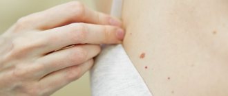
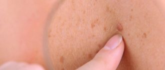
Let's start with what warts are in general. Warts, or papillomas, are dense, nodular, round or flat growths that most often form in 5 areas of the body:
- Palms
- Feet
- Armpits
- Genitals
- Face
You've probably heard many myths about warts. For example, that they arise from contact of the skin of the hands with frogs and toads, or that it is simply a superficial skin disease. In fact, papillomas of any type in children and adults have only one cause - the human papillomavirus (HPV). Warts are only an external manifestation of this disease.
Are warts dangerous during pregnancy?
About 90% of people around the world are already infected with HPV by default. But, like the causative agent of herpes, this virus can lie dormant for years. Therefore, the appearance of warts during pregnancy should not be taken as something extraordinary. But there is reason to wonder whether you are doing enough to strengthen your immune system.
Therefore, in the vast majority of cases, expectant mothers need not worry about this and postpone going to the dermatologist for the next 9 months. Exceptions include genital warts (condylomas). If they are located in the vagina or in the cervical area, then there is a real threat of infecting the baby with HPV during pregnancy or childbirth. In such cases, you will still have to consult a doctor. Also, do not put off solving the problem until later if the neoplasm looks atypical and more like a tumor.
Is it possible to remove warts during pregnancy on your own?
Many people are interested in whether it is possible to remove warts for pregnant women at home? Indeed, some folk remedies have proven themselves very well in solving this problem. But how they will affect the baby’s health is not known for certain. Therefore, in any case, it is impossible to do without prior consultation with a doctor. And if there are neoplasms in the intimate area, self-medication is strictly prohibited!
Do warts need to be removed and how can this be done at the Family Clinic?
Warts are a skin disease characterized by the appearance of benign neoplasms in the upper layer of the skin.
These peculiar tumors arise due to infection with the human papilloma virus. More than 150 types of this virus are known, many of which are very dangerous. Many people think what's wrong? A larger wart, a smaller wart - what difference does it make if they do not cause concern? Absolutely wrong judgment. Firstly, in addition to not being aesthetically pleasing, warts can cause pain, and secondly, there is always a risk of a benign lesion degenerating into a malignant one. Moreover, the longer warts are left untreated, the more they appear, and the likelihood of negative consequences increases.
Is it possible to remove warts with nitrogen during pregnancy?
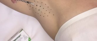
Can pregnant women remove warts with nitrogen in a clinical setting? There are no strict contraindications in this regard, however, some nuances of such procedures should be taken into account. On the one hand, cryodestruction is the cheapest and, most importantly, painless method of removing ill-fated formations. However, not all skin growths can be gotten rid of with its help. Thus, plantar warts are destroyed most effectively when treated with liquid nitrogen. However, it is impossible to influence the mucous membranes of organs (in particular, the uterus) in this way.
In addition, the cryodestruction method involves several procedures. And given that anesthesia is contraindicated at any stage of pregnancy, this only creates additional stress for the expectant mother’s body.
Can pregnant women have warts removed with laser?
The question of whether it is possible to remove warts during pregnancy using a laser is the second most popular question. However, there is no clear answer to this either. Laser therapy is considered a more effective and reliable method of getting rid of most skin lesions. Significant advantages of this procedure are accelerated recovery after surgery, compared to cryodestruction, as well as a low likelihood of relapses and scar formation at the site of wart removal. The downside is that it takes a long time for the tissue to heal after the plantar growths are removed.
Ways to remove warts
Often the patient ends up seeing a dermatologist after using folk remedies that did not produce results. In fact, you shouldn’t even start such therapy, because it can lead to active reproduction and malignancy of the formations. Our center uses the following methods of professional removal of warts, papillomas, and moles:
- Cryotherapy (liquid nitrogen);
- Radio wave method (using Surgitron or Fotek devices).
How else can you remove warts in pregnant women?
Methods that have proven themselves in removing skin lesions in pregnant women also include radio wave therapy and electrocoagulation .
The first is a targeted effect on the wart with a so-called radio knife. As a result, it is destroyed while maintaining the integrity of healthy tissue. The likelihood of relapse is negligible. A significant disadvantage is the risk of scar formation after removal of a large tumor. Therefore, the radio wave method is usually used to get rid of papillomas, which have a characteristic thread-like base.
The second one works on a similar principle. The only difference is that the excision of the wart is carried out with an electric scalpel, which literally evaporates it to the very root. In this case, the microvessels are welded together without causing bleeding. Electrocoagulation not only removes the wart, but also reduces to zero the likelihood of its reappearance or further growth. And when getting rid of benign tumors, it does not allow them to turn into a malignant form.
Recently, in Russia there has been an increase in the incidence of cervical cancer (CC) in young women of reproductive age, especially in the group of women under 29 years of age. As a cause of death in women under 30 years of age, cervical cancer accounts for 8.5% [4]. Among oncological diseases in pregnant women, it is 45%. According to S.I. Rogovskaya [10], among 1000 pregnant women, severe dysplasia and cancer in situ
, in 0.45 cases - invasive cervical cancer.
The main etiological factor of cervical cancer is the human papillomavirus (HPV), which was confirmed by the results of both epidemiological and molecular biological studies [2, 5, 8, 11, 14]. HPV is also the cause of dystrophic and malignant diseases of the vulva and vagina in women and the penis in men [6]. The likelihood of spontaneous elimination of HPV during carriage and the possibility of spontaneous regression of both subclinical and clinical forms of human papillomavirus infection (PVI) inclines a number of researchers to observational tactics [1, 3, 12]. However, HPV is not considered a normal representative of the vaginal biotope [7, 9].
Currently, there is no consensus on the effect of HPV on the course and outcome of pregnancy. It is known that pregnancy is a risk factor for the development of PVI and promotes active replication and persistence of the human papillomavirus. Research by A. Schneider et al. (1987) showed that the incidence of PVI in pregnant women is 2.3 times higher than that in non-pregnant women, while the amount of viral DNA in pregnant women is on average 10 times greater than the same amount in non-pregnant women. The number of cases of HPV transmission from mother to fetus, according to various researchers, ranges from 4 to 87%, which depends on the sensitivity of the diagnostic methods used [10, 13].
There is evidence that HPV infection of the genitals leads to an increase in spontaneous abortions, while HPV DNA is found in syncytiotrophoblast cells [12]. How the virus is transmitted from mother to fetus is unclear. Bloodborne transmission is unlikely because HPV is only occasionally found in white blood cells. Ascending HPV infection of the amniotic fluid and placenta is most likely. There are alarming reports about the detection of HPV in the amniotic fluid of pregnant women, about an increase in the frequency of papillomavirus lesions of the larynx and bronchi in children, which indicates their infection during pregnancy. Thus, HPV DNA is detected in 33% of newborns in nasopharyngeal aspirate, as well as in the amniotic fluid of HPV-positive women [13, 15]. Transmission is also possible through direct contact (skin contact), as well as during childbirth - infection of a newborn from an infected mother. The third possible route is infection during conception through sperm.
Respiratory tract papillomatosis, most often caused by HPV types 6 and 11, has a severe course in young children with a tendency to recur. In 75-87% of cases, signs of juvenile respiratory papillomatosis are recorded in the first 5 years of life, which is probably due to the functional immaturity of the immune system [16].
Features of the course of PVI during pregnancy.
Estrogens and progesterone increase HPV expression in the cervical epithelium and promote cellular proliferation and carcinogenesis, and during pregnancy there is an extremely high release of sex hormones. Due to increased vascularization, active metabolism in tissues, changes in vaginal microbiocenosis, and a decrease in the compensatory capabilities of the immune system, the risk of infection and the incidence of various infections increase. In this case, latent PVI can develop into sub- and clinical forms [10].
Pregnancy may be a risk factor for the development of PVI and contribute to active replication and persistence of HPV. During pregnancy, visible condylomas often recur, tend to grow, become loose, and can reach gigantic sizes.
Prevalence of PVI among pregnant women.
At the Moscow Regional Research Institute of Obstetrics and Gynecology, a survey of 700 consecutive pregnant women admitted to a specialized appointment for cervical diseases was carried out in order to identify PVI. During a screening examination of these pregnant women using the polymerase chain reaction method, clinical and laboratory manifestations of PVI were identified in 46 (67%) pregnant women.
The results of a survey of pregnant women with various diseases of the cervix showed that PVI is more often found in patients with cervical intraepithelial neoplasia (CIN) (95.5%), in pregnant women with complicated ectopia (32.4%), with leukoplakia (56%), after surgical treatment of CIN (42%), with polyps of the cervical canal (38.8%). It should be noted that even with an unchanged cervix, HPV was detected in every third pregnant woman (34%). Cytological signs of papillomavirus lesions of the cervix were observed in 12% of pregnant women with an unchanged cervix only in the second and third trimesters of pregnancy. In pregnant women with complicated ectopia, koilocytosis in combination with dyskeratosis of the stratified squamous epithelium of the cervix was detected in the first trimester in 7.4%, in the second trimester in 25.9% and in the third trimester in 31.5%. In pregnant women with cervical canal polyps, cytological signs of papillomavirus lesions of the cervix were detected in 20.8% of cases.
Despite surgical treatment of CIN at the preconception stage and follow-up of these patients, the number of cases of relapse of CIN during pregnancy was 4% against the background of PVI (presence of koilocytes).
In pregnant women with cervical leukoplakia, koilocytes were detected during cytological examination in 56% of cases. Most often, signs of papillomavirus lesions of the cervix were found in pregnant women with diagnosed CIN - in 73.3 and 77.7% of cases in the first and second trimesters, respectively.
The presented data once again confirms that the cytological research method currently remains the leading one in the diagnosis of cervical diseases. Unfortunately, there is still a misconception among gynecologists about the dangers of in-depth cytological examination of the cervix in pregnant women due to possible complications. At the same time, the high incidence of cervical diseases during pregnancy dictates the need for mandatory cytological examination of the ecto- and endocervix in pregnant women when registering them.
Genital condylomas of the vulva and vagina were identified during colposcopic examination in 62% of pregnant women. They were irregularly shaped fibroepithelial formations rising above the surface of the mucous membrane with finger-like or cone-shaped protrusions on a thin stalk, less often on a broad base in the form of a single nodule or in the form of multiple outgrowths resembling cauliflower or cockscombs. The surface of condylomas was covered with stratified squamous epithelium and often keratinized. Vessels were located in the underlying stroma. In some cases, inflammatory reactions, microcirculation disorders and edema occurring in the stroma contributed to the addition of a secondary infection. In 5.3% of pregnant women, cervical condylomas were detected in the form of a tumor with kidney-shaped papillae, evenly distributed over its surface and forming a repeating pattern. 9% of pregnant women had giant condylomas of the external genitalia and vagina, up to 7 cm in size.
State of local immunity.
The presence of benign cervical diseases and PVI was accompanied by a sharp decrease in local immunity, most pronounced in pregnant women with CIN. It was manifested by a decrease in the production of secretory immunoglobulin A (sIgA), which is the main indicator of immune defense, and an increase in the production of immunoglobulins A, M and G (IgA, M, G). The most pronounced changes in the content of immunoglobulins of all isotypes were observed in pregnant women with CIN. They were characterized by extremely low production of sIgA (2.2±0.5 μg/ml) and high level of IgM (24.8±0.9 μg/ml), which is considered as a marker of inflammation. The average IgA levels were 24.2±1.6 μg/ml, IgG - 1248.6±46.5 μg/ml. In healthy pregnant women, the level of sIgA was 12.2±0.5 µg/ml, IgA - 36.5±0.5 µg/ml, IgM - 2.6±0.5 µg/ml, IgG - 456.5±35 .5 µg/ml.
The results obtained confirm the connection between cervical diseases and impaired local immunity, which is of great importance for the selection of adequate immunocorrective therapy, and indicate that the degree of local immunity impairment is directly proportional to the severity of cervical disease.
Algorithm for managing pregnant women with PVI
Stage I - examination
— Diagnosis and treatment of other genital infections and vaginal dysbiosis.
— Extended colposcopy.
— Detection of HPV DNA with typing.
— Cytological examination (PAP test).
Stage II - determination of tactics
— Indications for observation: latent form of PVI, vestibular papillomatosis.
— Indications for treatment: genital warts of the vulva, vagina, cervix.
— The management tactics for pregnant women with CIN I should be watchful and expectant with dynamic colposcopic observation and cytological control, with final treatment of the cervix after childbirth.
— If there are signs of PVI and CIN I-III, anti-inflammatory treatment and correction of vaginal microbiocenosis are carried out, after which it is necessary to repeat the PAP test.
— If after treatment there are signs of PVI, CIN II-III in pregnant women, or if the results of colposcopic or cytological examination worsen, a cervical biopsy with histological examination and consultation with an oncologist are indicated.
— If CIN III is detected, a mandatory consultation with an oncologist is necessary; if CIN III is detected in the II-III trimesters, it is possible to prolong pregnancy with dynamic cytological and colposcopic monitoring once every 3 weeks, followed by treatment after delivery.
- Indications for cervical biopsy during pregnancy are atypical cytological and colposcopic patterns suspicious for cancer (heterogeneous surface, exophyte, erosion or ulceration and atypical vascularization).
Stage III - comprehensive examination and determination of management tactics in the postpartum period
based on colposcopy data, cytohistological re-evaluation of previous data.
Treatment of diseases associated with HPV during pregnancy must be carried out differentiated according to indications at any time, but preferably in the first trimester [10]. Before using destructive methods of treatment, it is recommended to conduct a comprehensive examination and treatment of concomitant inflammatory diseases of the genitals.
The methods of choice for the treatment of genital warts in pregnant women are radio wave therapy and the use of chemical coagulants - solcoderm, trichloroacetic acid. It is possible to use laser therapy, electrocoagulation, and surgical methods.
The radio wave method is the most acceptable from the point of view of gynecological oncology, since all removed material is available for histological examination. This fundamentally distinguishes it from laser and cryodestruction, in which the material is completely absent, and from electric knife treatment, in which tissue charring occurs. Removal of large genital warts of the vulva, vagina and cervix is carried out under local anesthesia using the radiosurgical method (“Surgitron”) using a radio loop in the “cut and coagulation” mode (power 2-4 units), the use of high-frequency waves (3.8 MHz) provides more gentle tissue incision and allows you to remove exophytic formations bloodlessly, painlessly, without traumatizing surrounding tissues and obtaining complete material for histological examination.
Due to the risk of adverse effects on the fetus during pregnancy, the use of cytostatic drugs is contraindicated, which, having antiproliferative activity, promote cell destruction, affecting both damaged and healthy cells.
A mandatory method of treating PVI in pregnant women is immunocorrective therapy. The use of interferons (IFNs) and their inducers is promising. IFNs are endogenous cytokines that have antiviral, antiproliferative and immunomodulatory properties. There is evidence of differences in the immune response during infection with high- and low-oncogenic types of HPV. In the presence of HPV types 16–18, there is a decrease in the production of α- and γ-IFN, an increase in the concentration of serum IFN, spontaneous production of IFN, leading to an imbalance in cellular immunity and, as a consequence, to a severe course of the disease.
During pregnancy, intravaginal, rectal and external agents, and systemic drugs are used. Interferon therapy is carried out from the second half of pregnancy. Viferon is the optimal drug for immunocorrection during pregnancy. It contains recombinant α2-interferon, as well as membrane-stabilizing components - α-tocopherol acetate and ascorbic acid. Viferon is an immunomodulator that affects the processes of differentiation, recruitment, functional activity of effector cells of the immune system, as well as the efficiency of immune recognition of antigen and increased phagocytic and cytolytic activity. To exclude the development of phenomena of refractoriness of effector cells to the action of IFN, systemic administration of the drug should be intermittent. In addition, the protective effectiveness of IFN has been proven in diseases caused by intracellular parasitic microorganisms (chlamydia, mycoplasma, etc.). Obviously, the effect in this case is also associated with the suppression of protein synthesis and activation of phagocytosis.
Viferon is available in the form of suppositories 150,000 IU, 500,000 IU, 1,000,000 IU and 3,000,000 IU for rectal use. Pregnant women are usually prescribed suppositories of 150,000 or 500,000 IU 2 times a day for 10 days. When using Viferon, a high concentration of interferon is created at the site of infection, which promotes rapid relief of subjective symptoms, reducing doses and duration of antibiotic therapy. The use of Viferon in the complex treatment of STIs in pregnant women has a positive effect on the immune system and increases the effectiveness of antimicrobial therapy.
Patients with PVI often experience a disturbance in the vaginal microbiocenosis - a sharp deficiency of lactobacilli, an excess of opportunistic microflora. There is a significant contamination with yeast-like fungi. In a large percentage of cases, there is infection with sexually transmitted microorganisms - chlamydia, genital mycoplasmas, etc. In the presence of a urogenital infection, antibiotic therapy is carried out after 12 weeks of gestation. Correction of vaginal microbiocenosis in pregnant women is carried out using local approved drugs. During pregnancy, therapy with glycyrrhizic acid, which has antiviral activity, is possible. Correction of vaginal microbiocenosis with the help of eubiotics is also necessary.
The issue of delivery in women with PVI is decided individually. Studies have shown that abdominal delivery does not reduce the risk of fetal infection (N. Sedlacek, S. Lindheim et al., 1989); cases of children born by cesarean section with laryngeal papillomatosis have been described.
Due to the high frequency of PVI in pregnant women and the participation of HPV in the processes of carcinogenesis, it is necessary to optimize the preconception preparation of women, including a comprehensive examination to identify HPV, carry out its typing, as well as treatment of HPV-associated diseases at the stage of pregnancy planning.
When planning pregnancy, subclinical forms of infection, as well as CIN, must be treated before pregnancy.
Carriage of HPV is not a contraindication to pregnancy.
It is not possible to completely cure a woman of virus carriage, therefore adequate preconception preparation is a sufficient measure to prevent exacerbation of PVI during pregnancy.
Safe removal of warts and papillomas at the Stoletnik MC
We have sorted out the question of whether it is possible to remove warts during pregnancy. If you are looking for where you can do this in Penza, we invite you to the medical office at the address: st. Chaadaeva, 95.
Highly qualified specialists and modern equipment are at your service. The procedure for excision of warts is performed surgically or by electrocoagulation. The extensive experience of our doctors and an individual approach to each patient guarantee a positive result when removing tumors of any complexity, without risks to the health of the mother and baby.
Make an appointment and consultation by phone: +7 (8412) 999-395, 76-44-20.
Care after removal
After exposure to liquid nitrogen, scars take one to two weeks to heal. It depends on the individual characteristics of the body and the degree of damage. If your doctor prescribes you to take any medications, you should take a course. The bubble that appears on the skin at the site of the session should not be wetted, punctured, and should not be touched at all. Sometimes treatment with potassium permanganate is required for some time after the procedure. The specialist will tell you about this in more detail at your appointment.
Literature
- Adler J. What the skin hides. 2 square meters that dictate how we live. – M.: Bombora, 2022. – 352 p.
- Gomberg M. A., Soloviev A. M. Treatment of warts and genital warts is simple and effective // Medical Council. − 2011.− No. 5. − P. 60−65.
- Molochkov A.V., Khlebnikova A.N., Lavrov D.V., Gureeva M.A. Genital papillomavirus infection. Tutorial. − 2010. − 10 p.
- Rogovskaya S.I. Human papillomavirus infection in women and cervical pathology. − M.: GEOTAR-Media, 2005. − P. 15–17.
- Yunusova E. I., Yusupova L. A., Mavlyutova G. I., Garayeva Z. Sh. Flat warts: features and treatment options // Attending physician (electronic edition). − 2016.− No. 5.