Mole or medical term Nevus.
This is a collective name for limited, benign formations of the skin or mucous membranes.
The development of which is based on melanocytes - specialized skin cells that produce melanin.
Fair-skinned people have an average of 20 to 30 nevi.
Some are congenital or develop after birth in young children.
With age, the number of moles may increase, which is not considered a pathology.
Acquired moles are associated with the accumulation of pigment-forming cells.
Such as melanocytes in the upper layers of the skin.
If we consider in detail, the nevus is located either in the epidermis, in the dermis (corium), or at the border between these layers of skin.
Such spots can vary in color, being light, dark brown and even black.
Some moles are flat, others are raised.
Intense and constant exposure of the skin to ultraviolet light, as well as hormonal causes, cause or accelerate the development of birthmarks.
In principle, the development of moles is a complex process.
Biological factors and the environment play an important role.
Nevus is usually benign.
Therefore, the reasons for surgical removal are mainly cosmetic.
Only if the spot is perceived as alarming, for example, its color or shape has changed dramatically, is a detailed examination prescribed to determine subsequent treatment.
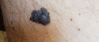
Black moles attract special attention.
They may appear against the background of hormonal changes during pregnancy and lactation.
Most patients consider the appearance of a dark nevus as an increased risk of developing skin cancer.
However, not every black mole on the skin is melanoma; however, they require consultation with a doctor.
First, the specialist will evaluate the clinical change in the formation by viewing it with the naked eye.
The next step in diagnosis will be dermatoscopy.
Dermatoscopy is a diagnostic procedure.
Used to differentiate skin lesions.
Allows you to determine whether a black mole is of epidermal, vascular or melanocytic origin.
Causes of moles
Most moles form in childhood and adolescence. Heredity and genetic predisposition play a key role. In adulthood, the appearance of new formations is facilitated by external and endogenous factors. The most important is hyperinsolation - regular and prolonged exposure of the skin to ultraviolet radiation.
Ultraviolet light is the main catalyst for the formation of melanin pigment in skin cells, and it is melanin that determines the color of the entire skin and its elements. The appearance of a black mole may be a consequence of too frequent and long exposure to the sun. Fans of artificial tanning are at risk, since solarium lamps are a source of intense ultraviolet radiation.
Changes in hormonal balance can contribute to the appearance of new nevi. For women, a risk factor is ovarian dysfunction (imbalance of sex hormones). Changes in hormonal levels can be triggered by taking oral contraceptives and hormone replacement therapy during menopause. The formation of black moles is possible during pregnancy and lactation.
Dangerous symptoms
Only a very few black moles pose a potential threat. The risk of malignant transformation to melanoma is low, but caution should be maintained. In any case, it is advisable to show the skin lesion to a specialist. You definitely need to see a dermatologist or oncologist if you have the following symptoms:
- The black mole increases in size. Even small growth should be alarming; a rapid increase in size is an unfavorable sign.
- The surface has changed (becomes “glossy” or loose).
- The formation peels off or bleeds.
- Unpleasant sensations appear in the form of pain, burning or itching.
- The shape has changed, the edge has become striated, uneven, asymmetrical.
- The color changes: an ordinary mole becomes black, bluish or dark brown.
Any dynamics should be alarming. There is no need to panic, but you need to make an appointment with a specialist. What you definitely cannot do is try to remove the tumor yourself using traditional medicine. Injury by mechanical or chemical influence is one of the main causes of malignant degeneration.
Why is the mole black?
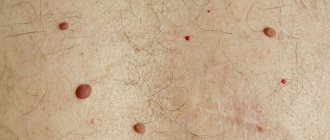
In order to get an answer to this question, you need to delve a little deeper into the structure of moles. As I wrote earlier, a mole (or pigmented nevus) consists of nevus cells. In the picture below, with black circles, I have indicated the nevus cells of an ordinary brown mole, which, as a rule, does not cause us concern.
The fact is that the color of a mole (black or brown) directly depends on how many nevus cells are in it, or more precisely, how much melanin (i.e. brown pigment) these cells have produced. The black color of a mole may appear due to the fact that its cells have produced too much melanin. A thick layer of this substance may appear black when viewed by the eye. If one of the moles on your skin has turned black or you just suddenly noticed it, don’t panic. She's probably fine. Look at this image:
There are three times more black dots (nevus cells) here than in the first one. The amount of pigment is also increased. This is why deep brown appears black.
Melanoma-dangerous nevi
Listed below are specific types of black moles that are melanoma-dangerous. This means that when exposed to unfavorable factors, they can degenerate into melanoma; the risk of malignant transformation is higher than in the case of ordinary nevi.
Dysplastic nevi. This type of nevi is characterized by the presence of melanocytic dysplasia, an atypical arrangement of melanocytes. Only a dermatologist can diagnose melanocyte dysplasia. The patient needs to know that a dysplastic nevus has a smooth surface. It does not rise above the skin, or only its central part rises. The shape is irregular, with uneven edges. The coloring is uneven, with black areas located in the center.
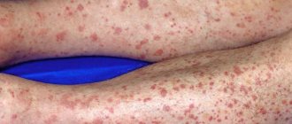
Blue nevus. Despite the name, the formation can be not only blue, but also black. Usually has the shape of a regular hemisphere and rises above the surface of the skin. The surface is smooth, the edges are even. Typical localization is the scalp, feet and hands, and gluteal region. The risk of malignancy increases after injury, including independent attempts at removal.
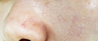
Nevus Ota. This type of tumor appears only on the face. Characteristic mainly for representatives of the Mongoloid race. The color is black or bluish. The differential sign is the presence of pigmentation of the sclera, iris or conjunctiva of the eye.
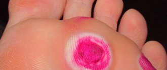
Borderline pigmented nevus. The neoplasm is formed in childhood. Subsequently, birthmarks increase in size throughout life, reaching one and a half to two centimeters in diameter. The differential feature is an uneven ring-shaped coloration, with a decrease in color intensity from the center to the periphery. The color is brown, darker in the central part.
Giant pigmented nevus. Refers to congenital. Increasing in size, such birthmarks reach gigantic sizes, up to 15 centimeters or more. A characteristic feature is an uneven surface with “potholes”, nodules and cracks. Hair growth from the nevus may occur.
For melanoma-dangerous moles, the risk of malignancy is higher than for ordinary skin tumors. Such nevi and birthmarks require observation by a dermatologist or oncologist. Malignant degeneration can be caused by exposure to external factors - mechanical or chemical damage, ultraviolet irradiation.
Types of dangerous black moles
A black, convex and flat mole does not always indicate an ongoing malignant process.
The following will describe moles that are melanoma-dangerous.
In the presence of favorable factors, they can degenerate into malignant, cancerous tumors.
Black moles that are considered potentially dangerous:
- Dysplastic nevus (dysplastic nevus)
Dysplastic nevus (nevoid tumor) is a pigment-type formation.
Has the ability to transform into atypical.
The presence of such stains may indicate a direct danger, which is determined using the so-called ABCD rules.
These rules mean:
- asymmetry of education (A)
- irregular or worn border (B)
- presence of more than two colors (C)
- spot diameter more than 5 mm (D)
Nevoid tumor does not tend to be inherited.
It develops equally in both men and women, and is diagnosed in 6% of the world's population.
Such a mole has a flat surface, less often it can rise above the level of the skin epidermis.
There is no typical location for this type of nevus.
A preliminary diagnosis can be made based on dermatoscopy.
Confirmation of the type of formation is possible only through histology after surgical removal of the mole.
The main treatment is excision of the tumor, followed by monitoring the patient every 3-12 months.
Photographic documentation of skin malignancy with overview photographs and microscopic images is especially important.
In this way, even the smallest changes can be detected and dangerous moles can be removed on time.
Particular attention should be paid if a black mole appears on the labia majora.
Although this phenomenon is rare, such a nevus can be malignant.
- Nevus Yadassona-Tits (blue/blue nevus)
Benign, rarely acquired, dermal, melanocytic tumor.
Consists of mature, pigmented, dendritic, spindle cells and/or epithelioid melanocytes.
Acquired blue nevi can also appear in adulthood.
Because of their color, they are often mistaken for malignant melanoma.
Possible widespread distribution.
But they are more often observed on the arms, legs and head, in the hair growth area (in more than 60% of cases).
Visually, the blue nevus has a regular shape and its surface is smooth.
Such formations are usually single, up to 1 cm in diameter (in rare cases, 2-3 cm in size have been found).
Although blue nevus has this name, it can be either dark blue or black.
The provoking factor for malignancy is injury to the mole or attempts to independently excise the tumor.
If the mole is small in size and is not subject to injury or growth, removal is carried out only at the request of the patient.
Since nevus Yadassona-Tits does not often develop into melanoma, dermatologists choose a wait-and-see approach.
If a black mole grows, it is advisable to prescribe removal of the tumor.
After excision of the nevus, its histological examination is mandatory.
- Nevus Ota (phakomatosis Ota-Sato)
The characteristic localization of the formation is the face; it often develops in patients of Asian appearance.
As a rule, a nevus is located in the cheek area and can affect the mucous membrane of the eye.
Such a black mole on the face has another name - oculocutaneous melanosis.
The formation is classified as melanomatous, but rarely progresses to an atypical tumor.
When diagnosing nevus of Ota, regular monitoring by a dermatologist is recommended.
It is believed that phakomatoses are progressive in nature and have a hereditary predisposition.
Surgical intervention to excise phakomatosis is rarely prescribed when signs of malignancy are detected.
To eliminate the formation in order to eliminate a cosmetic defect, a laser procedure is recommended.
There is currently no definitive research that shows the underlying cause of Nevus of Ota.
Some researchers believe that the phenomenon may be caused by a genetic mutation.
Others argue that hormonal factors or radiation can cause the development of phakomatosis.
Although more research is needed to confirm both options.
People with oculocutaneous melanosis, which has spread to the mucous membrane of the eye, are more likely to develop glaucoma.
This is because melanocytes, which cause hyperpigmentation, block the flow of fluid in the eye, thereby increasing intraocular pressure.
- Border pigmented nevus (border pigmented nevus)
A border nevus is a pigmented spot no larger than 1 cm.
The color scheme can vary from gray to brown and black.
Most often this mole is brown-black.
Borderline nevus can develop on any part of the skin.
As a preventive measure, it is recommended to remove the formation using laser equipment.
Nevus is benign.
It rarely develops into cancer, but experts recommend an annual examination by a dermatologist.
- Giant pigmented nevus (congenital/giant pigmented nevus)
A congenital pigmented nevus is considered gigantic if its size reaches more than 20 cm in diameter.
Giant congenital nevi can occur in people of any racial background and in any area of the body.
They are the result of localized genetic changes in the fetus that lead to excessive growth of melanocytes, which are responsible for skin color.
Congenital nevus may not be accompanied by any other signs.
And it can occur together with itching or increased dryness of the skin.
The color may be fairly uniform or consist of several shades, including shades of brown, black, red or blue.
The predominant number of patients have a black and red mole.
In approximately 5-10% of cases, giant congenital nevus is associated with neurocutaneous melanocytosis (excess pigment cells in the brain or spinal cord).
Characterized by neurological symptoms.
Such a nevus has an increased risk of transition from a benign formation to a malignant melanoma.
If possible, treatment includes surgery to remove the nevus.
In other cases, other therapy, such as laser dermabrasion, may be performed.
In most cases, when there are no neurological problems, the prognosis is good.
But it is necessary that the formation be regularly examined by a doctor.
There are also other types of nevi, for example, you can find a dark brown mole with black spots. Such a neoplasm is highly likely to be benign.
Often defined as a pigmented nevus of the papillomatous type.
During pregnancy, there are cases when a black mole appears on the nipple.
Usually after childbirth, during the period of normalization of hormonal levels, the formation disappears.
But in a mandatory case, you will need to consult a doctor if the black mole hurts and changes.
Diagnostics
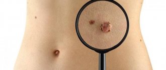
If you have a black mole on your body, it is not at all necessary to remove it, but you need to contact a specialist. The dermatologist will conduct a diagnosis, which consists of examining the neoplasm and studying it using optical or digital dermatoscopy (under magnification).
During the examination, the doctor evaluates the size of the tumor, its density, structure, consistency, surface character, symmetry, edge, color and other signs. As a rule, examination and dermatoscopy are sufficient to make a diagnosis. Biopsy and histological examination are carried out only before removal. In other situations, dermatologists prefer not to injure black moles unnecessarily.
Treatment: removal or observation?
Benign neoplasms of the skin do not require removal, but surgery can be performed for aesthetic reasons, as well as in situations where the nevus is located in an open area of the body, is exposed to ultraviolet radiation, or is injured by parts of clothing or jewelry.
Removal of benign black moles that are not melanoma-dangerous is carried out in the following ways:
- Surgical removal;
- cryodestruction;
- electrocoagulation;
- radio wave removal;
- laser removal.
Melanoma-dangerous nevi can only be removed surgically. The operation is performed in the presence of an oncologist and involves healthy skin. The removed tissues are necessarily sent for histological examination. Melanoma-dangerous nevi can also be removed in aesthetic medicine clinics if the medical institution has a dermatologist oncologist on staff. The operation is performed in a classical way; cryodestruction and other modern technologies are not used.
If malignant degeneration of the neoplasm is diagnosed, complex antitumor therapy is indicated, which includes surgical removal with wide coverage of adjacent tissues, radiation and chemotherapy.
After the procedure
After removing a nevus, it is extremely important to protect the skin from ultraviolet radiation. You can’t sunbathe in a solarium; you should be in the sun as little as possible. It is recommended to refrain from thermal procedures. It is impossible to remove crusts at the site of the surgical wound; the skin should be provided with maximum rest. After removing a mole on the face, it is not advisable to use decorative cosmetics.
You can learn more about the diagnosis of skin tumors and methods of their treatment at a consultation with a dermatologist at the Galaktika clinic (Moscow).
Dermatoscopy of moles
The main diagnostic measure is dermatoscopy.
Dermatoscopy is a visual examination of the skin performed by dermatologists.
Diagnostics is of great importance for the prevention and early diagnosis of skin cancer.
The study uses a device known as a dermatoscope.
This is a small instrument that can magnify the area of skin being examined.
The device also has a light source.
There are many types of dermatoscopes.
In simpler models, the diagnostician looks directly into the magnifying glass.
If the device model is more modern, the dermatoscope can be connected to a computer.
This will allow you to enlarge the image the required number of times.
The image can be downloaded and saved on your computer for later use or for sending to other healthcare professionals.
Dermatoscopy is a non-invasive imaging technique.
Allows you to see submicroscopic structures invisible to the naked eye.
Diagnostics is used to study pigmented and amelanotic skin lesions.
Dermatoscopy is also called epiluminescence microscopy, incident light microscopy, and skin surface microscopy.
It is the most commonly used imaging method in dermatological practice.