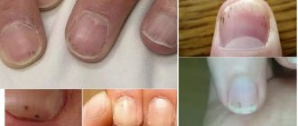
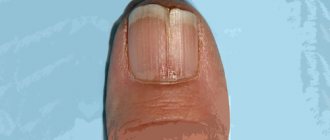
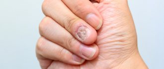
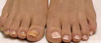
Blackened toenails or Melanonychia is a change in the color of the nail to dark brown or black. It may be diffuse or have the shape of a longitudinal stripe.
This problem is quite common; almost every person has encountered it.
Darkening can be the result of a minor injury, but often black spots on the nails are harbingers of dangerous internal diseases. Darkening of the nail plate is not always painful. The problem may not cause physical inconvenience to its owner.
Benign longitudinal melanonychia can occur in patients of all ages, including children, and affects both sexes equally. Darkening of the nails most often occurs in people with dark skin, especially in people with Fitzpatrick skin types V and VI.
Blackening of the nail beds may be due to genetic disorders, injury, medications, nutritional deficiencies, endocrine disease, connective tissue disease, inflammatory skin disease, tumor or fungal infection of the nails.
What causes melanonychia?
The nail plate is a hard, translucent structure made of keratin. Melanocytes are usually dormant in the proximal matrix of the nail, where it originates. Melanin is deposited in the growing nail when melanocytes are activated, resulting in the formation of a pigmented stripe - this is longitudinal melanonychia. Melanin deposition in the nail plate can be the result of 2 processes:
- Melanocytic hyperplasia;
- Activation of melanocytes.
Onychodystrophy (nail dystrophy) - symptoms and treatment
At the moment, there is no unified classification of onychodystrophies. Based on their origin, they can be divided into two groups:
- congenital onychodystrophy;
- acquired onychodystrophies.
Congenital onychodystrophies develop as a result of gene mutations and are inherited. The last group of diseases are in turn divided into:
- isolated onychodystrophy ;
- onychodystrophy, as part of other diseases.
Let's take a closer look at each group of diseases.
Isolated onychodystrophy
This subgroup of diseases includes: brittle nails, onychoschisis, Bo's furrows, onycholysis, onychogryphosis, leukonychia, the formation of longitudinal grooves and hyperpigmentation. Most often they develop as a result of ultraviolet irradiation, mechanical and chemical damage.
Brittle nails are the most common deformation of the nail plate. It is accompanied by breaking off the free edge of the nail with the destruction of all its layers. Occurs predominantly in women. The reasons for brittleness are frequent contact with hot water and abuse of manicure.
Onychoschisis is a transverse separation of the nail plate without signs of inflammation. With this disease, the nail grows correctly to the free edge, after which the nail plate splits into 2-3 layers. The most commonly affected nails are the index, middle and ring fingers. The reasons are repeated minor mechanical injuries, including improperly performed manicure, playing stringed musical instruments, etc.
Bo's groove (manicure onychodystrophy) is another common type of dystrophy. Characterized by the appearance of a transverse groove on the nail plate. It often forms on the nails of the thumb, index and middle fingers. The reason is a violation of the technique of removing gel polish using cutters.
Onycholysis is a common pathology in which the connection between the nail plate and the nail bed is broken. The separated part of the nail may take on a white-gray tint. The surface of the nail, as a rule, remains smooth, but if a fungal or bacterial infection is attached, the nail plate becomes uneven, rough and bumpy, thickened or brittle.
There are quite a few causes of onycholysis. These include:
- injury or compression of the nail;
- taking antibiotics such as tetracycline and fluoroquinolones;
- use of low-quality varnishes;
- avitaminosis;
- chronic dermatoses, diseases of the nervous, endocrine, digestive and cardiovascular systems;
- neurotic disorders (onychotillomania - the obsessive habit of destroying one's own nails using various devices, and onychophagia - the habit of biting nails and hangnails).
Onychogryphosis is a sharp thickening (hypertrophy) of the nail plate with the acquisition of a convex shape. As it grows, the affected nail begins to curl like a spiral or horn. The color of the nail plate becomes dirty yellow or brown.
The exact cause of onychogryphosis has not been established. The participation of external and internal factors is assumed. External triggers include various injuries to the nail apparatus, frostbite, wearing tight shoes, local infections and anhidrosis (impaired sweating). Internal triggers include diseases of the immune system (HIV), age-related endocrine disorders, chronic skin diseases, syphilis and varicose veins of the lower extremities.
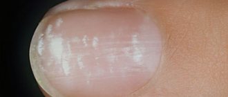
Leukonychia is the most common form of nail pigmentation, in which white areas of various shapes and sizes are observed in the thickness of the nail plate.
There are three forms of this disease:
- dotted (spotted) or stripe-like (changed areas disappear as the nail grows);
- subtotal (partial damage to the plate);
- total (persistent damage to the entire nail).
Causes of the disease: use of low-quality nail polish, illiterate manicure and pedicure, frequent contact with household chemicals, wearing tight shoes, lack of microelements (iron, calcium, zinc) and vitamins A, E, C, D.
Longitudinal grooves are superficial, weakly expressed single or multiple lines on the nails. It is observed both in healthy people and in cases of matrix dysfunction. The reasons are a lack of zinc (mainly in vegetarians), careless manicure and abuse, cuticle or matrix injuries, decreased immunity and frequent stress.
Nail hyperpigmentation occurs when pigments such as hemosiderin and melanin accumulate. As a result, the nails acquire a yellow or brown tint. Depending on the cause, there are two types of hyperpigmentation:
- medicinal - after taking tetracycline and resorcinol;
- chemical - after prolonged use of manicure varnishes.
Onychodystrophy, as part of other diseases
Onychorrhexis is a longitudinal separation of the nail. This pathology is observed in chronic dermatoses, constant contact with chemicals and solutions that dry out the nail plate.
Scleronychia is a special form of nail hypertrophy. It is characterized by the hardness of the nail plate, complete loss of elasticity and transparency. Leads to separation of the nail graft from the bed, similar to onycholysis. The nail becomes yellow-brown and the lunula disappears. The cause of the disease is endocrine disorders.
Trachyonychia is a rather unusual form of dystrophy. The nail plate is characterized by the absence of a lunula and the presence of small thin scales with a dull nail color. This pathology occurs in immunodeficiency.
Thimble-shaped nail wear - pinpoint depressions and pits on the surface of the nail plate, resembling a thimble. Most often, this nail dystrophy occurs with psoriasis, exfoliative dermatitis, alopecia areata and lichen planus.
Onychodystrophy of the “roof slats” type is nail dystrophy in the form of shallow longitudinal ridges or grooves located in parallel. Occurs in lichen planus and senile atherosclerotic microangiopathy - atherosclerotic lesions of small blood vessels in elderly people.
Nail patterns - erasing of the free edge of the nail due to constant scratching of itchy lesions. The edge of the nail plate itself becomes somewhat concave. Such dystrophy occurs in chronic itchy dermatoses - neurodermatitis and eczema.
Hapalonychia is a rare form of onychodystrophy in which softening of the nail plate occurs. Such a nail easily bends and breaks off, and cracks form on the free edge. The cause of this pathology is a violation of sulfur metabolism during the formation of keratin in the nail.
Congenital onychodystrophy
Onychomadesis is the separation of the nail plate from the nail bed from the side of the ridges. The process occurs quite quickly and acutely, sometimes accompanied by pain and inflammation. Once matrix function is restored, a healthy nail plate grows. With relapses, atrophy of the nail bed may develop, which leads to complete loss of the nail. This type of onychodystrophy can be inherited and first appears after a finger injury.
Koilonychia is a saucer- or cup-shaped depression on the surface of the nail. The nail plates of the hands on the index and middle fingers are most often affected, but sometimes all nails are involved in the pathological process. The exact cause of the disease is unknown; the role of a hereditary factor is assumed. Cases of koilonychia occurring against the background of anemia, thyrotoxicosis, Cushing's disease, typhoid fever and other diseases have been recorded.
Anonychia is the congenital absence of one or more nails. This pathology is rare and is a hereditary anomaly. It can simultaneously occur with a violation of the structure of the hair, the functioning of the sweat and sebaceous glands and other developmental defects.
Platonychia is a flattening of the nail plate, the absence of its natural convexity. This type of dystrophy is quite rare and is an anomaly.
Micronychia are short nail plates on the fingers, less often on the toes. Can be inherited. Sometimes it is a sign of other diseases (for example psoriasis).
Hippocrates' nails are a genetic disease of the nail apparatus, characterized by convex nail plates that resemble “drumsticks.” The nails look rough, but they are also quite fragile. The exact cause of this pathology is unknown [1][4][5][7].
Classification
Melanocytic hyperplasia refers to an increase in the number of melanocytes in the matrix. This can be a benign or malignant process. In this case, benign hyperplasia or malignant hyperplasia develops.
Nail melanoma most often affects the thumbs and index fingers.
Melanocyte activation is an increase in the production and deposition of melanin in nail cells (onychocytes) without an increase in the number of melanocytes.
Pathogens can cause irregular melanonychia because they stimulate inflammation by activating melanocytes.
Also, exposure to chemicals can cause nails to darken.
A spot on the nail is an indicator of a problem
Sometimes a specialist just needs to look at the color and nature of the spot to determine what disease caused it:
- White stripes appear in patients with diseased kidneys and dysfunction of the gastrointestinal tract.
- Yellow formations may indicate poor nutrition and skin diseases.
- Green and brown are due to fungal infection or penetration of Pseudomonas aeruginosa.
- Violet are visualized with a severe hematoma.
- Brown colors are typical for patients with kidney failure. They also appear due to malnutrition or lack of protein and folic acid.
- Black ones indicate serious liver problems.
- Dark brown ones develop with vitamin deficiency, diabetes, and pregnancy.
Leukonychia - why the nail hurts: causes of toenail pathology
Leukonychia - pain in the nail
Leukonychia disease is accompanied by the appearance of small white spots on the nail bed. The degree of damage is individual - from isolated cases to multiple manifestations. Why does my nail hurt? The causes of this toenail pathology are varied. These include:
- Impact of external factors
- Traumatic situations
- Chemical lesions
In addition, internal pathologies lead to the development of the disease - metabolic disorders, infectious diseases, lesions in the central nervous system.
Table of contents
- Etiology and pathogenesis
- Clinical manifestations
- Principles of treatment
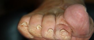
Onychomycosis (fingernail fungus, toenail fungus) is a fungal infection of the toenails or fingernails that affects any component of the nail complex, including the base, bed or plate. The disease can be accompanied by pain, discomfort, and changes in the appearance of the nails, which in some cases leads to serious physical and professional limitations, anxiety for patients and a decrease in their quality of life.
In our company you can purchase the following equipment for the treatment of onychomycosis:
- AcuPulse (Lumenis)
In the USA, onychomycosis of the nails occurs in approximately 2–13% of the population, in Canada - in 6.5%, in England, Spain and Finland - in 3-8%. According to a systematic review of 1914 articles, the prevalence of onychomycosis is as follows:
- general population - 3.22%;
- children - 0.14%;
- elderly people - 10.28%;
- patients with diabetes mellitus - 8.75%;
- patients with psoriasis - 10.22%;
- HIV-positive patients - 10.40%;
- patients on dialysis - 11.93%;
- after kidney transplant - 5.17%.
Onychomycosis accounts for about half of all nail pathologies and is the most common nail disease in adults. As for the affected areas, statistically, the legs are affected much more often than the arms.
The main forms of onychomycosis are:
- Distal lateral subungual onychomycosis
- Proximal subungual onychomycosis
- White superficial onychomycosis
- Endonyx-onychomycosis
- Candidal onychomycosis
Often patients have a combination of these subtypes, or total dystrophic onychomycosis - damage to the entire nail complex.
