The article was prepared by a specialist for informational purposes only. We urge you not to self-medicate. When the first symptoms appear, consult a doctor.
Red moles are benign neoplasms that grow from blood vessels. Red moles are also called hemangiomas.
According to statistics, red moles most often appear in childhood. They are observed somewhat less frequently in the adult population.
Until now, scientists have not decided whether hemangioma should be considered a vascular tumor or a pathology of a congenital nature. However, recent data indicate that red moles are the result of proliferation of the vascular endothelium, which allows hemangiomas to be considered vascular tumors.
Prevalence and localization
According to available statistics, hemangiomas are detected immediately after the birth of a child. This happens in 87% of cases. Girls are at risk; they account for about 70% of all diagnosed red moles. By the way, it is hemangiomas that make up up to 48% of all soft tissue formations that are detected in childhood.
A red mole can be found in any part of the body, but 80% of all such defects are localized in the upper part of the body. It is also possible to detect hemangiomas, although extremely rarely, on the bones, in the brain, lungs, and liver.
- 95% of all hemangiomas are simple formations;
- 3% – cavernous formations;
- 2% – mixed type formations.
Forecast
Possible consequences of the disease
Hemangiomas can cause necrosis (death) of tissue, so the lesion becomes an entry point for infection. The addition of a purulent process can cause sepsis. With hemangioma of bones, their destruction is possible. In addition, the process can contribute to blood clotting disorders and the formation of blood clots. Malignancy is possible - degeneration of hemangioma into cancer.
With timely consultation with a doctor, the neoplasm is successfully treated in 100% of cases Source: What a pediatrician needs to know about infant hemangiomas. Zakharova I.N., Kotlukova N.P., Roginsky V.V., Sokolov Yu.Yu., Zaitseva O.V., Maykova I.D., Idrisova G.R., Pshenichnikov I.I.: Medical Council , 2016.
Causes of red moles
The reasons for the development of hemangiomas have not yet been precisely established. It is unknown why they appear most often in the face area. Scientists attribute this to the fact that it is here that there is an abundant vascular network.
Childhood
Researchers agree that red moles are a consequence of pathology in the formation of vascular tissue, which occurs due to disturbances in the process of fetal development.
Vascular tissue passes through all parts of the fetal body along a chain of pericytic cells at the time of organ formation. When there is a lack of oxygen to any organ or tissue of the fetus, pericytic cells quickly react to hypoxia and try to eliminate it. To do this, using the production of special proteins, they attract a large number of pericyte cells to the problem area. Cells begin to form new areas of blood supply, eliminating existing hypoxia. Sometimes, due to imperfections in the mechanism, the reproduction of specific proteins does not stop even when oxygen starvation has already been eliminated. As a result, the vascular tissue continues to grow, forming a hemangioma.
Red moles are also called vascular hyperplasia, which indicates that they are a consequence of a disruption in the growth process of vascular tissue. Answering the question of how this happens is quite problematic, since it is necessary to carefully monitor how the intrauterine development of fetal tissue occurs. And the only data available are those obtained from the study of stillborn babies or aborted fetuses.
Adult patients
- The appearance of red moles in adulthood is explained by hormonal imbalances in the human body. This often occurs during gestation, during menopause, while taking medications with hormones, when using oral contraceptives, or due to pathologies of the endocrine system.
- It is possible that viral infections, ultraviolet rays, radiation, and chemical agents can provoke the growth of hemangioma.
- A lack of ascorbic acid can affect the condition of vascular tissue.
- If the capillaries are regularly damaged, this can provoke the appearance of hemangiomas.
- Vascular hyperplasia may indicate diseases of the internal organs, but the location of red moles does not indicate a problem in the area where they are most concentrated.
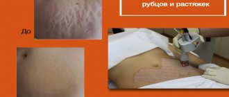
Treatment methods
Treatment methods for angioma are selected after examination. If the formation is flat, it is easier to remove than a convex one. No major pain relief is required before the removal procedure. A local anesthetic may be used. After the procedure, the affected area may bleed slightly.
After completion of the procedure, a rehabilitation period is recommended, during which exposure to sunlight on the affected area is limited.
There are many ways to remove angioma.
- Application of X-rays
. They help identify and reduce the mole. If it is small, it can be completely eliminated after a course of therapy. At the moment, the method is rarely used due to the risk of malignant degeneration. - Surgical removal
. After the procedure, the mole is completely gone. A connective tissue scar may appear in its place. Therefore, the method is used for localization in invisible areas. After the procedure, rehabilitation methods are used. For example, antibacterial powders or ointments, a ban on swimming for 1-2 days. - Cauterization with carbon dioxide
. The technique is applicable for small formations located on the upper layers of the epidermis. Therapy for large moles is not carried out using this method, since its lower layers may remain intact, so it will form again. - Chemical sclerosis
. Suitable for capillary type moles. A substance is injected subcutaneously that eliminates the effect of blood vessels. The mole stops receiving blood and gradually disappears. - Cryodestruction
. The mole is cauterized with liquid nitrogen. Due to the low temperature, the walls of the capillaries are destroyed, and a crust gradually forms. Healthy tissue will form in its place. The technique is applicable when angioma penetrates to the deep layers of the epidermis. - Coagulation
. With its help, large and small formations are removed. There are no scars left after the procedure. We will use infrared, radio wave, and light methods.
Removal is recommended if the aesthetic appearance is compromised and there is a risk of malignant degeneration.
Red moles in newborns
During the neonatal period and infancy, red moles in children are very noticeable, but they often disappear by the age of 5.
Such benign tumors do not pose a danger if they:
- They do not itch, do not hurt, do not cause any concern;
- They do not become larger, for example, in a few months they do not increase in size by 3 times or more;
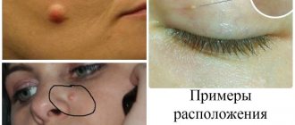
- Not located under the eye, on the nose, on the face or on the genitals.
It is known that hemangiomas can rapidly grow along the periphery, this is especially true for the first months of a child’s life. Therefore, in 10-12% of cases they are removed, since there are indications for this. The fact is that as the mole grows, it will destroy nearby tissues, causing defects not only cosmetic, but also functional. This is relevant in the case when hemangiomas are located on the ears, under the eyes, etc. Normal functioning of organs becomes impossible due to compression by the overgrown formation.
Diagnostics
The exact causes of a red mole have not been established, but methods for its diagnosis and treatment have been developed at a high level.
Types of examination to establish the correct diagnosis:
- Taking an anamnesis - the doctor asks questions about the existence of a similar mole among the patient’s family members, about the possible reasons for its appearance and changes;
- Referral to a more specialized specialist - dermatologist, surgeon, oncologist, endocrinologist, gastroenterologist, neurologist;
- Dermatoscopy – the doctor conducts an external examination of the birthmark;
- Digital dermatoscopy is a digital image of a mole taken using a dermatoscope and displayed on a computer screen. The possibility of such magnification and examination of the birthmark simplifies the study;
- Biopsy is a method for diagnosing malignant neoplasms; if this procedure is carried out correctly, the result is 100%. Types of biopsy: Puncture - a piece of tissue is taken with a needle in a limited amount under anesthesia;
- Total excision is also a therapeutic procedure in which tumors are removed or used for histological testing;
- Ultrasound;
Features of red moles in adults
Red moles, as primary formations, do not appear in adulthood. They arise from previously undetected vascular proliferations. Most often, they are treated before the child enters school, so if a hemangioma is present in adulthood, this means that it was either not treated, or the tumor is located on internal organs.
The most dangerous location of a red mole is considered to be the vertebral body. As the tumor grows, the structures of the spine will weaken, which contributes to the occurrence of fractures.
Classification of red moles
Morphology of hemangiomas
Red moles can be capillary or cavernous. Capillary hemangiomas are represented by layers or a group of vessels that are closely pressed to each other. Each of the vessels has a wall made of a membrane, one or several layers of cells, similar in structure to epithelial cells. The lumens of these small fused vessels are filled with blood cells. Sometimes fused capillaries form lobules separated by thin stroma.
As for cavernous moles, they are formed by cavities of different sizes and shapes. The cavities are represented by a layer of endothelial cells, which in their structure resembles the endothelium of capillaries. A rupture of the cavity septum and the formation of papillae in the resulting lumen are possible.
Location of red moles
- Simple hemangiomas are located on the surface of the skin.
- Cavernous or cavernous hemangiomas are located under the skin.
- Combined hemangiomas have the cutaneous and subcutaneous parts.
- Mixed moles contain elements of other tissues, for example, nervous or connective. These are hemangiomas such as angiofibromas, hemlymphangiomas, etc.
Origin of red moles
- Acquired moles are those that appear during a person’s life. However, they can have exclusively percutaneous localization. Complex forms of hemangiomas cannot be acquired. Often discovered during surgery, they are present in the human body from birth, but have not been diagnosed.
- Congenital red moles are detected either during the newborn period or in infancy.
Course of the pathology
Simple moles do not pose a threat in terms of disrupting the functioning of certain organs.
In turn, complex moles can affect the following organs and systems:
- On the organs of vision, on the brain, on the ears;
- On the vertebrae, but it is difficult to remove such formations, since they are located in hard-to-reach places;
- On large vessels or on vascular nodes.
Prevention education
To prevent the formation of angioma, prevent multiple formation of moles, damage and malignant degeneration, it is recommended to use the following rules:
- prevention of skin damage in the area of angioma;
- drink plenty of fluids to restore water and electrolyte balance and remove toxic products;
- consumption of vegetables, fruits, and other healthy foods high in vitamins K, C, E, D;
- daily bowel movements;
- no visits to the solarium or tanning in the open sun;
- the use of creams with a high degree of protection against ultraviolet rays, SPF of at least 50;
- daily skin hydration.
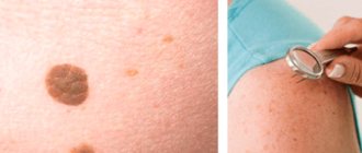
Despite the absence of danger from moles, it is necessary to monitor them constantly. Usually they are of poor quality, but with prolonged exposure to a damaging factor they can develop into skin cancer. If there is a risk, it is better to periodically consult a dermatologist. He will record every mole on the body, promptly identifying an increase during a re-examination.
If a mole spoils the aesthetic appearance, causes discomfort, or becomes dangerous, it can be removed in a timely manner. Many methods have been developed that can do this painlessly.
Features of red moles
Hemangiomas are somewhat different from other formations. Features of red moles are as follows:
- The first 90 days after the baby is born will be characterized by accelerated growth of the mole;
- In premature babies, hemangiomas grow 2 or 3 times faster than in children born on time;
- Small red moles may disappear on their own in the first five years of a child’s life;
- Only simple hemangiomas can resolve spontaneously; other forms of moles require intervention;
- It is impossible to predict what will happen to the mole after it has regressed or after growth has stopped.
Symptoms of red moles
- A simple mole. A simple red mole most often looks like a red spot; its size can vary. The spot rises somewhat above the skin. If you press on the formation from the edge, while capturing a healthy area of skin, the mole will become lighter and smaller. When the pressure stops, the shape, color and size return to the same. In the first 4 months after birth, the mole will grow around the periphery. You can understand this if you make a stencil of it on paper and apply it again after 2-3 weeks.
- Cavernous hemangioma. A cavernous red mole is located under the skin. The skin above it remains unchanged. A mole can be in a capsule or without it. If there is no capsule, then the boundaries of the formation will not be clear. The color of the mole is blue; the vessels that feed the mole may appear through the skin. Just like a simple hemangioma, the cavernous formation becomes smaller in size when pressure is applied to it. Sometimes the skin that is located above the mole has a higher temperature. Education does not pulsate. When palpated, you can feel the lobules of which it consists. Cavernous formations on the neck, near the ears, and on the head are dangerous, as they grow quickly and are capable of penetrating into nearby tissues.
- Combined hemangioma. A combined red mole has both a subcutaneous and a supracutaneous part. Moreover, the portion that is located under the skin is most often larger compared to the supercutaneous part.
- Mixed red mole. A mixed formation has elements not only of vascular tissue, but also of other tissues - lymphoid, nervous, connective.
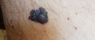
Precancerous neoplasms (precancrosis)
Precancerous neoplasms are those that, under the influence of congenital or current causes, have acquired a tendency to malignant degeneration. As a rule, these are chronic conditions that are observed in a person for a long time.
Thus, precancerous tumors are dangerous neoplasms on the skin that can lead to the development of oncological processes. These include:
- Actinic keratoma is a keratosis in which dry crusts and scales appear on the skin of older people. When they peel off, slight bleeding may occur.
- Xeroderma pigmentosum is a hereditary tumor that develops due to increased sensitivity of the skin to ultraviolet radiation. Rarely encountered, these are pigmented spots that become warty growths.
- The cutaneous horn is a cone-shaped tumor that looks like a horn. Has a yellow or brown color. occurs in exposed areas of the body that are regularly subjected to friction or pressure. Typical for older people
- Bowen's disease is an intraepidermal cancer. Without treatment, it can transform into invasive skin cancer. In the early stage, Bowen's disease appears as a small reddish-brown spot measuring 2-50 mm. has a flaky surface and raised, uneven edges. After removing the scales, a weeping but not bleeding surface remains.
Spontaneous problem resolution
Red moles go away on their own if they are superficial or simple. This is possible in no more than 15% of cases. Most often, formations that are localized on closed parts of the body regress.
At first, the tumor becomes less bright and decreases in size. After six months, the mole becomes a pale pink spot located at skin level. In this case, the skin over it atrophies. After another 3-4 years, only a small depigmented area remains on the skin.
Complications
Hemangiomas (red moles) can disrupt the functioning of organs that are located next to them. This is especially dangerous if they grow in the liver, near the eyes, or in the brain.
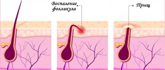
- Inflammation and the appearance of ulcers on the mole are possible. As the pathological process fades, spontaneous disappearance of the mole is not excluded.
- Bleeding from a mole as a result of its traumatization. Bleeding is dangerous if there is a large mole, if it is a cavernous or combined formation localized on the internal organs.
- Infection.