Causes Symptoms Diagnostics Treatment Advantages of treatment at MGK Prices
Xanthelasmas of the eyelids look like small single or multiple flat yellowish plaques located at the inner corner of the upper (usually) or lower eyelid. Xanthelasmas are benign formations that are not prone to malignancy; their appearance is associated with general disorders of lipid metabolism.
What is Xanthomatosis?
A xanthoma is a skin lesion caused by the accumulation of fat in macrophage immune cells in the skin and, less commonly, in the layer of fat under the skin.
Some types of xanthoma indicate lipid disorders (eg, hyperlipidemia or high blood fats), where they may be associated with an increased risk of coronary heart disease and sometimes pancreatitis.
Xanthomas are classified into the following types depending on where they are found on the body and how they develop.
General information
Xanthelasma (xanthoma) of the skin of the eyelids is a benign formation in the form of a flat, slightly raised yellowish plaque on the skin.
The predominant localization of the pathological formation is the inner corner of the upper eyelid. Formations can be either multiple or single, symmetrical, varying in consistency and shape - soft/hard, even flat or nodular. Eyelid xanthelasmas are a type of xanthomas that appear on the eyelids when they are absent elsewhere on the body. Histologically, eyelid skin xanthoma consists of xanthoma cells (histiocytes), which contain intracellular fat deposits, that is, they contain vacuoles that are filled with partially esterified cholesterol . They are located predominantly in the upper reticular layer of the dermis/perivascular region, less often in the area of the appendages. These formations are not prone to malignant degeneration and do not pose a threat to life.
Xanthelasma of the eyelids (a type of xanthoma) is the most common manifestation of xanthomatosis. It occurs more often in females (1.1%) and much less frequently in males (0.3%) aged 15-75 years with a peak at the age of 30-50 years. Xanthomatosis is a disease characterized by multiple deposits of lipid substances, mainly in the skin (elbows, buttocks, thighs), less often in tendons, bones, dura mater), caused by hyperlipidemia .
Eruptive xanthoma
- Lesions usually appear as seedlings of small red-yellow papules
- They most often occur on the buttocks, shoulders, arms and legs, but can occur throughout the body
- Rarely the face and inside of the mouth are affected
- Lesions may be tender and usually itchy
- Lesions may resolve spontaneously within a few weeks
- Associated with hypertriglyceridemia (increased levels of triglycerides in the blood) often in patients with diabetes mellitus (diabetes mellitus)
Symptoms
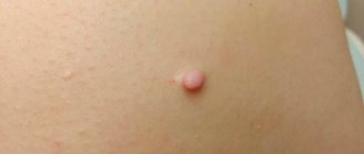
Xanthelasma is a light orange/yellow plaque that slightly protrudes above the unchanged surface of the skin, with a predominant localization on the upper eyelid (at the border of the fixed/moving part), sometimes at the inner corner of the eye. On palpation, the formation has a soft consistency and is painless. They can be single/multiple, located on one or both eyelids. In some cases, xanthelasmas merge to form a solid yellow stripe with an uneven contour or lumpy elements.
Characterized by slow, steady development, a tendency toward slow fusion of neighboring elements and the absence of spontaneous healing. There are no subjective sensations on the part of the patient. The formation can grow to the size of a large bean, but does not transform into a malignant neoplasm. In cases where xanthelasmas are symptoms of xanthomatosis, they can also be located on the lower eyelid and other parts of the body: on the face, elbow/knee joints, neck, buttocks, etc.
The resulting xanthelasmas do not regress on their own and persist throughout life. In some cases, their number continues to gradually increase. Below is a photo of xanthelasma.
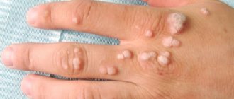
Common xanthoma
- Xanthoma-like lesions are caused by a rare form of histiocytosis.
- Lipid metabolism is normal.
- The skin lesions usually consist of hundreds of small yellowish-brown or reddish-brown bumps that are usually evenly distributed on both sides of the face and torso. They can be especially hard on the armpits and groin.
- Small bumps may join together to form sheets of thickened skin.
- In 30% of affected people, the mucous membranes of the mouth, respiratory tract or eyes (mucous membranes) are affected. Warty plaques in the mouth are called verruciform xanthoma.
- 40% of affected people develop diabetes insipidus, a condition that results in an inability to control water loss (leading to constant thirst and excessive urine production). This occurs due to the overgrowth of histiocytes on the lining of the brain (meninges).
- May affect internal organs (eg liver, lungs, kidneys, etc.)
- Self-limiting and eventually improves on its own, but may persist for many years.
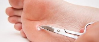
Recovery after xanthelasma removal
The recovery period after xanthelasma removal usually takes one week. During the specified period of time, it is not recommended to use decorative cosmetics to prevent infection of the wound. You should also temporarily refrain from taking hot baths, visiting baths, saunas, and avoid other types of effects that dilate blood vessels. Without eliminating the causes that cause the formation of cholesterol formations, a possible complication after removal of xanthelasma may be its recurrence.
What causes xanthoma?
There are several main disorders in which xanthoma is caused by a disorder of lipid (fat) metabolism. Because lipids are insoluble in water, they combine with proteins to form compounds called lipoproteins. Lipoproteins transport lipids and cholesterol in the blood to various parts of the body. Based on their size and weight, common lipoproteins are classified as chylomicrons, very low-density lipoproteins, low-density lipoproteins, and high-density lipoproteins (Fredrickson classification). They all play a role in maintaining the metabolic functioning of the body.
Changes in lipoproteins may result from a genetic defect (eg, primary hyperlipoproteinemia) or from an underlying systemic disorder such as diabetes mellitus, hypothyroidism, or nephrotic syndrome. These underlying diseases can cause elevated levels of certain lipids and lipoproteins, which then manifest as cutaneous xanthoma.
Monogenic familial hypercholesterolemia: type IIa
Mutation in the LDL (Low Density Lipoprotein) receptor
- High LDL level
- Total cholesterol in heterozygotes is 9-14 mmol/l
- Total cholesterol in homozygotes is 15-30 mmol/l
Polygenic hereditary hypercholesterolemia type IIIA
Wide range of results
Polygenic familial combined hyperlipidemia: type IIb
- Mixed genetic and life history causes
- Elevated total cholesterol levels
- Elevated triglyceride levels
- Low HDL (High Density Lipoprotein) Cholesterol
- Elevated LDL cholesterol levels
- LDL may be normal levels but denser and more likely to cause atheroma (sebaceous cyst)
Moderate hypertriglyceridemia
- Often also associated with high blood pressure, obesity, diabetes, metabolic syndrome, high insulin levels, high uric acid levels
- This may be due to alcohol or medications such as systemic steroids, isotretinoin, acitretin
- Triglycerides 2_10 mmol/l
- Often associated with low HDL cholesterol levels
Severe hypertriglyceridemia: Types 1 and V
- Mixed genetic and life history causes
- Diabetes
- Familial LPL (lipoprotein lipase) deficiency
- Triglycerides > 10 mmol/l
- Elevated total cholesterol levels
- Raised chylomicrons
- Elevated LDL cholesterol levels
Wide beta hyperlipoproteinemia: type III
- Rare mutation of the apo E gene
- Triglycerides 5-20 mmol/l
- Total cholesterol 7-12 mmol/l
The reason for the appearance of xanthoma with normal levels of fat in the blood is currently unclear.
Prevention
There are no exact recommendations for prevention of xanthelasma.
Need to know! Experts can only give general recommendations that will help patients get out of the risk group and avoid the appearance of such formations with age:
- monitor your weight and introduce more healthy foods into your diet;
- add foods containing fiber , and also eat up to three hundred grams of vegetables and fruits daily;
- Replace animal fats with vegetable fats , but consume them in reasonable quantities;
- drink at least 1.5 liters of water per day ;
- exercise regularly ;
- try to give up smoking and alcohol ;
- control metabolism , if necessary, visit a nutritionist and endocrinologist.
What examination is required to treat xanthoma?
A skin biopsy may be required to confirm the clinical diagnosis of xanthoma.
Appropriate blood and urine tests and x-rays are performed to determine the cause of abnormal lipoprotein levels, if present. The risk of cardiovascular disease, including heart attacks, peripheral vascular disease and stroke, increases with elevated levels of certain lipoproteins. It is important to identify contributing factors so that appropriate therapy can be instituted.
Contraindications
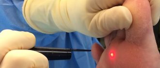
If you have xanthelasma of the eyelids, you should consult a specialist before removing them with a laser. Laser therapy should absolutely not be carried out if the following disorders are detected in the patient:
- herpetic type skin lesions;
- infectious diseases;
- diabetes mellitus;
- diseases of the cardiovascular system;
- dysfunction of the thyroid gland;
- oncological diseases;
- neurological disorders;
- diseases of the hematopoietic system.
It is also not recommended to carry out the procedure during pregnancy and lactation.
During the initial examination, the doctor will reliably assess the patient’s physical capabilities and make a decision on the possibility or impossibility of the operation.
How does xanthoma heal?
The primary goal of treating xanthoma that is associated with an underlying lipid disorder is to identify and treat that lipid disorder. In many cases, treatment of the underlying disorder will reduce or resolve the xanthoma. In addition, treating hyperlipidemia will reduce the risk of heart disease, and treating hypertriglyceridemia will prevent pancreatitis. Lipid disorders are treated with changes in diet and lifestyle, with or without medications.
Dietary measures should include:
- Prepare most dishes from vegetables, salads, grains and fish
- Minimize saturated fat (found in meat, butter, other dairy products, coconut oil, palm oil)
- Minimize your intake of simple refined sugars found in carbonated drinks, sweets, cookies and cakes
- If you are obese or overweight, aim to lose weight slowly by reducing your calorie intake and increasing exercise.
Very effective medications may also be prescribed. These may include:
- Statins are pharmaceutical drugs designed to combat high levels of cholesterol in human blood (HMG-CoA reductase inhibitors). Such as simvastatin and atorvastatin reduce the production of cholesterol by the liver, resulting in lower LDL cholesterol, increased HDL cholesterol and a mild reduction in triglyceride levels. Treatment should be monitored by regular blood tests to check lipid levels and ensure that liver and muscle enzymes are normal, as statins sometimes cause problems, especially when given at higher doses.
- Fibrates, such as bezafibrate, may be added to reduce triglyceride production by the liver, lower triglyceride levels, and increase HDL cholesterol levels. They may cause gastrointestinal side effects.
- Ezetimibe may be added in high-risk patients or those who do not tolerate higher doses of statins. It reduces the absorption of cholesterol from the intestines, lowering total and LDL cholesterol.
- Nicotinic acid lowers cholesterol, LDL cholesterol and triglycerides, and increases HDL cholesterol. At therapeutic doses of at least one gram per day, it causes redness of the skin. Its analogue, acipimox, is better tolerated.
- Cholestyramine and colestipol are rarely used because they are not as effective as the drugs listed above and are poorly tolerated.
Surgery or locally destructive techniques may be used to remove xanthomas that do not resolve spontaneously or with treatment of the underlying cause. Advanced xanthoma that affects vital organ functions can be treated with chemotherapy drugs or radiation therapy.
Advantages of xanthelasma treatment and prices at MGK
By contacting the Moscow Eye Clinic, you can be assured of a quick and reliable diagnosis of xanthelasma of the eyelid and its effective treatment. Removal of xanthelasma of the eyelid can only be performed by a specialist with appropriate professional skills. To avoid possible complications, this procedure should be entrusted to an experienced doctor. At the Moscow Eye Clinic you will be able to undergo all the necessary tests, based on the results of which the attending physician will recommend you the most effective treatment methods. The Clinic employs specialists with extensive professional experience who enjoy well-deserved respect both among colleagues and among patients.
Removal of xanthelasma of the eyelid is performed on an outpatient basis under local anesthesia. During the operation, sterile instruments and disposable consumables are used, which eliminates the risk of infectious complications.
Author:
Yakovleva Yulia Valerievna 5/5 (1 rating)
Honey. portal: