Moles Benign tumors
- Seborrheic keratosis
- Moles (pigmented nevi)
- Other benign skin tumors (papillomas, hemangiomas, granulomas, etc.)
So, now I will dwell in more detail on the most common benign skin tumors (I will write a separate article about malignant ones).
As I already said, the classification of skin tumors is very extensive. And it is needed mainly for very smart professors, in order to give students bad marks on exams. In my daily work I am guided by the principle of oncological vigilance , dividing everything that I see into “cancer-hazardous” and “cancer-safe” . Depending on the degree of “danger”, I choose the method of removal and decide on the need for histological examination .
Attention! The presented photographs and micrographs are not absolutely typical for the listed diseases. All skin tumors may look alike to a non-specialist. Do not try to diagnose yourself based on what you see. If you have any doubts about a particular formation on the skin, be sure to consult a specialist!!! ps: all tumors shown in the pictures were successfully removed and subjected to histological examination (i.e. all diagnoses are final).
The most harmless from the point of view of an oncologist are tumors that arise from cells of the superficial layers of the skin that do not contain pigment (that is, not moles ). Safe non-pigmented skin tumors include papillomas, warts, seborrheic keratosis (once again, I’ll make a reservation that we are talking only about the most common tumors ). It is not interesting to write about papillomas - they are small growths of soft consistency on a thin stalk, similar in color to the skin. Removed by electrocoagulation.
But we can dwell on seborrheic keratosis in more detail - it is painfully often similar to pigmented tumors and even melanoma .
Seborrheic keratosis
The causes of their occurrence are not fully understood. But given that these tumors are more common in older people in open areas of the body , it can be assumed that ultraviolet radiation is harmful. An important factor is the degree of skin pigmentation: the lighter it is, the higher the risk of benign and malignant tumors . As a rule, it is represented by one or several grayish-brown plaques with a rough surface . It grows slowly. It usually does not cause discomfort, although it may be accompanied by minor itching.
| As astrologers say, a typical representative of the sign is a tumor in the form of a gray plaque with a peeling surface. It appeared several years ago and is slowly increasing in size. Upon examination and dermatoscopy, there is a picture of keratosis. The tumor was removed by electrocoagulation. Histological conclusion: seborrheic keratosis. |
| The same. In principle, it is not necessary to send for histology. And so everything is clear. However, the histological conclusion is seborrheic keratosis. |
| A more interesting case is that a darker area is visible along the periphery of the tumor. All other clinical signs and dermatoscopy data indicate seborrheic keratosis. The tumor was removed, the base was treated with an electrocoagulator. Histological conclusion: seborrheic keratosis. No further treatment is required. Recommendations include protection from ultraviolet radiation and repeated treatment if similar tumors occur on other areas of the skin. |
| An even more interesting case. The patient came forward on her own after visiting an oncologist, where a diagnosis of “suspicious melanoma” was established, and surgery involving wide excision under anesthesia in a hospital was recommended. Yes, it looks like a “bad mole” in appearance. After dermatoscopy, I was diagnosed with seborrheic keratosis. The tumor was removed on an outpatient basis without suturing using electrocoagulation. Histological conclusion: seborrheic keratosis. The time spent by the patient for examination and treatment was 45 minutes. I can’t say exactly how much nerves I saved. |
| And this is a really difficult case. Upon examination and dermatoscopy, there is more evidence for a pigmented nevus. The history includes fairly rapid growth (5-6 months), itching, and discomfort in the tumor area. All the reasons for making a diagnosis of “suspicious melanoma.” An operation was performed - excision of the tumor. Histological conclusion: seborrheic keratosis. |
This is such an absolutely harmless, but sometimes difficult to diagnose sore, seborrheic keratosis . Briefly again:
- appears more often on the skin of exposed areas of the body in older people;
- outwardly it looks like a plaque with a dry, scaly surface from grayish to brown, itching may bother you;
- lesions are often multiple;
- does not pose a danger to life and health;
- removed by electrocoagulation.
I have everything about seborrheic keratosis. If you have any questions, write to
Seborrheic keratosis. (Synonyms: keratopapilloma, senile keratosis, keratoma, basal cell papilloma) is a tumor that grows very slowly and can form over several years and is the most common skin tumor of epithelial origin. This disease is not associated with the human papillomavirus and is associated with sun exposure absent.
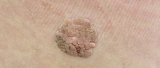
At first, a keratoma is a limited yellow or brown spot, which gradually increases in size. The surface of the spot is gradually covered with gray-yellow crusts, which are easily removed. Over time, the lesion begins to protrude above the surface of the skin, the crusts become more dense and riddled with cracks. Keratoma can be on a variety of parts of the body, except the palms and soles. The lesions can be single or multiple. Keratomas, which are located in places of greatest friction, pose a particular danger as they are constantly injured (the tumor becomes inflamed, bleeds, pain and itching appear).
If the keratoma begins to bother you: it increases in size, turns red, or begins to itch, you should consult a doctor. Under no circumstances should you remove any keratoma yourself! The optimal treatment is laser removal of keratoma. A dermatologist is a specialist whom you should consult for advice. After a preliminary examination (dermatoscopy), a dermatologist will select the optimal treatment for you. Seborrheic keratosis has many clinical variations. One of the types of seborrheic keratosis is melanoacanthoma. Melanoacanthoma is a keratosis with a high content of brown pigment, with melanin found almost exclusively in keratinocytes. This form of keratoma is very often confused with melanoma during routine examination. But with a dermoscopic examination, it is easy to detect the main clinical signs (comedone-like structures, milia-like cysts, medullary structures), which make it possible to correctly diagnose and differentiate these signs from melanoma. Conclusions: in general, keratomas are safe tumors, since they are benign and only pose a danger to the patient’s life. in case of malignant degeneration into squamous cell carcinoma. Thus, it is very important to monitor the condition of the tumor and identify possible signs of its transformation into cancer.
| Keratoma before removal | Keratoma after removal |
The article was prepared by dermatologist of the Tiger Cub Children's Medical Center Bedretdinova Rushaniya Shamilovna
Nevi (aka moles)
My first article about moles is on the website of the PLASTES center https://www.plastes.ru/nevus.html Here I want to pay more attention to the issues of additional diagnostics and the safety of removing pigmented skin tumors.
We will immediately remove the questions about why moles appear on the skin and why melanomas arise from them from consideration as uninteresting and, in our case, idle. Let's start the conversation right away from the moment the mole for some reason began to bother me.
Here is a typical example of a letter from my correspondence with patients:
“...I don’t know what to do and what to do? Very scary. I am 33 years old. And all my life I have lived with a mole about 1 cm in diameter. It is swollen and with villi, almost black in color, but with uneven edges. On the thoracic region. Never bothered me. And starting in June of this year, the mole itself began to itch around the mole, as if it was pulling inward. All this is tolerable. I turned to our oncologists, they said that as long as it looks like a benign tumor, it should be removed (literally from the head physician of the N-sky oncology dispensary). The consultation lasted no more than 5 minutes. Visual inspection, without any equipment. I asked to prescribe tumor markers, they refused, they said we didn’t see any point. Nothing was really explained. Very scary. I read about melanoma. What should I do? The general condition is strange: it seems like a cold, but something is wrong. A little later I was told that at the consultation they identified my mole as a congenital pigmented nevus. Are there any guarantees that the outcome of the surgical intervention will be good??? I was really very frightened by the stories of friends that people died after removing a mole...”
Well, what can I say? I perfectly understand the state of mind and fear of this girl. There are several such letters a day, and such patients have to be constantly reassured and explained the same thing.
The most important thing is to understand: until the tumor is removed and subjected to histological examination, there will be no diagnosis!!! And no additional research methods (tumor markers, dermatoscopy, CT, PET, etc.) will ever give us a definite answer to the question of whether it is melanoma or not.
Another question is how to remove the tumor to minimize the risk of adverse consequences. This is where the main “terrible secret” lies - in the method of removal. In order not to let my thoughts wander, I will immediately reveal this secret. If I have even the slightest suspicions and doubts, I insist on COMPLETE SURGICAL EXCITION OF THE NEOPLASM .
Now I will explain why. I completely agree that any intervention in a tumor can lead to rapid uncontrolled progression (growth of the tumor itself and the appearance of metastases). That is why ALL SUSPICIOUS MOLES SHOULD BE REMOVED SURGICALLY, IMMEDIATELY AND COMPLETELY WITH THE HEALTHY SKIN AROUND!!! For exactly the same reason, IF THERE ARE SUSPECTS, YOU CAN NOT DO A PARTIAL BIOPSY, YOU CANNOT DO ELECTROCOAGULATION OR LASER REMOVAL!!!
See how simple everything turns out to be. That's where all these scary stories come from. The doctor simply underestimated the degree of danger, did not attach importance to suspicious symptoms and, instead of complete excision, performed coagulation (with electricity or laser - it makes no difference). The tumor was “disturbed” and began to grow. If the same tumor had been immediately and completely removed, then no further unfavorable sequence of events would have occurred.
However, please note that I am not saying that complete excision is an absolute guarantee of safety. Even after complete surgical excision, the tumor can progress into metastases. But only if it really was melanoma. As you already understand, it is possible to find out whether it was melanoma or not only after a histological examination.
Next, I will present several cases when I insisted on complete surgical excision of pigmented tumors.
| In general, nothing unusual. This is what “calm” moles look like under a microscope. I posted this illustration to show that a dermatoscope is not the most accurate diagnostic device. The indication for excision in this case was a feeling of discomfort and itching in the area of the mole. Histological conclusion - pigmented nevus. |
| A young woman has a pigmented nevus in the décolleté area. Over the past few months, he has noticed a change in color (it has become more intense) and an increase in the size of the mole. Even these two signs are enough to make a diagnosis of “dysplastic nevus” (restless mole) and surgical excision. And, despite the danger of a rough scar, I had to have surgery. Histological conclusion: dysplastic nevus. That is, pre-melanoma. The result of the operation is shown in the image on the far right. There is no more tumor. There are also dangers of degeneration into melanoma. I don’t know what would have happened if the operation had not been performed or the woman had applied later. I also cannot say what would have happened if electrocoagulation had been performed instead of surgery. Perhaps this would become a provoking factor for tumor transformation. |
| This case also illustrates well my approach to choosing a method for removing “moles.” This is the spot that appeared on the young man’s skin on the plantar surface of his foot. It appeared suddenly; there was nothing in this place before. This spot does not cause any discomfort. What to do? If you think that by looking through a deramtoscope, I will immediately make the correct diagnosis - you are mistaken. I have never seen such a picture before. Guided by the principle of cancer vigilance, which says “if the diagnosis is not clear, think about cancer,” I insisted on surgical excision. The histological answer was unusual - pinpoint hemorrhage. Apparently, a microtrauma occurred, which the patient did not even remember. You may think that the operation was performed in vain. To which I will answer your question: what if it turned out to be melanoma? Once again, I will note the main thing - we will never know the exact diagnosis until we remove the tumor. And in such situations it must be removed immediately and completely. |
These are just a few examples from my practice. If I have time, I will add to this gallery. Finally, a few points about moles:
- The majority (99.9%) of moles do not pose any danger to life or health. They can be safely removed in different ways: electro- or laser coagulation, surgery. It all depends on the size and location.
- If a mole bothers you (no matter how), be sure to see an experienced doctor. In such cases, it is preferable to perform complete surgical excision with mandatory histological examination.
- You can ask questions at
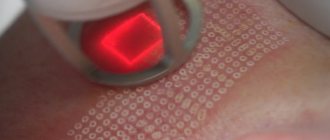
Removal of keratoma using laser
Laser keratoma removal is a modern method of treating benign skin tumors caused by excessive keratinization and proliferation of the upper layers of the epidermis. You can get rid of the characteristic plaque, growth or crust in one visit to the medical center.
To effectively and safely solve the problem, we use the latest generation carbon dioxide laser. Its light energy instantly evaporates (removes) skin cells, simultaneously disinfects the site of exposure and coagulates (glues) its vessels. The procedure is quick, painless and without complications.
The price for keratoma removal in our clinic is from 250 rubles. You can sign up for the procedure online or by phone - call!
What are keratomas, where do they come from and what are they?
Keratomas are single or multiple neoplasms resembling plaques, growths, crusts or spots that rise above the surface of the skin. Most often, mature and elderly patients (40 – 60 years and older) encounter them. The appearance of keratomas is caused by disruption of keratinization processes and pathological division of cells in the upper layer of skin.
The main reason for the appearance of keratomas is the natural aging of skin cells. With age, their adaptation mechanisms change, which, against the background of a decrease in local immunity, leads to spontaneous proliferation of epidermal cells. In essence, the skin becomes very susceptible to the negative effects of various factors and ceases to control its response to them.
Various external and internal environmental factors can provoke uncontrolled cell division and the appearance of tumors.
- Excessive exposure to ultraviolet rays.
- Endocrine disorders.
- Vitamin A deficiency.
- Decreased production of sex hormones.
- Long-term use of medications.
- Regular trauma to the skin, for example, rubbing with clothing.
- Skin contact with chemicals, etc.
The resulting keratomas are benign in nature. Some of them demonstrate spontaneous self-destruction and go away on their own, others grow slowly, and others can become malignant. Based on the clinical manifestations and prognosis of development, experts distinguish several types of keratomas.
- Senile keratoma is the most common neoplasm, often called senile keratoma. The pathology appears on open areas of the skin and goes through several stages of development: from a dark spot to the formation of a soft, scaly tumor, which can go away on its own or transform into a horny keratoma with subsequent malignancy.
- Horny keratoma - so-called. cutaneous horn, which is quite rare. Most often, the pathology appears on the face or genitals. The neoplasm looks like a dark-colored, dense tubercle that rises above the surface of the skin. At any moment it can transform into a malignant tumor.
- Seborrheic keratoma is a slow-growing growth that occurs on the upper body or scalp. From a small yellowish spot, it turns into a loose scaly formation with crusts that periodically peel off. There are no cases where seborrheic keratoma goes away on its own or turns into a malignant tumor.
- Follicular keratoma - a neoplasm appears around the hairs and their follicles, most often on the face and scalp. The pathology looks like a small flesh-colored nodule with a flaky surface. It is not prone to malignancy, but can relapse even after laser removal.
- Solar keratoma is a precancerous growth that most often appears on the face. It has the appearance of multiple, small scaly papules, which over time turn into dense, rough brown plaques. Sometimes solar keratoma self-destructs and after a while recurs in the same place. But most often it transforms into a malignant tumor.
- Angiokeratoma is a pathology formed by epidermal capillaries and externally resembles a dark, scaly vascular mole. The neoplasm can be single or multiple, spreading over the surface of the body or genitals. There were no cases of spontaneous resolution or transformation of angiokeratomas into a malignant tumor.
It is important to understand that keratomas are not a harmless flaky skin formation, but a potentially dangerous pathology that requires treatment. There is no need to delay with him! Laser removal of keratomas will help solve the problem and maintain health.
Indications and results of laser keratoma removal
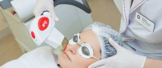
If you encounter potentially dangerous symptoms, you should immediately consult a dermatologist. After collecting an anamnesis, visual examination, and additional research, the specialist will make an accurate diagnosis and, if necessary, prescribe laser removal of the keratoma.
The procedure is carried out on an outpatient basis and after complete healing of the skin does not leave any traces - the benign neoplasm is removed without a trace.
Indications
- Changing colors or sizes
- Further spread over the surface of the skin
- Bleeding
- Soreness
- Accidental injury, etc.
Contraindications to the procedure
- Photodermatosis;
- Viral diseases in the acute stage, including herpes;
- Pregnancy, breastfeeding;
- Diseases, injuries, inflammations of the skin at the site of laser exposure;
- Oncological pathologies, etc.
You can get acquainted with the full list of contraindications during a consultation with a doctor at our clinic in Moscow. An experienced specialist will comprehensively study your problem, conduct differential diagnostics and decide on the advisability of using a laser.
How is the procedure performed?
Removal of keratoma on the face, body or scalp is carried out simultaneously - in 1 visit to the doctor. The light energy of a CO2 laser acts in a targeted manner - only on neoplasm cells, to a strictly specified depth and without damaging surrounding healthy tissue.
Instantly vaporizing the keratoma, the laser beam disinfects the impact site and seals the blood vessels. The procedure is carried out sterile and bloodless. All painful sensations are neutralized by the use of local cooling and anesthesia.
Unlike cryodestruction with liquid nitrogen, laser light energy completely removes the pathological formation, which prevents its further growth and reduces the risk of relapse.
Recovery after keratoma removal
After surgical removal of a tumor with a laser, a small scab forms on the skin - a protective crust that cannot be torn off, wet, scratched or subjected to any impact. New healthy skin is formed under it and this process must not be disrupted!
Therefore, during this period, we recommend that you refuse any kind of thermal and water treatments - visiting a bathhouse, sauna, solarium, etc. After 5 - 7 days (sometimes longer), the crust will fall off on its own, and you will see new skin, which also needs to be protected from aggressive influences, especially from ultraviolet rays.
A dermatologist at our clinic in Moscow will tell you in more detail about how the recovery period goes and how to properly care for the treated area of skin. The doctor will answer all your questions, calculate the cost of treatment for keratomas and help you get rid of them forever. Appointments are by appointment only - plan your visit in advance and call us.
Other neoplasms
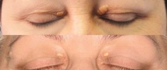
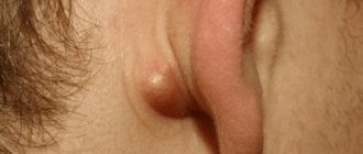
Very often we have to deal with papillomas . As a rule, these are small (2-3 mm) growths on the skin, with a thin base, soft consistency and pale pink color. They are usually located in the area of skin folds ( axillary and groin areas, submammary folds, neck ). Apparently, the cause of their occurrence is skin irritation in conditions of high humidity. Papillomas are not dangerous in terms of cancer . They can be easily removed using an electrocoagulator . Some comrades manage to “grow” papillomas to absolutely mind-boggling sizes, fearing the adverse consequences of removal. Do not be afraid! Here are some examples of papillomas that could have been removed much earlier.
Inflammatory tumors, the so-called pyogenic granulomas, . They are indeed very ominous. A red tumor appears on the skin , which grows very quickly and bleeds . There is only one solution to the problem in such situations - removal with histological examination.
Something else needs to be said about hemangiomas . These are tumors consisting of altered blood vessels . As a rule, they are congenital and can reach quite large sizes. In adults, hemangiomas often appear on the skin in the form of small red spots. Special treatment for small hemangiomas is not required. Removal is possible for cosmetic reasons.
BLESS YOU!!!