How to recognize lentigo?
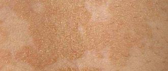
Treatment of this problem should begin with a correct diagnosis. The main symptom is a thickening of the stratum corneum of the skin, so that lentigo spots in most cases rise slightly above the surface. These spots are located both on the face and on the body.
Other characteristic symptoms:
• bright color of all shades of brown; • round or oval shape, clear boundaries; • visual sensation of swelling; • size from 1 to 3 mm, in rare cases the spot grows up to 2 cm; • severe itching with lentiginosis.
What is the clinical picture
When pigmentation is detected on the skin, most people do not associate it with any disease. Moreover, rarely does anyone know about lentigo and what it is.
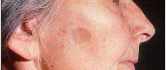
This is due to the fact that the spots do not cause discomfort. The only thing the patient notes is a change in their color from light to darker (as in the photo).
Sometimes the pigmented areas may thicken slightly or become bulging.
Such changes signal the degeneration of this neoplasm from benign to malignant. Then you need to urgently consult a doctor for diagnosis.
If your dermatologist has looked at the spots and determined that they are benign (not cancerous) and cannot turn into skin cancer, then he can begin to treat them. Sometimes we simply freeze the lesions with liquid nitrogen on the day of presentation. Laser treatment can make dark skin even darker if the wrong type of laser was used.
Dermatological surgeon Amy Paul
Causes
Lentigo is nothing more than an accumulation of excess melanin - a coloring pigment. These spots can either be inherited or appear unexpectedly in adulthood. Unfortunately, doctors have not yet fully studied this disease.
The following reasons for its appearance have been precisely defined: 1. Old age. Lentigo on the face is very common in people over 70 years of age, sometimes these spots begin to appear after 40. 2. Young age. The so-called juvenile lentigo in children and adolescents is known. Its causes are unclear; with the onset of adulthood, the spots disappear on their own. 3. Skin injuries. Occurs at the site of superficial wounds. 4. Excessive insolation. Excess ultraviolet radiation always has a negative effect on the skin, solar lentigo is proof of this. 5. Lentiginosis, or congenital lentigo, is inherited and manifests itself not only on the face, but also on the body together with other unpleasant pathologies: heart disease, congenital deafness, etc. 6. Malfunctions of the liver and intestines. There are cases where lentigo spots disappeared after surgical operations on the gastrointestinal tract.
What are the types of lentigo (5 options)
There is the following classification of lentigo:
- Solar – occurs as a result of prolonged exposure to the sun. People with fair and sensitive skin types are more susceptible to it. The appearance of this type of pigmentation is perceived as a primary sign of photoaging. The spots appear mainly on the neck, arms and face. This type of lentigo is not considered a disease, but rather a cosmetic defect.
- Simple (adolescent or juvenile) - it has nothing to do with ultraviolet radiation and skin aging. Most often it makes itself felt in adolescence or immediately after childbirth in women. Lentigines appear on the skin and mucous membranes in the form of spots up to 10 mm in size.
- Senile - they are also called liver spots. They are dark brown washouts (up to 1 cm in diameter) with uneven contours that appear on the face, neck and arms. People over 50 years of age are more susceptible to this disease if they previously did not particularly adhere to sun protection.
- Melanotic - these are ink-colored spots that appear randomly on the skin, which is more exposed to direct sunlight. They have a teardrop shape.
- PUVA - appears six months after undergoing therapy against psoriasis. The spots have unclear boundaries and resemble freckles in appearance. The torso is more susceptible to damage.
Chloasma also occurs. Women often encounter it during pregnancy or menopause.
Dermatologist Michelle Green
Lentignosis of the mucous components is characterized by a scattering of flat pigment spots of a light cream color and with smooth contours.
Such a disease can be acquired at the genetic level by inheritance or due to certain third-party factors.
Even knowing the signs of each type of lentigo, you should not make a diagnosis yourself. This is only within the competence of the relevant specialist. Therefore, there is no need to postpone your visit to him.
Treatment of lentigo on the face
The connection between lentigo and certain diseases of internal organs and the risk of developing skin cancer is alarming. Therefore, if freckles and moles rarely pose a danger to our health, then if lentigo occurs on the face, treatment should begin immediately.
First of all, you need to reconsider your lifestyle: avoid direct sunlight, apply creams with a high level of SPF protection to your skin, and do not skimp on skin care in terms of nutrition and hydration. If necessary, a dermatologist will prescribe the use of certain medications.
In addition, it is worth following a diet that excludes possible allergens. You should also check your gastrointestinal tract and put it in order; if you are overweight, you will have to mercilessly say goodbye to it.
Is it possible to cure this disease and how?
It is necessary to treat lentigo when the spot is too large, is constantly exposed to mechanical and solar influence and spoils the appearance with its presence.
Typically treatment comes down to the following steps:
- regular whitening of pigmented areas on the skin with special medications;
- constant application of protective creams against UV radiation to the skin;
- superficial exfoliation of the epidermis using various cosmetic procedures.
Don't worry if the spots disappear and then return after a few days or weeks. The pigment usually consists of several layers, so peeling occurs layer by layer.
Anesthesiologist Laura Rome
As a last resort, they resort to surgical intervention. Then the lentigo is cut out from the skin surface.
During the operation, the specialist captures part of the healthy tissue so that the stain does not appear again in the future. The remaining scar is easily removed plastically.
Arbutin, azelic acid and licorice extract have a good effect as whitening agents.
They contain ascorbic and kojic acid, retinoids and hydroquinone. The latter component is present in modern drugs, but in minute quantities. All other components are absolutely harmless and easily tolerated.
Laser destruction
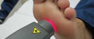
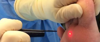
For those who want to get rid of lentigo without subsequent scars, laser therapy is recommended.
With it, the beam captures and destroys only the neoplasm, and does not affect healthy tissue in the neighborhood. This procedure is painless and is performed without local anesthesia.
If you have persistent solar lentigo/sun spots, the best combination is the Fraxel Dual laser and Yag laser.
Dermatologist Michelle Green
During the session, the patient may feel a slight tingling sensation on the skin.
Immediately after pigmentation has been reduced, redness may appear in the areas treated with the laser. If you are prone to allergies, slight darkening may appear in these places.
Before laser therapy, the patient must undergo a series of examinations to exclude existing contraindications.
Phototherapy
If the diagnosis of malignant pigmentation is excluded in the patient, then a phototherapy method may be offered.
This means exposing problem areas to a light beam. This stimulates the natural synthesis of collagen, thereby increasing the regenerative capabilities of tissues and rejuvenating them at the cellular level.
I have better results with the Q-swithced laser. I have used the Cutera Laser Genesis with great success. For patients who want a more conservative approach, a combination of hydroquinone and retin-A creams is suitable. IPL phototherapy is effective in some patients, but it is still superior for treating melasma.
Plastic surgeon Temp Patterson
Peeling
People with light age spots are recommended to resort to chemical peeling of the skin. The fact is that, in addition to removing darkened areas, adjacent healthy skin is also affected.
The following types of peelings are often used:
- retinoic;
- TCA peel;
- ABR peeling;
- pyruvic;
- azelaic.
This procedure is performed with local anesthesia. This is followed by a short recovery period of 10-12 days. As a result, the skin structure is completely restored and pigmentation disappears without a trace.
However, in addition to the beauty treatment, you should also follow a proper skin care regimen to brighten your skin.
Plastic surgeon Grant Stevens
There are many different types of peelings, but experts often praise these options:
- Peeling Cosmelan. The peeling itself is brown in color and has a thick consistency. Contains azelaic, phytic, kojic, ascorbic acids, as well as arbutin. The product brightens the skin well. First, in the clinic, the patient is applied a mixture of Cosmelan 1, which should remain on the skin for about 8-12 hours. At home, the mask is removed with cleansing cosmetics. 2 days will pass, after which the patient independently applies the Cosmelan 2 mixture every day. This must be done throughout the year.
- Jessner peeling. This name is given to a patented product that contains salicylic, lactic acids and resorcinol. This is a medium peel. The effect is amazing. The product will not only help remove pigmentation, but will also eliminate many other problems (acne, wrinkles, etc.).
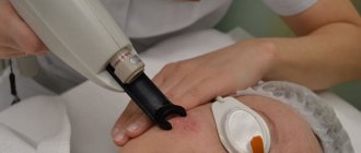
Lentigo – treatment with the M22 device
Modern methods of getting rid of lentigo include photothermolysis - treatment with IPL light or laser radiation. The essence of this method is the pulsed effect of light beams on the skin in the affected area, during which excess melanin is destroyed without affecting healthy areas of the skin.
It is generally accepted that laser copes with this problem better. However, modern light devices, for example, the M22 multi-module platform, demonstrate a high rate of removal of various age spots. For each type of hyperpigmentation, including lentigo, a special program is provided, which significantly increases the effectiveness of M22 compared to other photosystems. M22 rays penetrate the skin, destroy excess melanocytes and keratinized cells, cause the synthesis of collagen and elastin fibers and other active substances. These processes promote rejuvenation and rapid regeneration of damaged areas, lightening of spots and smoothing of the relief. After just a few sessions, the spots cease to be noticeable and the skin acquires a healthy color.
Lentigo therapy at home
You can treat lentigo yourself at home if it is clearly established that the pathology is benign.
All folk recipes are based on the use of natural lightening agents:
- lemon juice;
- hydrogen peroxide;
- ammonia;
- rowan;
- calendula;
- black currant;
- parsley;
- almonds;
- honey
Here are some productive recipes for homemade masks:
- The stems, leaves and roots of parsley are finely chopped to a paste. Then add honey. The resulting composition is applied to problem areas and, after leaving for half an hour, washed off with warm water.
- Place the lemon peel in boiling water and keep it warm for about 30 minutes. Then let the water sit and wipe the stains up to 3 times a day.
- Grind almonds in a coffee grinder and mix them in equal proportions with oatmeal. Add 5-7 g of milk powder and flavor with a small amount of water to form a mass similar to sour cream. Use this thickener to scrub the affected areas for about 5 minutes and wash off any remaining residue. Do this no more than 2 times a week.
- Dilute dry white clay (1 tbsp) with warm water until smooth and add a little lemon juice. Apply the composition to age spots and wash off after 20-25 minutes. This mask is used every day.
The suggested masks should be done in the evening, when you are not planning to go outside. This is due to the fact that after the procedure you cannot expose the skin to UV exposure.
As a treatment for lentigo, it is recommended to use pharmaceutical ointments with a whitening effect:
- zinc;
- sulfuric;
- Clotrimozole;
- salicylic.
Among the creams, the following brands are distinguished: Skinoren-gel, Melanativ, Clearvin. They should be applied to the skin twice a day.
I recommend using a combination of whitening cream (hydroquinone 4%) and tretinoin (vitamin A). But it will take months of treatment to achieve results. The laser is much faster.
Plastic surgeon Sam Naficy
Contraindications
It is not dangerous to remove lentigo with a laser, although there are a number of contraindications:
- oncological diseases;
- pregnancy and breastfeeding;
- blood diseases;
- diabetes;
- skin damage in areas of increased pigmentation.
Diseases from the list may not be absolute contraindications, but consultation with your doctor is required. During pregnancy and lactation, laser lightening is not recommended, because the spots that arise often go away on their own after the baby finishes feeding, as hormonal levels change.
Treatment of skin melanoma
Cutaneous melanoma (MC) is a malignant tumor of neuroectodermal origin that develops from transformed melanocytes located primarily in the skin. In addition to the cutaneous form of melanoma, which occurs in more than 90% of cases, there are also extracutaneous forms of the tumor, which include melanoma of the mucous membranes (gastrointestinal tract, genitals, nasal cavity and sinuses), melanoma of the membranes of the brain and spinal cord and uveal melanoma. Due to the pronounced predominance of the skin form in the structure of this disease, the latter is the most studied in terms of prognosis and treatment options [1].
Surgery
Primary melanoma
Definitive surgical removal of the tumor should preferably be performed within 4–6 weeks of initial diagnosis [3]. A mandatory step in the treatment of local CM is adequate excision of the primary tumor within healthy tissue. The choice of indentation is based on the results of a morphological study, namely the thickness of the primary tumor [1].
Table 1 | Recommendations for choosing an indentation when removing primary cutaneous melanoma [1, 2]
Lentigo maligna
Lentigo melanoma (lentigo maligna, Dubreuil's limited precancerous melanosis), a flat intraepidermal malignant tumor, is a slow-growing melanoma in situ that usually occurs in areas exposed to ultraviolet light, such as the face. In general, the choice of margin should take into account the cosmetic and functional consequences of the operation, and micrographic excision guidance can be used to preserve more tissue, especially in the facial area [3].
For acral localizations of melanoma (skin of the feet and hands, subungual bed), modified resection options can be used to maximize the preservation of limb function. It is not recommended to routinely perform prophylactic lymphadenectomy, chemotherapy or radiation therapy to unchanged regional lymph nodes, as well as to the area of the removed primary tumor. For tumor thickness > 1 mm, the standard diagnostic method is sentinel lymph node biopsy (SLNB), followed by regional lymphadenectomy if it is involved; this procedure is carried out only in specialized institutions. If it is not possible to perform SLNB, regional lymph nodes should be examined as thoroughly as possible, using ultrasound to navigate to a suspicious lymph node, followed by fine-needle puncture and cytological examination of the obtained material.
In the absence of micrometastases in the sentinel lymph nodes, no further lymph node surgery is required [3]. But with stages IIB and IIC melanoma (thickness more than 4 cm), it has a high risk of progression and recurrence.
For micrometastases in sentinel lymph nodes
Studies have confirmed that radical lymph node dissection does not improve survival prognosis. However, if micrometastases are detected during sentinel lymph node dissection, radical lymph node removal is usually recommended. The potential benefits of complete lymph node dissection should be considered for patients with sentinel lymph node tumors greater than 1 mm in diameter [3].
Clinically identified lymph node metastases
When melanoma metastases to lymph nodes are diagnosed (clinically or using imaging techniques), radical lymph node dissection is considered the standard treatment [1, 3].
Skin metastases
Treatment of cutaneous melanoma metastases is primarily surgical, but if the lesions are numerous or extensive and cannot be removed, systemic therapy should be considered. For multiple lesions on the limbs, isolated perfusion of the limbs with melphalan and TNF-α is used as a palliative method. At stage III in patients with satellite/transit metastases, the procedure can have a therapeutic effect, as evidenced by the data obtained on 5- and 10-year survival (40 and 30%, respectively). Isolated limb infusion is a modification of this method [3].
Distant metastases
If this is technically possible and reasonable (for example, in oligometastases), it is worth considering complete surgical removal of distant metastases. The use of this tactic is most effective for patients with normal values of tumor markers (LDH and S100B protein). In the presence of melanoma metastases to the brain, both stereotactic radiation therapy and surgery are considered equally effective.
Many studies suggest that resection of single or multiple metastases may be associated with a favorable outcome for stage IV patients. Neoadjuvant therapy followed by surgical resection of metastatic lesions may be considered. The value of cytoreductive interventions must be considered critically as there is no evidence that they improve survival prognosis. In some clinical situations, it makes sense to use cytoreductive surgery for palliative purposes, especially in combination with postoperative radiation therapy for local disease control [3].
Radiotherapy
Radiation therapy for the primary tumor is rarely used. However, patients in whom surgery would cause severe disfigurement may receive radiotherapy for curative purposes [3].
Regional lymph nodes
The role of adjuvant radiotherapy to draining lymph nodes after removal of primary melanoma has not yet been established. Adjuvant radiotherapy after lymphadenectomy may be considered for patients at high risk of regional recurrence. If lymph node dissection is not completed or the metastatic lymph nodes fail, radiotherapy to the regional lymph nodes may be recommended. However, the clinical value of using such treatment tactics has not been proven (except for alleviating symptoms) [3].
Preventive postoperative radiation therapy to the area of distant regional lymph nodes can be carried out in case of massive damage, characterized [1]: • involvement of 4 or more lymph nodes in the tumor process; • germination of metastasis beyond the capsule of the lymph node; • the size of the affected lymph node is more than 3 cm.
When planning radiation therapy in routine practice, an irradiation regimen of ROD 2–2.5 Gy to SOD 45–60 Gy per operated lymphatic collector should be used [1].
Skin metastases
Metastases that are too extensive for surgical treatment can be controlled with radiotherapy alone [3].
Bone metastases
Radiotherapy is effective in the palliative treatment of patients with melanoma metastases to bone. The main indications are pain, increased risk of bone fracture, and spinal canal compression with or without neurological symptoms [3].
Brain metastases
Melanoma has a strong tendency to metastasize to the brain. Patients with such metastases have a life expectancy of only 3–5 months after diagnosis. Symptom control in the short term can be achieved with dexamethasone by reducing secondary edema. With radiation therapy, the severity of neurological disorders can be reduced in 50-75% of cases, which is usually associated with a general improvement in condition. Headaches are relieved by radiotherapy in approximately 80% of cases. Both stereotactic single-dose radiotherapy and surgical resection are suitable only for single or few (usually 3–5), not too large lesions (up to 3 cm in diameter) [3].
Adjuvant therapy
Adjuvant therapy is offered to patients without evidence of macroscopic metastases but at high risk of microscopic metastases. Adjuvant drug therapy significantly reduces the quality of life, so the presence of indications and the need for its use should be carefully weighed and discussed with patients. In published studies, adjuvant therapy was used primarily in patients with tumors greater than 1.5 mm in thickness or, according to AJCC criteria, in patients with stage II and III melanoma [3].
Adjuvant cytotoxic chemotherapy
A number of controlled trials of adjuvant chemotherapy in stage II and III patients have failed to demonstrate any therapeutic benefit. There are currently no indications for adjuvant systemic chemotherapy for melanoma other than controlled trials [3].
Adjuvant immunotherapy with interferon-α
Interferon-α is the first agent in the adjuvant therapy of melanoma to show significant improvement in disease-free survival both clinically and in some prospective randomized trials, and also has an effect on overall survival, although it has significant toxicity. Results from several meta-analyses showed a significant improvement in disease-free survival (hazard ratio 0.82, p < 0.001) and a significant but less significant improvement in overall survival (hazard ratio 0.89, p < 0.002) [4]. Adjuvant interferon is proposed in some European countries for resected stage II or III melanoma with a high risk of recurrence, based on a proven reduction in disease-free survival [3].
Systemic therapy for unresectable (stage III) and metastatic (stage IV) melanoma
The main indications for systemic therapy for melanoma are unresectable regional metastases and the presence of distant metastases (stage IV). Systemic therapy has two main goals: 1) prolongation of survival; 2) reduction in tumor size or reduction of symptoms [3].
Targeted therapy
45% of patients with cutaneous melanoma have the V600 activating mutation of the BRAF gene, for which several highly selective inhibitors have been developed. Vemurafenib and dabrafenib are approved for the treatment of melanoma in the United States and the European Union. Vemurafenib is administered orally, the current standard dose is 960 mg twice daily; Dabrafenib is available as an oral medication, the standard dosage is 150 mg twice daily. Minor side effects have been reported: arthralgia, fatigue, cutaneous side effects such as photosensitivity (with vemurafenib), development of epithelial tumors and, in rare cases, the appearance of new primary melanomas. The development of secondary resistance to BRAF inhibitors over varying time courses is common. The current standard of care for patients with BRAF mutations is a combination of BRAF and MEK inhibitors. Melanomas that arise in sun-protected areas have cKIT mutations, and the cKIT inhibitor imatinib mesylate is used for this type of tumor. A study in patients with stage II melanoma found an objective response in 23% of patients with cKIT mutated melanoma [6]. NRAS mutation is found in 15–20% of cutaneous melanoma cases. There are currently no direct NRAS inhibitors [3].
Immunotherapy
Blockade of CTLA-4 and PD-1 molecules expressed by lymphocytes eliminates the suppression of immune responses and leads to prolonged activation of lymphocytes, which allows the destruction of tumor cells. Such immunostimulation is nonspecific and may lead to immunologically mediated toxicity. The anti-CTLA-4 antibody ipilimumab is approved for the treatment of melanoma in the US and EU. It is currently used in a course of four intravenous infusions at a dose of 3 mg/kg over 3 weeks. Some patients may develop serious autoimmune reactions, including skin rash, colitis, thyroiditis, hepatitis, hypophysitis, etc. The response rate to ipilimumab is only about 15%, but in such patients with stage IV melanoma, previously treated with other drugs, it was observed long-term remission [3].
The study and introduction of anti-PD-1 antibodies has changed the role of ipilimumab, which in the future will no longer be considered as first-line therapy, but will likely be used in combination with anti-PD-1 antibodies. PD-1 antibodies (nivolumab and pembrolizumab) are approved for the treatment of unresectable metastatic melanoma in the USA and Europe [3].
Chemotherapy
Currently, chemotherapy may be considered for stage 2 and 3 patients who are refractory to immunotherapy and targeted therapy. For advanced melanoma, a range of drugs with comparable efficacy are available for systemic chemotherapy. Chemotherapy can cause tumors to regress and associated symptoms to decrease. The longest monotherapy is dacarbazine. According to recent studies, remission rates are in the range of only 5–12% [3].
Carrying out polychemotherapy with the inclusion of dacarbazine, platinum derivatives, nitrosourea and vinca alkaloids leads to an increase in the effectiveness of treatment, but does not improve the overall survival of patients in comparison with mono regimens. Polychemotherapy can be used in selected patients with metastatic disease with ECOG status ≤ 2. The following are the most commonly used treatment regimens [1].
Table 2 | Polychemotherapy regimens used in the treatment of metastatic melanoma of the skin [1]
Sources:
- Aliev M.D. et al. Clinical recommendations for the diagnosis and treatment of patients with skin melanoma // Association of Oncologists of Russia. Electronic resource. URL: https://oncology-association. ru/docs/recomend/may2015/21vz-rek. pdf. – 2014.
- Schadendorf D. et al. Melanoma //Nature reviews Disease primers. – 2015. – T. 1. – P. 15003.
- Garbe C. et al. Diagnosis and treatment of melanoma. European consensus-based interdisciplinary guideline–Update 2016 //European Journal of Cancer. – 2016. – T. 63. – P. 201-217.
- Mocellin S. et al. Interferon alpha adjuvant therapy in patients with high-risk melanoma: a systematic review and meta-analysis //Journal of the National Cancer Institute. – 2010. – T. 102. – No. 7. – pp. 493-501.
- Eggermont AMM et al. Adjuvant ipilimumab versus placebo after complete resection of high-risk stage III melanoma (EORTC 18071): a randomized, double-blind, phase 3 trial //The lancet oncology. – 2015. – T. 16. – No. 5. – pp. 522-530.
- Guo J. et al. Phase II, open-label, single-arm trial of imatinib mesylate in patients with metastatic melanoma harboring c-Kit mutation or amplification //Journal of clinical oncology. – 2011. – T. 29. – No. 21. – pp. 2904-2909.
What types of hyperpigmentation exist?
Freckles (ephylides ) are a genetically determined skin condition. These are small round spots, 1-2mm in diameter. As a rule, they are characteristic of blondes, brown-haired and red-haired people and appear or intensify when exposed to ultraviolet radiation on the skin of the face, hands, and body unprotected from UV rays. In winter, they may become less bright or disappear altogether.
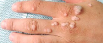
Lentigines are oval-shaped spots, slightly protruding above the skin with a diameter of 2 to 20 mm. Typically located where the skin is most exposed to UV radiation
- Face
- Hands
- Neckline and shoulder area
- Upper back
What determines the success of treatment
The result of hyperpigmentation treatment depends on how deep the pigment lies.
Based on the depth of the pigment, all types of hyperpigmentation can be divided into two large groups. It is this classification that allows us to understand whether our treatment will be successful or not.
Epidemal hyperpigmentation is pigmentation with clear boundaries, sometimes protruding above the surface of the skin, usually dark brown in color. She is the one who responds well to treatment
Dermal hyperpigmentation is a dull light brown pigmentation, sometimes with a grayish tint, that does not have clear boundaries. This type of pigmentation is practically untreatable.
Mixed hyperpigmentation - pigmentation has areas of dark and light shades at the same time. Treatment in this case has a partial positive effect.