What are the causes of acrochordon?
The exact reasons for the appearance of acrochordon are still unknown.
Predisposing factors for its occurrence are considered
|
Histological examination reveals a thinned epidermis over a loose connective tissue stroma.
Acrochordons
Acrochordons are small, soft, benign skin growths, often located on a thin base (“pedicle”).
- Acrochordons are the most common neoplasm of adult skin.
- Acrochordons are most often harmless, but can cause aesthetic dissatisfaction in the patient.
- Acrochordons tend to occur on the skin of the eyelids, neck, armpits, groin folds and under the breasts.
- One person can have from one to hundreds of acrochordons.
- Almost every person experiences the appearance of acrochordons during their lifetime.
- Middle-aged and obese people are most prone to developing acrochordons.
- Obesity is associated with the appearance of acrochordons.
- Treatment methods for acrochordons include cryodestruction with liquid nitrogen, laser destruction, and surgical excision.
WHAT ARE ACROCHORDONS?
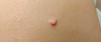
Acrochordons (syn. fibroepithelial polyp, skin papilloma, soft fibroma) are benign skin neoplasms, flesh-colored, soft consistency, located on a thin base. The size can vary from 2mm to 1cm. They are harmless; one person can have from one to hundreds of acrochordons. Men and women are equally susceptible to their occurrence. Obesity is also associated with the development of acrochordons. Some elements may resolve spontaneously.
WHY DO ACROCHORDONS APPEAR?
The exact cause of acrochordons is unknown.
WHERE DO ACROCHORDONS APPEAR MOST OFTEN?
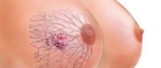
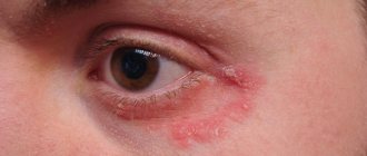
Acrochordons can occur on almost any part of the skin. However, the most common areas are: the skin of the neck, axillary areas, eyelids, groin folds, buttock folds, and in women, the skin under the mammary glands.
WHO HAS A TENDENCY TO ACROCHORDONS?
Acrochordons are more common in middle-aged people and tend to become more common before age 60. Children and teenagers are also susceptible to acrochordons, especially in the armpits and neck. Acrochordons are more common in overweight people. Increased hormone levels during pregnancy can lead to the appearance of acrochordons. Acrochordons, as a rule, are not associated with any diseases, but there is a relationship between their appearance and increased levels of glucose and cholesterol in the blood.
IS THERE A RISK OF ACROCHORDON DETERMINATION INTO MALIGNANT EDUCATION?
Acrochordons are generally not cancerous and will not become malignant if left untreated. If skin tumors similar to acrochordons bleed, grow quickly, or change color, you should consult a doctor.
ARE ACROCHORDONS CONtagious?
No. There is no convincing evidence that acrochordons are contagious.
WHAT CAN ACROCHORDONS BE DIFFERENTIATED WITH.
As a rule, diagnosing acrochordons is not difficult. However, there are skin neoplasms similar to them. These are dermal nevi (“moles”), fibromas, warts and keratomas. Very rarely, skin cancers such as basal cell carcinoma, squamous cell carcinoma, and melanoma can mimic acrochordons. Sometimes a skin biopsy is used for a more accurate diagnosis.
WHAT COMPLAINTS DO ACROCHORDONS CAUSE IN PATIENTS?
With the exception of a cosmetic defect, acrochordons usually do not cause complaints from the patient. However, when injured by elements of clothing, they can bleed, be painful, and change color. They can also fall off on their own if the blood flow to the acrochordon is disrupted due to trauma.
IS IT POSSIBLE TO REMOVE ACROCHORDONS USING EXTERNAL PREPARATIONS?
As a rule, acrochordons are removed by physical methods, such as cryodestruction with liquid nitrogen, laser destruction, and surgical excision. It is not recommended to remove acrochordons yourself, because... this may pose a risk of developing a secondary infection.
IS IT POSSIBLE TO PREVENT THE APPEARANCE OF ACROCHORDONS?
No. Unfortunately, there are no effective methods for preventing acrochordons.
Koval Yu.G.
You can get help information and also sign up for removal by calling: 956-70-86
How does acrochordon proceed?
In general, the acrochordon is a benign neoplasm that is not prone to malignancy (malignancy) and proceeds without complications or harm to health and mainly represents a comedic problem. With trauma (shaving, clothing, jewelry), acrochordons can come off with minor pain and bleeding. No dangerous Complications for injury to the acrochordon have not been described. In some cases, the leg of the element is torsed with its necrosis and death, which also does not threaten health.
What are the signs of acrochordon
Acrochordons are often found in the folds (for example, in the armpits, inguinal folds) of the neck, eyelids. The usual localization is the torso (abdomen, back). Acrochordons have been described in the vulva, scrotum and perianal area. There are several types of acrochordons.
| Small acrochordon Presented with papules 1-2 mm in width and height, located mainly on the neck and armpits. The color is often flesh-colored or light brown. The elements are soft to the touch and painless. |
| Filiform acrochordon Presented in the form of thread-like formations about 2 mm wide and 5 mm long, they can be located on a thin stalk and “hang” over the surface of the skin. Most often light and dark brown in color. Soft on palpation and painless. |
| Large acrochordon Large-sized soft fibroids with a diameter of up to 1 - 2 cm, often with a “warty” surface. They can either fit tightly to the skin or be on a stalk. The color is flesh-colored, yellow, light white and dark brown. In some cases, the surface is compacted (keratinization). |
Acrochordon of internal organs
The acrochordons of the larynx, large intestine, and rectum have been described, including “giant” ones up to 15-20 cm in length.
Birt-Hogg-Dube (BHD) syndrome
A rare autosomal dominant disease caused by a combination of acrochordona and skin tumors (fibrofolliculomas and trichodiscomas) with other neoplasms, especially malignant ones (tumors of the gastrointestinal tract and kidneys).
Coxsackie virus: “who” it is and how dangerous it is
The signs, for example, of rotavirus infection are well known to everyone. But what if an intestinal infection, in addition to (or instead of) nausea, vomiting and diarrhea, “gives” herpetic sore throat, rash, damage to the nervous system and a number of other serious disorders? The disease is widespread and, at the same time, not “known” enough. So, meet the Coxsackie virus.
One “family” - one character
The Coxsackie virus got its name “in honor” of a small town located in New York state. Where in 1948, researchers Gilbert Dalldorf and Grace Sickles isolated it while examining the feces of a child with polio. Scientists were looking for a way to treat the latter and a new pathogen was “discovered” by accident.
It was called “Coxsackievirus type A” and belongs to the group of enteroviruses. Type B was isolated a year later by Dr. Melnick and his colleagues. As a result of infection of mice with material from children suffering from serous meningitis. Today, serotypes A and B are also divided into subtypes. And the Coxsackie virus type A has at least 23 “variants”, and the serotype B has at least 6.
True, they all have common properties and features characteristic of all representatives of the large family of enteroviruses. And this is also: polio virus, ECHO viruses and a number of enteroviruses that have only an “ordinal” name (68-101).
A distinctive feature of the family is its stability in the external environment.
Enteroviruses:
- are not destroyed by 70% alcohol, 5% Lysol and 3% phenol,
- resistant to changes in acidity (pH from 3 to 10),
- when frozen, they remain active for several years,
- at a temperature of +4 + 6 – several weeks,
- at room temperature – several days.
They tolerate repeated freezing and thawing very well. But they are still sensitive to chlorine solutions (with some “reservation”), and absolutely cannot tolerate temperatures above 50C (death within 30 minutes) and direct ultraviolet irradiation.
Enteroviruses demonstrate a special “predilection” for the tissues of the respiratory, digestive, nervous and muscular systems. Due to which the corresponding symptoms are caused.
“Where” can you find the Coxsackie virus
The main mechanism of infection by the virus is fecal-oral. But for some types the airborne route is also characteristic. The source is always a sick person or an asymptomatic virus carrier.
The release of the pathogen into the environment (the period of “infectiousness”) lasts:
- 3-7 days (from the moment of infection) from the nasopharynx,
- and 3-4 or more weeks - with feces.
Infection most often occurs:
- when eating contaminated food (usually vegetables and fruits)
- water (drinking, when swallowing water from a swimming pool, natural sources),
- and also through “dirty hands”, in contact with household items contaminated with the virus.
The virus “prefers” warm, humid climates. Therefore, it circulates almost constantly in the tropical and subtropical regions. Which causes a high risk of infection at the “resort”. Whereas in temperate latitudes, the risk of the disease is observed mainly in summer and early autumn. Although, according to the latest data, amid the covid pandemic, the seasonality of Coxsackie infection has shifted. And now the “chance” of infection remains until November.
Children are more susceptible to infection than others due to frequent violations of personal hygiene rules and immune system characteristics. And also “overcrowding” in kindergartens and schools.
While adults more often experience the infection in a mild or asymptomatic form. However, there are frequent exceptions here.
How to suspect an infection
The entrance gates of infection, as already noted, are always the mucous membranes of the respiratory or digestive systems. During the incubation period (as the virus “accumulates”), which is an average of 4-6 days, the infected person does not experience any symptoms.
And only after the end of the “incubation” does Coxsackie begin to spread to other tissues of the body, simultaneously provoking the launch of immune mechanisms.
The onset of the disease, if it is not asymptomatic, is often accompanied by:
- a sharp rise in temperature to 39-40C,
- symptoms of intoxication (headache, body aches, etc.),
- abdominal pain,
- nausea and vomiting (including repeated),
- the appearance of painful herpes-like rashes in the oropharynx (herpetic sore throat)
- as well as a pinpoint red rash on the palms and soles (mouth-palm-sole symptom).
Blisters and rashes may appear on other parts of the body. And the presence of bubbles is always accompanied by severe pain. The rash, at first glance, may resemble chickenpox. However, chickenpox is characterized by a special staged pattern of rashes: first, papules (convex formations on the skin) appear, then they turn into blisters, which eventually open to form ulcers. All “this” is accompanied by itching. And only then does healing occur.
The rash caused by Coxsackie infection is characterized by a dense surface and severe pain. The bubbles do not open. And over time they dry out, leaving dark spots. Patients often experience intense muscle pain. Conjunctivitis may also occur.
At the same time, this “classic” manifestation of infection is more typical for the Coxsackie virus type A. In some cases, the only symptom, for example, may be only a “strange”, prolonged fever or signs of acute respiratory viral infection with an unusually high temperature.
And Coxsackie type B infection, among other things, affects the liver, pancreas, heart and pleura (the lining of the lungs), causing corresponding symptoms and complications.
Dangerous complications
Fortunately, infections caused by the Coxsackie virus do not often cause complications. But at the same time there may be:
- meningitis,
- encephalitis,
- myocarditis (heart damage),
- pericarditis (inflammation of the lining of the heart),
- pleurisy (inflammation of the serous membrane of the lungs),
- organ failure due to severe dehydration,
- hepatitis,
- convulsions and paralysis
Complications are more often observed against the background of immune deficiency or general weakening of the body (due to chronic diseases or other factors). Which makes newborns, infants, the elderly, cancer patients, HIV-infected and some other groups of patients especially vulnerable.
Diagnosis of Coxsackie infection
Laboratory detection of the Coxsackie virus is based on only two tests:
- PCR test of feces to detect the virus itself (RNA)
- or a blood test for IgM antibodies to the virus.
In this case, the first option is used as the main one. Since, let us remember, the excretion of the pathogen in feces continues for about 3-4 weeks from the moment of infection.
Whereas antibody testing is often used as an additional test. When it is necessary to carry out differential diagnosis with other infections that provoke meningitis and other pathologies. And confirmation of the diagnosis is an increase in the level of antibodies by at least 4 times in the period between 4-5 and 14 days of illness.
And after an infection, persistent, and according to some data, lifelong immunity remains.
How to treat Coxsackie virus
As such, there are currently no drugs against the pathogen itself. And all therapeutic measures are aimed at eliminating symptoms.
Among the medicines used:
- "general" antiviral,
- antipyretics,
- antihistamines,
- sorbents,
- drugs that restore water and electrolyte balance (Regidron and others),
- local antiseptics
- and antibiotics, in case of bacterial infection.
At the same time, self-medication for Coxsackie infection is extremely undesirable. If you suspect an infection, and especially if you have a confirmatory PCR test, you must immediately consult a doctor.


