In all countries of the world, superficial mycoses of the skin (SMS) are an urgent interdisciplinary problem. They are registered in 20% of the world's population [1]. Dermatophytosis predominates among MVP, affecting 10% of the population [2, 3]. Dermatophytosis takes second place in the structure of morbidity after pyoderma [3]. The predominance of the incidence of skin mycoses in large cities makes it possible to call them “diseases of civilization” [4, 5]. An analysis of the results of the Achilles project (1988-1997), in which several European countries and Russia took part, showed that 35% of patients who consulted a dermatologist had one or another mycosis [6-8]. Dermatophytosis of the feet, compared to other localizations taken together, is recorded 3 times more often [9, 10]. Much less attention is paid to mycoses affecting large folds [9, 11—14]. However, mycoses of large folds (MCF) are a significant interdisciplinary problem in medicine.
Analysis of the incidence of the population of Russia on the ISS is practically impossible. What is this connected with? In the ICD-10 classification there is only athlete's foot (B35.6). The traditional approach to MCS, which existed before the introduction of this classification into practice, included athlete's foot, rubromycosis and candidiasis of large folds. Each of the nosological forms has its own epidemiological and clinical features, a range of trigger factors that influence the course and chronicity of the process and, naturally, features of the choice of treatment tactics [2-4, 15-17]. Many years of experience in the system of postgraduate professional education have shown that dermatologists currently register rubromycosis of large folds in the section “Other dermatophytosis” (B35.8), and candidiasis of large folds - in the section “Candidosis of other localizations” (B37.8). Sometimes, among mycoses of large folds, only rubromycosis of the inguinal-femoral region and candidiasis of large folds are distinguished [18].
Inguinal dermatophytosis is a collective term that unites largely clinically similar skin lesions caused by various pathogenic and opportunistic fungi. It has even been proposed to distinguish the following variants of inguinal dermatophytosis (dermatomycosis): epidermophyton inguinal ( Epidermophyton floccosum
), inguinal rubrophytosis (
Trichophyton rubrum
), inguinal dermatomycosis (
Trichophyton mentagrophytes var. interdigitale
), candidiasis of the groin (yeast-like fungi of the genus
Candida
), mold mycosis of the groin (
Aspergillus
spp
., Scopulariopsis brevicaulis
) and mixed dermaphytic-candidiasis or dermaphytic-mold mycosis of the inguinal region [11, 12]. This is explained by the fact that the etiological basis for the diagnosis is important for treatment, since not all topical antifungal drugs are equally effective against various pathogens of mycoses. However, candidiasis and mold mycosis do not belong to dermatophytosis and this classification has not found application in practical mycology. It is important to note that specialists studying the problem of skin mycoses, citing the classification of dermatophytosis according to ICD-10, indicate nearby the previously accepted synonym/equivalent of the disease. With inguinal athlete's foot, in particular, two nosological forms of the disease are synonymous/equivalent. These include epidermophytosis and rubrophytosis of large folds [19].
The cause of inguinal dermatophytosis is various pathogens, which are recorded unequally often: Epidermophyton floccosum
- in 35-40% of cases;
Trichophyton mentagrophytes var.
interdigitale - 20-25%;
Trichophyton rubrum
- in 15-20% [1, 11, 12]. Candidal mold fungi, as an independent etiological factor in damage to large folds, are detected in 3-5% of cases [1, 9, 11, 20]. However, verification of mycoses pathogens is not carried out in most dermatological institutions, not to mention city clinics and somatic hospitals of related specialties.
A number of authors include lesions of the pubic region, caused by zoophilic trichophytons, as inguinal epidermophytosis. Thus, over a 10-year period in the Andijan region of Uzbekistan, during an examination of 2022 patients with trichophytosis, lesions in the pubic area were detected in 248 (12.3%) cases [21]. When examining 286 patients with a similar pathology, a spectrum of pathogens was established. In 97.7% of cases it was Tr . verrucosum
, in 2.3% -
Tr .
mentagrophytes var. gypseum .
Clinically, an infiltrative-suppurative form of trichophytosis was detected in 62.2% of patients [22]. In these publications there are no indications of damage to the inguinal folds. On the other hand, in 2005, with zooanthroponotic microsporia in 4 women, mycosis was localized in the pubic region, spreading to the inguinal folds and inner thighs [23]. The results of studies carried out in the Republic of Kyrgyzstan indicate that in case of inguinal dermatophytosis, various pathogens were more often sown: Tr . verrucosum
(52.2%),
Tr . rubrum
(25%),
Epidermophyton floccosum
(20.6%),
M. canis
(2.2%) [24].
It is significant that the author describes clinical variants of damage to the inguinal folds. The superficial-spotty variant predominated in cases of infection with Epidermophyton floccosum
(76%), infiltrative (85.7%) and infiltrative-suppurative (67.9%) - in cases of
Tr
.
verrucosum
.
When isolating Tr.
rubrum, these forms were recorded in 8, 14.3 and 28.3% of cases, respectively.
The author points out that the superficial spotted form when Tr . verrucosum
was the initial stage of the infiltrative process (16%).
It can be assumed that Tr . verrucosum
is a significant etiological factor for athlete's foot in the republics with developed livestock farming, as well as among rural residents [24].
Another side of the problem of inguinal dermatophytosis is that the process often involves various large folds - axillary, intergluteal, inguinal-scrotal, folds under the mammary glands in women, between the overhanging part of the abdomen and the pubis in obese people. Data on the involvement of these localizations in the literature are fragmentary and are in the nature of descriptions of clinical cases. Currently, most articles and even guidelines describe only athlete's foot (dermatophytosis inguinalis) [25–27]. At the same time, manuals and monographs published 10-15 years ago provide differential diagnostic criteria between epidermophytosis and rubrophytosis of large folds [3, 4, 28].
The choice of treatment tactics for MCS depends not only on the etiological factor and the specific activity of the topical antimycotic. When large folds are affected, the process often has a multifocal nature. An important role in the epidemiology of MCS caused by Tr. rubrum
and
Tr.
mentagrophytes var. interdigitale , play foci of fungal infection on the feet, especially in men. It should also be remembered about the possibility of a hidden course of mycosis in the scrotum, which is affected almost constantly and is clinically characterized by mild erythema and slight infiltration without pronounced peeling [4]. However, data on the frequency of lesions in other areas of the skin during MCS are not fully covered in the literature, although the effectiveness of therapy directly depends on the sanitation of these lesions. For example, according to V.V. Karpov [29], during a survey of 57 male military personnel in 2004-2005. with damage to the inguinal folds and pubic region, tinea pedis and onychomycosis were found in all patients. Most often, with dermatophytosis of large folds, the process is localized in the groin area (48.7-73.6%) [3, 28, 30]. The incidence of isolated fungal infections of the groin area is only 10% [12].
To determine the type of pathogen of MCS, a bacterioscopic examination is carried out, which makes it possible to distinguish true mycelium (dermatophytes) and pseudomycelium (candidiasis). Cultural research, even in specialized laboratories, according to foreign authors [2, 31], allows one to obtain reliable information in 50% of cases, and in domestic ones - only in 36%. This is explained by technical errors in collecting the material, its transportation to the laboratory, the preliminary use of topical antifungals independently or as prescribed by a doctor without following treatment regimens, and the use of hygienic skin care products in everyday life containing drugs with an antifungal effect.
Inguinal dermatophytosis may resemble banal or candidal intertrigo, the vesicular form of Darier's disease, chronic familial pemphigus Gougereau-Hailey-Hailey [16], eczema, exudative erythema, limited neurodermatitis, intertriginous psoriasis [32].
Choosing a treatment strategy for MCS is not an easy task. Ointment-based preparations are rarely used, since the rashes are localized in areas with high humidity of the skin. MCS can be complicated by secondary infection and mycotic eczema, especially with concomitant diabetes mellitus. In this regard, mycosis proceeds torpidly and is more difficult to treat [33]. Therefore, during treatment, it is advisable to use topical single drugs that simultaneously have a pronounced antimycotic, antibacterial and anti-inflammatory effect. One such drug is sertaconazole cream ( Zalain
) [25, 34, 35].
High lipophilicity of sertaconazole ( Zalaina
) leads to its accumulation in the deep layers of the skin, ensuring that an effective therapeutic concentration is maintained in it for 48 hours after application. Resistance of mycotic pathogens to the drug has not been registered. Relapses of the disease after a full course of treatment are practically absent. The drug does not have a systemic effect, does not cause side effects and is well tolerated by patients. The duration of its administration is determined individually depending on the characteristics of the clinical case, on average it is 2-4 weeks. Approved for use in children without age restrictions [36, 37].
In this publication we will talk about epidermophytosis of large folds (ECF). The involvement of dermatologists and mycologists from various regions of the country made it possible to study the problem taking into account gender characteristics, previous therapy, structure and concomitant pathology, topic of the process, combination with mycoses of other localizations, the presence of mycids, etc.
The purpose of the study, based on the results of a multicenter study in the Russian Federation, was to study the features of the course of ECS and provide a multifactorial analysis of the effectiveness of its treatment with the topical antimycotic (TA) sertaconazole ( Zalain
).
Causes
In most cases, athlete's foot is diagnosed in men. Cases among children and adolescents are extremely rare.
The causative agent of the disease (fungus) is transmitted through contact and household contact. You can become infected with athlete's foot, for example, in a bathhouse or after using someone else's personal hygiene products.
Factors that provoke the development of the disease include:
- high temperature and humidity of the environment;
- excess weight;
- skin microtraumas;
- increased sweating;
- sedentary lifestyle.
Material and methods
The work was carried out as part of a multicenter study “Study of real practice in the treatment of patients with dermatomycosis in the Russian Federation”, conducted in 2012–2013. under the patronage of the pharmaceutical plant EGIS (Hungary) simultaneously in 50 regions of the Russian Federation. 97 medical and preventive institutions of various profiles, 174 doctors are involved. To unify the research, an original version of the questionnaire was developed. Instructions have been prepared for filling out the questionnaire in the form of a multimedia presentation containing a detailed analysis of the differential diagnostic criteria for epidermophytosis, rubromycosis and candidiasis of large folds with photographs of patients. The questionnaire also asked people to express their own opinion about the peculiarities of the course of mycosis in the patient. In total, doctors filled out 5025 questionnaires for patients with MVP. There were 2784 patients with dermatophytosis, including 735 (26.4%) with dermatophytosis of large folds. Patients with ECS predominated ( n
=615; 83.7%). One of the conditions of the experiment was the selection of patients with minimal damage to the nail plates, when the KIOTOS index (clinical index for assessing the severity of Sergeev’s onychomycosis) allowed only local therapy for mycosis of the feet.
The work was carried out in stages.
Stage I. Processing questionnaires of pacemaker patients filled out by doctors using Excel. Of the 615 questionnaires, 235 (38.2%) contained complete data. The sample was representative to obtain reliable data.
Stage II. Studying the features of the course of ECS in Russia.
Stage III. Multivariate analysis of the effectiveness of ECS treatment with sertaconazole ( Zalain
).
Statistical processing of the research results was carried out in the laboratory of mathematical theory of experiment, Faculty of Biology, Moscow State University. M.V. Lomonosov using the statistical software package Stastica 6.0. The method of correlation analysis (Spearman coefficient) was used. Those values of correlation coefficients for which p
was less than the significance level (α=0.05), i.e. these coefficients were statistically significantly different from zero.
Prevention
To avoid the possibility of inguinal athlete's foot, you need to follow the rules of personal hygiene, especially in saunas and swimming pools. Use a personal towel, washcloth, and underwear.
If a person has suffered this disease, his household items, linen and clothing must be thoroughly disinfected.
If symptoms of inguinal athlete's foot appear, SM-Clinic specialists recommend not delaying going to the doctor and not delaying treatment. You can make an appointment in St. Petersburg by calling the phone number listed on the website.
Inguinal dermatophytosis: etiology, clinical picture, modern treatment options
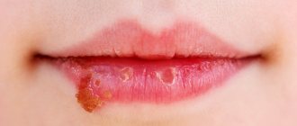
Modern ideas about dermatomycetes - causative agents of fungal infections. The causative agents of dermatomycosis - dermatomycetes - are ubiquitous in nature. They belong to lower plants, making up different species depending on their preferred habitat. All of them are relatively related microorganisms, with individual morpho-physiological characteristics and ecological association [17]. Dermatomycetes are pathogens that attack keratin-containing tissues of humans and animals - skin: hair, nails, without penetrating into deep tissues. Therefore, the diseases they cause are classified as superficial mycoses or dermatymycosis. Dermatomycetes actively invade and destroy keratin-rich structures due to the activity of their enzyme systems, in particular keratinases. Once in the soil, infected keratin-containing tissues of humans and animals are slowly destroyed (hydrolyzed), acting as a reservoir of pathogenic fungi. Studies of the enzymatic activity, primarily proteolytic, of a number of dermatomycetes conducted in the last decade have formed new ideas about the pathogenicity factors of these microorganisms. It was found that dermatomycetes produce several types of proteases that can destroy various protein structures, in particular, γ-globulins, fibronectin, α1ρ1keratin and fibrillar protein - collagen. Representatives of the genus Microsporum, in particular Microsporum canis, have 6 proteases with different molecular weights. It has been suggested that enzymes with molecular weights of 122, 63, 25 kDa are induced during the transition of the saprophytic form of the fungus to the parasitic one [2]. New methods for diagnosing fungal infections A large role in identifying and identifying the type of pathogenic fungi is given to laboratory methods for diagnosing dermatomycosis. In general clinical practice, to confirm the diagnosis of mycosis, as well as to carry out a differential diagnosis with other dermatoses, the microscopic examination method is most often used. Before microscopy, affected objects (skin scales, hair, vesicle covers) are pre-treated with a 10% solution of potassium hydroxide or a 25% solution of sodium hydroxide to clear the preparation. Therefore, this diagnostic method is often called the CON test. Recently, a new method of microscopy of native preparations using calcofluor white staining has been developed and put into practice. This reagent is used as a bleaching agent in the paper and textile industries. The combined use of calcofluor white with KOH makes it possible to identify both young and mature hyphae of the fungus. According to specialists from the Research Institute of Medical Mycology named after. P.N. Kashkin, the use of the calcofluor white staining method for the microscopic diagnosis of dermatomycosis allows for a 10% increase in the detection of fungal infections compared to the standard KOH method [2,16]. To establish the type of pathogen, the material is inoculated on Sabouraud's nutrient medium, followed by identification of the isolated culture. However, even if all the rules for collecting material are observed, with good laboratory equipment and high professionalism of laboratory staff, the number of detected positive results of cultural research is very small. Thus, according to foreign literature, the proportion of positive cultural studies barely reaches 50%; in domestic studies, the pathogen cannot be isolated in 64% of cases. This is due to technical errors (violation of the rules for collecting material, its transportation, etc.), but most often due to previous (usually local) antifungal therapy [5]. In recent years, all over the world, including in our country, a new method of gene diagnosis of mycoses of the skin and nails has been actively developed and introduced into the practice of medical institutions: direct DNA diagnosis of dermatophytosis. Currently, a domestic PCR diagnostic system has been developed and tested using a pair of species-specific primers Trichophytom rubrum and Trichophytom mentagrophytes var. interdigitale – the main causative agents of fungal diseases in humans. Now, by taking material from the affected nails, the doctor can record the presence/absence of a fungal infection with almost absolute accuracy within 24 hours. In addition, this method immediately determines the type of pathogen based on the genetic material of the fungus found in the scraping. A comparative study of various methods for identifying dermatophytes in patients with typical clinical signs of onychomycosis, conducted in 88 patients at the Sechenov Moscow Medical Academy of Skin Diseases Clinic, found that the DNA diagnostic method of onychomycosis is the most sensitive. The sensitivity of the PCR analysis was 93.6%, the KOH test was 83.9%, and for the culture method it was only 39.2%. It has been shown that DNA diagnostics is a more advanced method for identifying fungal infections compared to those traditionally used in the practice of dermatological institutions. Increasing the efficiency of diagnosing dermatomycosis in clinical practice in the future will be associated with the widespread use of this method, which is significantly superior to existing regulated methods [4,5]. Inguinal dermatophytosis - modern diagnostic and therapeutic options It is well known that among dermatomycosis the first place is occupied by mycosis of the feet with onychomycosis. For many years, they have surpassed other infectious skin lesions in prevalence and incidence among adults. According to WHO, every fifth person on the planet suffers from a fungal infection of the feet. A study of the epidemiology of dermatophytosis conducted by domestic dermatologists at the Medical Center of the UDP of the Russian Federation showed that the share of onychomycosis is 77.75% among all cases of dermatophytosis. Dermatophytosis of the nails (mainly on the feet) was recorded more than 3 times more often than dermatophytosis of other localizations combined. A large number of publications in the domestic and foreign press are devoted to this problem. Much less attention is paid to superficial mycoses affecting large folds, most often affecting the groin areas [3]. According to the literature, intertriginous (inguinal) mycosis of the skin in the general structure of fungal infections of the skin can reach 10%. The clinical picture of the disease is very characteristic and has clear diagnostic signs. The rashes are represented by sharply demarcated, sometimes slightly raised spots of a pinkish-brownish color of round or oval shape with fine-plate peeling on the surface. The rash is characterized by gradual peripheral growth and merging of nearby lesions, as a result of which the lesions take on polycyclic scalloped outlines. In this case, the lesion can extend beyond the inguinal fold, spreading to the skin of the thighs, buttocks, abdomen, and in men to the scrotum and penis. The central zone of the lesion gradually turns pale, peeling becomes barely noticeable and the inflammation undergoes regression. At the same time, the peripheral zone is represented by a brightly hyperemic, edematous inflammatory ridge protruding above the surface of the skin (cord symptom). When the process worsens due to increased friction and sweating, especially in the summer, small blisters, papules, crusts may appear on the surface of the lesion, especially along the periphery, and itching intensifies (Fig. 1). Without treatment, the disease takes on a long-term, protracted nature, worsening in the summer. The use of corticosteroid ointments in the form of self-medication, which is so common nowadays, or as prescribed by a doctor who has not recognized mycosis, leads to a temporary subsidence of inflammatory phenomena, and mycosis takes on a sterile, protracted character. Both various pathogenic dermatomycetes and conditionally pathogenic fungi of the genus Candida and mold can take part in the development of this pathology. The most common causative agents of inguinal mycoses are Epidermophyton floccosum, Trichophytom mentagrophytes var. interdigitale and Trichophytom rubrum. Opportunistic fungi are often detected in association with dermatophytes. According to a number of authors, inguinal dermatomycosis in 35–40% of cases was caused by Epidermophyton floccosum, in 20–25% by Trichophytom mentagrophytes var. interdigitale and 15–20% – Trichophytom rubrum. As independent causes of damage to the inguinal folds; candino-moldy fungi accounted for 3–5% [6,7]. Conducted at the Sechenov Moscow Medical Academy of Skin Diseases Clinic by Professor A.A. Haldin and his colleagues laboratory studies using the cultural method in 30 patients with mycosis of the inguinal folds revealed the following results. Of the 26 examined men, 14 (46.8%) were found to have Epidermophyton floccosum, 8 (26.6%) were found to have Trichophytom mentagrophytes var. interdigitale, in 4 (13.3%) – Trichophytom rubrum. All 4 (13.3%) women examined showed an association of dermatophytes with fungi of the genus Candida. In 27 patients, athlete's foot inguinal was discovered for the first time during examination at a clinic hospital, where patients were being treated for other dermatoses (psoriasis, atopic dermatitis, seborrheic dermatitis, parapsoriasis, rosacea, necrobiosis lipoidica). In three patients, where Epidermophyton floccosum was identified as the etiological agent, the skin process was unilateral. The authors regarded this circumstance as a certain pathomorphosis of the disease. With other pathogens, the clinical picture was of a classic nature and no specific features were identified. In the vast majority of patients, fungal infection of the inguinal folds was isolated. Only 4 out of 30 patients had inguinal dermatomycosis combined with mycosis of the feet. In two women with diabetes and obesity, dermatomycosis was also observed under the mammary glands. In only one patient suffering from atopic dermatitis, the fungal infection caused by Trichophytom rubrum was widespread with lesions on smooth skin and feet [8]. The frequent detection of dermatophytosis infection with damage to the groin and feet in patients with atopic dermatitis is indicated in many domestic and foreign publications. In foreign literature, a special term “atopic–chronic–dermatophytosis syndrome” has been proposed to denote this combination. A study by Russian dermatologists who observed 26 patients with atopic dermatitis in combination with mycosis of the inguinal folds and feet showed that the addition of a fungal infection contributes to a more torpid course of the underlying dermatosis and reduces the effectiveness of the therapy [13]. Clinicians are well aware that fungal infections of only smooth skin without involving nail plates and hair in the pathological process can be cured using only external antimycotics. Currently, antimycotics represent one of the largest groups of dermatological drugs - more than 100 items and more than 20 dosage forms [15]. The doctor faces the difficult task of choosing a highly effective and safest antimycotic. Today, these characteristics are fully met by terbinafine, which belongs to the class of synthetic antifungal drugs of the allylamine group. Compared to antimycotics of other groups, it acts at the earliest stages of sterol metabolism of the cytoplasmic membrane of fungal cells, inhibiting the enzyme squalene epoxidase, which leads to the death of pathogenic fungi. Terbinafine has a wide spectrum of antimycotic action. In therapeutic concentrations, the drug acts in two ways - fungistatically and, to a greater extent, fungicidal against dermatophytes, molds and some dimorphic fungi, and therefore the sanitizing effect is achieved with a lower concentration of the drug. The activity of terbinafine against yeast-like fungi, depending on their type, can be fungicidal and fungistatic. The high therapeutic efficacy of this antimycotic is combined with high safety, since it has the lowest minimum inhibitory concentration (MIC) against dermatophytes compared to systemic antimycotics of the azole group [11,12,18]. The number of external antifungal drugs used in modern medical practice is constantly increasing. The pharmaceutical market is constantly updating the range of antifungal agents, mainly due to the emergence of new analogues of existing antifungals and new dosage forms. The doctor always faces the difficult task of choosing a highly effective, maximally safe and optimal drug in terms of pharmacoeconomic indicators for a given patient. In each specific clinical situation, the doctor must choose an antimycotic depending on the clinical symptoms, duration of the disease, the presence of concomitant pathology, age, create a stable motivation in the patient to comply with the drug regimen, and also assess the patient’s financial capabilities. One of the possible ways to reduce treatment costs is to use generics. Currently, the arsenal of external antimycotics has been replenished with the domestic generic terbinafine (trade name of the drug Termicon, pharmaceutical) in the form of a spray. The main indication for the use of the dosage form of terbinafine in the form of a spray is fungal infections of large folds, and especially the inguinal folds. When using the spray, a uniform distribution of the active substance over the surface of the skin is ensured, while maintaining the normal functioning of the skin and the functioning of its sebaceous and sweat glands. Unlike fat-based products, when using Thermikon in the form of a spray, there is no greenhouse effect. In addition to the antifungal effect, Thermikon spray has a drying, antipruritic and anti-inflammatory effect. In this regard, it manifests itself most actively during the acute period of the disease, reducing the severity of redness, swelling, maceration and itching of the skin. The drug has a pronounced gyroscopic effect, so its use is especially effective in cases of fungal infection of folds or in the presence of lesions with a weeping surface. Therefore, an additional indication for the use of the drug is the prevention of possible skin infections with increased hyperhidrosis, obesity, diabetes, as well as in people actively involved in sports, visiting swimming pools, fitness centers, saunas and massage rooms. Thermikon spray can be used by adults once or twice a day, depending on the indications. Before applying the drug, it is necessary to clean and dry the affected areas. The drug is sprayed onto the affected areas in an amount sufficient to thoroughly moisturize them, and, in addition, applied to adjacent areas of both affected and intact skin. The duration of treatment and frequency of use is 7 days, 1 or 2 times a day. A decrease in the severity of clinical manifestations is usually observed in the first days of treatment. Employees of the department of skin and venereal diseases of the medical faculty of MMA named after I.M. Sechenov conducted a comparative study of the effectiveness of terbinafine in the form of 1% dermgel and 1% spray in the treatment of fungal infections of large skin folds. Complete mycological cure was established in all 30 patients who used both 1% spray and 1% gel, regardless of the type of pathogen. However, in patients using 1% spray, the regression of clinical symptoms (especially itching and maceration) was somewhat faster than that when using 1% dermgel. It has been confirmed that a weekly course of external use of 1% terbinafine spray, when used once a day, is sufficient to completely clean smooth skin from fungal infection. The drug was well tolerated by patients; no general or local effects were recorded during its use. The rapid subsidence of inflammatory symptoms (swelling, hyperemia, maceration, itching) observed during treatment once again confirmed the opinion that terbinafine has an anti-inflammatory effect [10]. Among the domestic manufacturers of external antifungal drugs, it should be noted pharmaceutical, which since 2004 has been producing the drug Termicon (terbinafine) in the form of a spray.
References 1. Kotrekhova L.P., Raznatovsky K.I. Etiology, clinical picture, treatment of dermatitis in patients with diabetes mellitus. Problems of medical mycology – 2005. – T.7, No. 4. – pp. 13–18. 2. Elinov N.P., Vasilyeva N.V., Raznatovsky K.I. Dermatomycoses, or superficial mycoses of the skin and its appendages—hair and nails. Laboratory diagnostics. Problems of medical mycology–2008.–Vol. 10–No. 1.–P. 27–34. 3. Sergeev A.Yu., Sergeev Yu.V. What does research into the epidemiology of dermatomycosis teach the clinician? Advances in medical mycology, 2003, volume No. 2, pp. 154–155. Moscow, National Academy of Mycology 4. Sergeev V.Yu., Sergeev A.Yu. Genes, molecular methods and new concepts in the diagnosis of fungal diseases.//Problems of medical mycology, 2006–Vol.8–No.2–P.85. 5. Tsykin A.A. Onychomycosis: DNA diagnosis, improvement of combination therapy. Author. diss. for the scientific degree Ph.D. degrees honey. Sci. M. 2008.–24p. 6. Tarasenko G.N. Modern aspects of practical mycology Ros.zhur. leather and veins Bol. – 2006. – No. 6. – S. 49–61 7. Sergeev V.Yu., Sergeev A.Yu. Dermatophytes: new in the diagnosis, therapy and prevention of the most common human mycoses. Consilium medicum. – Dermatology. – 2008. – No. 1. – P. 30–35 8 Haldin A.A., Sergeev V.Yu. Izyumova I.M. Modern ideas about inguinal dermatophytosis: etiology, epidemiology, clinical picture and effective therapy. Ross.zhur. leather and veins Bol. – 2005. – No. 5. – S. 43–48 9. Evans E.G., Seaman R.A. James I.G.//Clin. dermatol. and Venus. – 2004.–№1–P.103–106. 10. Khaldin A.A., Tsykin A.A., Izyumova I.M. Clinical and etiological effectiveness of 1% Lamisil spray in the treatment of fungal infections of large skin folds. Ross.zhur. leather and veins Bol. – 2007. – No. 1. – S. 56–61 11. Bonifaz A. Saul A.//Klin. dermatol. and Venus. –2003.–№2–P.75–78 12. Potekaev N.S., Potekaev N.N., Rukavishnikova V.M. Lamisil 10 years in Russia. Moscow, - “Medical Book”, 2003. 13. Potekaev N.N., Sherina T.F. On the issue of the association of dermatoses and mycoses of the skin. Ross.zhur. leather and veins Bol. – 2004. – No. 6. – S. 55–57 14. Stepanova Zh.V., Novoselov A.Yu., Vorobyov I.A. and others. Results of a clinical study of 1% Terbizil cream in the treatment of mycoses of smooth skin. Consilium medicum. – Appendix “Dermatovenereology”. – 2004. – P. 5–7. 15. Sergeev Yu.V., Shpigel B.I., Sergeev A.Yu. Pharmacotherapy of mycoses. M. – “Medicine for All”, 2003. 16. Manual of Clinical Microbiology 8th ed. American Society for Microbiology.–Washington, DC, 1991.–1364 p. 17. Hay R. Literature review//JEDV.–2003.–Vol. 19, Suppl. 1–P.1–7. 18. Rosen T. Schell B.–J., Orengo I. //Int. J. Dermatol. –1992.–Vol. 126/– P.56–60.
Pathogenesis
When they get on the skin and folds, fungi “settle” in the upper layers of the epidermis, destroy collagen, so these areas lose their elasticity and become loose. Dermatophytes, fixing themselves in the epidermis, produce keratinase, an enzyme that destroys human keratin. As a result of this, the stratum corneum of the skin is destroyed, and with the rapid proliferation of fungi, an inflammatory process develops. It is characterized by its progression and spread to the periphery of the lesions, where inflammation is most pronounced.
There are cases of spontaneous recovery of patients if the rate of epidermal restoration prevails over the rate of fungal reproduction, and the body actively copes with the pathogen. Everyone's susceptibility to fungal infections is not the same. Dermatophytes do not cause disease if the barrier function of the skin inherent in a healthy body is preserved, which includes acidity and specific protection.
The macroorganism produces a number of antimicrobial and antifungal factors into the blood and secretes onto the surface of the skin, and their deficiency predisposes to the development of fungal diseases. Also, the manifestation of the disease does not occur if the integrity of the skin is preserved - in this case, long-term carriage of the pathogen is observed.
Concomitant diseases play an important role in the development of fungal infection. It has been noticed that inguinal athlete's foot very often occurs against the background of diabetes mellitus , vegetative-vascular dystonia , hepatitis , and cancer. Severe fungal infections of the folds occur in HIV-infected people. It often happens that changes in skin folds serve as a “signal” of diabetes mellitus. This is due to the fact that the bactericidal activity of the skin in diabetes mellitus is lower than in healthy individuals.
Fungus in the groin: treatment, drugs effective in men, inexpensive
No. 1 – Mikozolon ointment
For inguinal fungus, Mikozolon ointment is applied to the affected areas twice a day for a week. It is an antifungal, antiseptic and anti-inflammatory agent designed specifically to relieve inflammation and itching due to mycotic infections.
Mycozolon inhibits the growth of dermatophytes and other fungi, and also blocks the proliferation of bacterial flora. It has an anti-inflammatory effect and relieves allergic manifestations.
Epidermophyton floccosum - the causative agent of athlete's foot
The fungi Epidermophyton flocosum are anthropophylls. They cause disease only in humans. Most often, the superficial layer of smooth skin is affected: most often the inguinal folds, less often the intergluteal folds, folds in the lower abdomen and under the mammary glands.
Microscopy of skin scales taken from the affected areas reveals short (2 - 4 µm) branching intertwined mycelium and rectangular arthrospores arranged in chains. When growing on Sabouraud's nutrient medium, the colonies are round in shape, initially flat or dome-shaped, later folded-lumpy with depressions in the center. The surface is velvety-mealy or leathery with radial folds. Initially grayish-brown, lemon-olive or yellowish-green, later white.
Under microscopy, in mature cultures one can see septate mycelium with blunt-pointed macroconidia, which are arranged in the form of clubs or banana bunches in bunches of 3-5 pieces, growing directly from hyphae. Chlamydospores appear in old crops. Microconidia are absent.
Rice. 3. View of colonies of the fungus Epidermophyton flocosum at different stages of growth.
Rice. 4. Microscopy of mature cultures of the fungus Epidermophyton flocosum.
Diet
Patients with inguinal dermatophyte can stick to a common table, and with candidiasis, products that provoke the development of a fungal infection are excluded for a period:
- Sweets, sweet drinks, confectionery, sugar, condensed milk, jam, jam, white rice, dried fruit, limited to pasta and potatoes.
- Yeast baked goods, it is better not to use yeast bread during treatment.
- Kvass, kefir, sauerkraut and other pickled vegetables, cheeses, soy sauce.
- Alcoholic drinks (until complete recovery). Drinks obtained by fermentation (beer, wine, champagne) are completely excluded.
- Nuts and dried fruits, which often have invisible mold.
Symptoms
Inguinal dermatophytosis is characterized by an acute onset of the disease. The lesions are pink-red in color and are sharply limited from healthy skin. The surface is peeling. The lesions reach 15 cm in diameter and can unite. Swelling occurs, papules, pustules, vesicles and scales appear.
Since fungi affect keratin-containing tissues (hair, skin, nails), one of the first signs may be the appearance of a hairless area with scales and a rash. There may be lesions with the formation of crusts. Pain and itching appear. Discomfort increases while moving.
Inflammation of the skin most often affects the inguinal folds, the surface of the thighs, and the scrotum, without affecting the genitals.
In children
In infants, candidiasis of the mucous membranes and skin most often occurs. Candida lesions of the inguinal-femoral folds, intergluteal folds and axillary folds are very common.
Infection of a child with this type of fungus is possible when passing through the birth canal. A gradual decrease in antimicrobial immunity leads to the development of smooth skin candidiasis. A fungus in a child’s groin manifests itself as bubbles that burst open, merging into red lesions. Along the edge of the lesion there is always a strip of exfoliating epidermis, and the surface of the lesions themselves is shiny and constantly moist. In the depths of the folds there are painful cracks. A feature of candidiasis of the folds is its tendency to spread. Around large lesions, dropout lesions quickly appear (they look like small erosions), and in weakened children, lesions spread to the abdomen, thighs and buttocks, sometimes the skin of the legs and feet is involved.
Tips on how to get rid of fungus faster
Tip #1
An important condition for the treatment of dermatophytosis is the fight against sweating. You can reduce the negative impact of sweat by daily washing the skin folds with cool water. An alternative option is to wipe the skin with a damp towel and then dry thoroughly.
Tip #2
Dermatologists advise wiping the skin in the folds with one of the products from the following list:
- Salicylic alcohol 2% or tannin alcohol 1%;
- Amikazole;
- Levaril;
- Batrafen;
- Tolmitsen.
After treatment with salicylic or tannin alcohol, sprinkle the skin with boron powder.
List of sources
- Belousova T.A. Inguinal dermatophytosis: etiology, clinical picture, modern treatment options. RMJ 2008; 23: 333: 1555-1558.
- Lukasheva N.N. Features of clinical diagnosis of dermatophytosis. Conc Med (Dermatology) 2007; 2: 24-28.
- Belousova T.A., Goryachkina M.V., Gryazeva T.M. Principles of external therapy for dermatoses of combined etiology. Conc Med (Dermatology). - 2011; 2: 16-20.
- Bonifaz A., Saul A. //Clinical dermatology and venereology. —2003. - No. 2. - P. 75-78
- Leshchenko V. M. Fungal skin diseases. In the book: Skin and venereal diseases (a guide for doctors). Ed. Yu. K. Skripkina, V. N. Mordovtseva. M., 1999. T. 1. P. 257-311.
Drugs of systemic – general – action
Tablets are rarely needed, since most patients recover mainly through the use of ointments and creams. General drugs are prescribed in case of extensive lesions and the development of complications.
Fungoterbin tablets are prescribed for common fungal processes, which are accompanied by a bright clinic
The basis of systemic treatment are azoles and allylamines:
- Ketoconazole;
- Itraconazole (1 time per day, 2 weeks);
- Fluconazole;
- Fungoterbin;
- Terbinafine (1 time per day, 2-3 weeks).
At-risk groups
Men are more susceptible to dermatophytosis inguinalis than women. The following also increase the risk of infection:
- history of fungal infections;
- weakened immunity;
- disruption of the endocrine and digestive systems;
- stressful conditions;
- climatic conditions (high humidity and air temperature);
- close-fitting clothing;
- presence of chronic diseases;
- increased sweating (hyperhidrosis);
- frequent visits to public baths and saunas;
- obesity;
- genetic predisposition.
Price issue
Inexpensive but effective - how much do anti-fungal drugs actually cost?
| Name | Average price, rub | Volume, g |
| Mycozolon | 77-88 240 | 15 30 |
| Travocort | 830-1030 | 15 |
| Clotrimazole | 94-199 | 20 |
| Terbinafine | 85-88 240 | 15 30 |
| Triderm | 750-852 | 15 |
| Ecodax | 223 RUR | 10 |