Factors causing the disease
This disease is chronic and can be transmitted at the genetic level. However, among the reasons for its appearance or exacerbation, doctors name several factors:
- weakening of the immune system and increased infections;
- exposure to third-party chemicals (medicines, household and industrial preparations);
- the body’s own synthesis of dangerous components due to stressful situations;
- existing diseases (allergies, gastrointestinal disorders, damage to the mucous membrane, diabetes).
Accurate identification of the most likely causes of the disease subsequently determines the most effective method of treating lichen ruber.
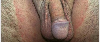
Genital lesions in LP
Therapy for genital lesions with LP is fundamentally similar to treatment tactics for lesions of the oral mucosa. The main goal of therapy is to prevent or minimize the formation of scarring, synechiae, vaginal stenosis in women and phimosis in men. TGCS are used as the first line of therapy, while standard antipruritic drugs are used to relieve itching. Initially, an intensive regimen is used, usually clobetasol 2 times a day for 1-2 months to limit the inflammatory process, followed by a transition to a maintenance regimen - using the drug 2-3 times a week. The above treatment tactics are appropriate in the treatment of lichen sclerosus, and their effectiveness has been confirmed in a number of clinical case reports. To relieve friction and pain, it is possible to use intravaginal applications of emollients. If the anal area is affected, topical corticosteroids in the form of foam or suppositories can be used. In case of disease refractory to therapy, as well as severe damage to the genitals, systemic oral corticosteroids can be used in a course with a gradually decreasing dosage and transition to topical maintenance therapy. Topical retinoids have a pronounced irritant effect and are usually poorly tolerated in the erosive form of the disease. There are a number of retrospective studies and case reports with successful use of topical calcineurin inhibitors. Despite a burning sensation when applied to erosive lesions, topical calcineurin inhibitors are better tolerated than topical retinoids and are widely used in the treatment of LP affecting the genitals.
Follicular form of LP
The goal of therapy for the follicular form of LP (lichen planopilaris) and its variants is to stop the inflammatory process as early as possible to minimize the death of epithelial stem cells of the hair follicle through inflammation-induced apoptosis, as well as control of concomitant symptoms of the disease until the spontaneous occurrence of clinical remission. Strong and very strong topical corticosteroids are used as the first line of therapy in clinical practice. Intralesional administration of corticosteroids is practiced by many specialists, despite the lack of sufficient data in favor of this technique, as well as the risk of developing scalp atrophy with prolonged use of drugs. Many doctors prefer twice daily use of strong and very strong topical corticosteroids for 6-8 weeks, with a gradual transition to use 3 times a week or as required. With an aggressive, rapidly progressive course of the disease, systemic corticosteroids (30-80 mg/day in terms of prednisolone) are often prescribed, and in some cases, systemic cyclosporine (3-10 mg/kg/day), although there is insufficient data on the effectiveness of drugs in these groups. In the presence of pronounced inflammatory changes, in addition to topical therapy, a course of prednisolone with a gradual dose reduction (starting from 40 mg/day) may be prescribed, and hydroxychloroquine may be prescribed simultaneously. It is believed that the latter helps to reduce the immune response to self-antigens and also modulates T-cell-mediated gene expression. Its immunomodulatory effects as well as its good safety profile may be useful in the treatment of follicular LP, making hydroxychloroquine a preferred treatment option for lichen planopilaris. In case of intolerance to hydroxychloroquine or if there are contraindications to its use, tetracycline antibiotics, which are second-line drugs, can be used. Also, second-line drugs include mycophenolate mofetil and cyclosporine.
Nail damage
Despite significant functional and cosmetic impairments associated with nail lesions in LP, evidence-based data are currently insufficient. The goal of therapy is to prevent or minimize permanent scarring (as with the follicular form of LP), and it is also important to stop the inflammatory process at the earliest possible stage to achieve optimal results. Topical and systemic corticosteroids are used as the first line of therapy, as well as their intralesional administration; systemic use of drugs is indicated when several nail plates are affected. According to the literature, the use of alitretinoin appears to be effective. For many dermatologists, including the authors of the article, the use of systemic corticosteroids (in the form of pulse therapy or a short course with a gradual dose reduction) is preferable to intralesional administration of corticosteroids, due to the convenience and effectiveness of this technique. Also, there is evidence of an increase in the effectiveness of topical therapy with strong and very strong topical corticosteroids when applied under occlusion.
Proven methods for treating lichen ruber
Therapeutic methods are the main method of treatment and depend on the stage of the disease. At the same time, an integrated approach is being implemented aimed at blocking both the causes themselves and the unpleasant consequences of deprivation:
- impaired functioning of the immune system, which is the cause of the manifestation of lichen, is leveled with the help of immunosuppressants;
- the main means of treating lichen planus are hormonal drugs that reduce the supply of components to the foci of immune inflammation that cause the development of ulcers;
- an integral stage of recovery procedures will be the administration of anti-allergenic drugs and vitamin A, which promotes the effective healing of eroded layers of the skin or mucous membrane;
- in severe forms of the disease, antibiotics and synthetic interferons and interferonogens are prescribed in the treatment of lichen ruber.
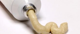
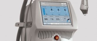
In addition to taking medications, their analogues in the form of ointments are used, as well as physiotherapeutic procedures (UV and laser irradiation, magnetic therapy). As part of an integrated approach, folk remedies based on natural substances are effective.
Our center’s specialists will be able to determine the optimal and effective treatment. They have the appropriate equipment and sufficient experience to make a detailed diagnosis of the factors causing lichen planus.
Symptoms of lichen planus in adults
Signs and symptoms of shingles last for weeks or months, and periodic relapses can occur for years, flaring up and dying down. The appearance of the lesions depends on their location.
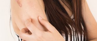
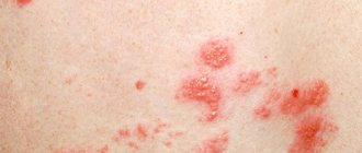
In cutaneous LP, the lesions can be present anywhere on the skin, usually on the wrists, legs, palms and soles or trunk, and are 2 to 4 mm in diameter with angular edges, purple in color, and distinctly shiny under transverse light. These lesions tend to be symmetrically distributed and may also coalesce into rough, scaly patches. In rare cases, blisters may form. Moderate to severe itching is common, common, and difficult to treat.
There are several variants of cutaneous LP, which can present in different ways. The lesions may become large, scaly and warty (lichen hypertrophis), especially on the lower legs. New spots may appear at the site of minor damage to the skin, such as a superficial scratch (Koebner phenomenon). Sometimes degeneration (atrophy) of the skin can occur while lesions persist (lichen lichen), and some patients experience a lack of sweating due to degeneration of the sweat glands (anhidrosis).
Skin diseases
What diseases do dermatologists treat: KP’s guide to skin ailments
Unusual darkening (hyperpigmentation) or lightening (hypopigmentation) of the skin may occur in areas where wounds have healed.
Between 50 and 70% of patients experience symptoms related to the mucous membranes, moist, pink skin that lines the inside of the mouth, vagina, and esophagus. LLP on mucous membranes may appear as red, painful ulcers or lesions that have a reticulate, white pattern. Oral symptoms often occur before skin lesions develop. Oral symptoms may appear first, consisting of dryness and a metallic taste or burning sensation in the mouth, which may be the only sign of the disease.
Hair loss, although not common, can be one of the consequences of LP, which is called planopillaris. When hair loss occurs, it may affect small patchy areas of the scalp (atrophic symmetrical alopecia) or cause bald patches (frontal fibrosing alopecia). If tinea is not treated, hair loss becomes permanent due to scarring.
Nail lesions are present in 10 to 25% of patients with LP and tend to appear as roughness, vertical ridges or cracks, and thinning of the nail. This can eventually lead to scarring of the nail.
Trouble swallowing or pain when swallowing may indicate esophageal ringworm. It is important to treat esophageal disease because over time it can lead to a narrowing of the esophagus, called an esophageal stricture.
Some cases of the cutaneous form resolve over time, while the oral, genital, nail, and esophageal forms are more persistent and may worsen over time. Patients with LP have an increased risk of squamous cell carcinoma, especially of the oral mucosa, and should be monitored periodically.
Experimental and Promising Therapeutic Strategies
Key Points
- Various drugs, such as sulfosalazine, mycophenolate mofetil, azathioprine, griseofulvin, and adalimumab, have been used to treat cutaneous and follicular forms of LP, as well as lesions of the mucous membranes.
- Most of the listed methods have not been introduced into clinical practice due to an unsatisfactory risk-benefit ratio.
- A prerequisite for the success of future therapeutic options is the identification of the molecular mechanisms of the pathophysiology of the disease.
Skin lesions in LP
There is an abundance of experimental agents that have not entered routine clinical practice due to an unsatisfactory risk-benefit ratio or unreasonably high cost. An example of such a drug is sulfosalazine, which has not entered into widespread clinical practice, despite the presence of a convincing evidence base and the absence of serious side effects. In early clinical trials, griseofulvin appeared to be a promising drug for the treatment of LP, however, it was subsequently studied only in sporadic studies, and its effectiveness was shown to be lower compared to hydroxychloroquine. Antifungal drugs such as itraconazole and terbinafine are thought to interact with inflammatory cytokines. They have also been studied and shown to be somewhat effective, but no more rigorous studies have been conducted. Systemic immunosuppressive drugs, such as azathioprine and methotrexate, have been used in severe generalized forms of LP, however, due to fairly high toxicity, indications for their use are limited. Comparable efficacy of topical calcipotriol and strong topical corticosteroids was shown in a randomized clinical trial, however, other data on the use of the drug are contradictory. It is believed that thalidomide may be effective in the treatment of LP due to its immunomodulatory effects on multiple cytokines and immune pathways, including interferon-gamma, but no studies have been conducted on the use of the drug. There is evidence of the effectiveness of adalimumab in localizing LP lesions on the skin. However, blocking tumor necrosis factor alpha does not always lead to recovery; the role of this cytokine in the development of lichenoid inflammation has not been fully determined. Phosphodiesterase 4 is a key enzyme in the formation of cyclic AMP in many cells of the immune system. Apremilast, an oral phosphodiesterase 4 inhibitor, has been shown to be effective in the treatment of cutaneous LP.
Damage to the mucous membrane of the oral cavity and genitals in LLP
Since the appearance of lesions of the skin and mucous membranes is based on common molecular mechanisms, there is a significant overlap of therapeutic techniques used for lesions of different locations. The effect and effectiveness of drugs such as sulfosalazine, azathioprine, mycophenolate mofetil, hydroxychloroquine, TNF-alpha inhibitors and other biological drugs, and topical thalidomide in lesions of the mucous membranes and genitalia were studied. In addition, pharmacognostic agents such as purslane and curcuminoids have been shown to be effective in a number of clinical cases. However, larger-scale studies of these drugs have not been carried out, and they have not found widespread use in clinical practice. The list of experimental drugs used to treat lesions of the oral mucosa in LLP would be incomplete without indicating such techniques as PUVA, extracorporeal photochemotherapy, intralesional injection of bacillus Calmette-Gerer, which, despite their obsolescence, are used only in refractory cases, allow us to better understand molecular mechanisms underlying the pathogenesis of the disease. There is evidence of the effectiveness of photodynamic therapy in the treatment of genital lesions in LP, however, this technique has not been widely used in clinical practice and is considered experimental
Follicular form of LP
Despite the fact that data on the molecular mechanisms of the pathogenesis of the follicular form of LP are very scarce (perhaps even more scarce compared to other lichenoid dermatoses), there is an example when studies of gene expression in this disease provided grounds for the use of experimental techniques in clinical practice. practice. The involvement of peroxisome proliferator-activated receptor-gamma in the etiopathogenesis of follicular LP has allowed clinicians to use pioglitazone, a peroxisome proliferator-activated receptor-gamma agonist, to treat refractory cases. However, soon doubts arose about the accuracy of the data obtained from genetic research, as well as conflicting outcomes of therapy for the follicular form of LP with the use of pioglitazone. Similar to pioglitazone, finasteride has been successfully used in the treatment of frontal fibrosing alopecia after misinterpretation of the clinical results obtained when eliminating the component of androgenetic alopecia that existed in a patient with frontal fibrosing alopecia. This statement was later refuted in clinical practice. These examples demonstrate how important it is in the modern era of evidence-based medicine, after carefully conducted molecular studies, to study the pharmacodynamics of the active substance and the possibility of targeted delivery and use of drugs.