General information
Dermatophytosis (dermatophytosis) - trichophytosis, epidermatophytosis inguinal, rubrophytosis, tinea pedis, microsporia, scab (favus) are widespread mycoses of the skin and its appendages.
Dermatophytes are highly contagious diseases that can be transmitted from person to person, from animal to person, or from soil to person. There are three genera of parasitic dermatophytes: the genus Trichophyton (22 species), the genus Microsporum (9 species) and the genus Epidermophyton (2 species). These fungi are united under the common name - dermatophytes, which parasitize the stratum corneum of the epidermis, hair, and nail plates. Based on the natural reservoir of dermatophytes/source of infection for humans, the following are distinguished:
- anthropophilic dermatophytes - whose habitat is humans;
- Zoophilic dermatophytes - live on wild/domestic animals and birds with the possibility of transmission to humans;
- geophilic / hydrophilic dermatophytes —whose habitat is soil/water.
Below are the main causative agents of dermatomycosis and the diseases they cause.
Dermatophytosis in humans occurs in almost all countries, and the predominance of certain pathogens, their localization and frequency depend on the lifestyle of the population and environmental factors. Some dermatophytes are endemic only in certain areas, while others are widespread. Among the most common fungal diseases, the most common is dermatomycosis of smooth skin ( microsporia , trichophytosis , mycosis of the feet (hands) , lichen versicolor ). Superficial dermatophytosis of smooth skin is characterized by predominant damage to the superficial stratum corneum of the epidermis and hair.
Depending on the location, the following are distinguished: dermatophytosis of the smooth skin of the trunk ; dermatophytosis of the beard/mustache area ; dermatophytosis inguinalis ; dermatophytosis of the scalp ; dermatophytosis of the feet (skin/nails of the foot area are affected), etc.
Dermatophytes are characterized by different abilities to selectively damage/utilize different keratin-containing structures of human skin. Thus, trichophytons equally affect the stratum corneum of the epidermis, nails, vellus/long hair ( scab , trichophytosis ); Epidermophyton infects exclusively smooth skin (dermatomycosis inguinal); microsporum - skin/hair, less often - nails. These features are primarily due to the different abilities of different types of dermatophytes to process keratin breakdown products, as well as existing differences in the types of keratin.
A feature of dermatophytes is also the difference in virulence. Thus, in terms of the degree of contagiousness/prevalence for humans in the Russian Federation, Trichophyton rubrum is in the lead, followed by Microsporum canis, T. mentagrophytes and Epidermophyton floccosum.
General characteristics of dermatophytes
The optimal temperature for mushroom growth varies between 25-30°C (lower than human blood temperature). The most important condition for the development/growth of dermatophytes is the presence of a moist environment. Dermatophytes tolerate low temperatures well and die at high temperatures, and are resistant to UV rays. The optimal environment for them is neutral/slightly alkaline; acidity shifts, especially towards the sour side, have a negative effect on mushrooms. Dermatophytes feed on nitrogenous/carbon-containing substances, which are widely present in the human body (di/mono-sugars, nitrogen salts, amino acids, etc.). Fungi secrete a large number of enzymes, which are considered to be significant virulence factors.
The age of the patient plays a significant role in the development of the mycotic process, which is due to age-related changes/fluctuations in the acid-base balance of the skin, as well as the chemical composition of the sebaceous secretion. Thus, in children under 1-2 years of age, a pronounced acidic sweat reaction is observed, which gradually decreases, approaching neutral at the age of 5-10 years. Accordingly, the incidence of trichomycosis in this age period reaches its peak. During puberty, the acidity of sweat increases again.
In adulthood, the sweat reaction in different areas of the skin varies widely. For example, sweat on the scalp, back, and chest has an acidic reaction, while sweat on the interdigital folds has a neutral/slightly alkaline reaction. Differences in the chemical composition of skin sebum in children and adults are also significant. In addition, the hair of adults contains fatty acids that have a fungiostatic effect. Trichomycosis (microsporia, trichophytosis, favus) can affect long scalp hair and, when fungi spread to smooth skin, vellus hair and are observed mainly in children. At the same time, mycoses of the feet and groin area (athlete's foot/rubrophytosis) are more common in adults.
Next, trichophytosis will be directly discussed
Trichophytosis (ringworm, trichophytosis) is one of the types of dermatophyte infections, which is a highly contagious fungal disease of humans/animals, manifested by damage to the skin and its appendages (hair/nails). Caused by fungi of the genus Trichophyton. Some species of dermatophytes parasitize exclusively on humans (anthropophilic), while others can parasitize both humans and animals (zoophilic).
The group of anthropophilic trichophytons is represented by the most frequently occurring Tr. Tonsuraus/Tr. violaceum, and the group of zoophilic trichophytons is Tr. Verrucosum/Tr. mentagrophytes v. gypseum. Anthropophilic fungi cause superficial trichophytosis. At the same time, there is a predominant location of the fungal elements inside the hair with a minimal reaction from the skin; the damage is superficial.
Zoophilic trichophytons, parasitizing animals, cause deep infiltrative-suppurative trichophytosis. They are characterized by the location of fungal elements predominantly around the hair/in the epithelium of the hair sheath. They occur with the formation of an inflammatory perifollicular infiltrate with purulent melting of the hair follicles and sometimes the surrounding connective tissue.
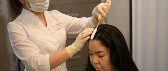
Diagnostics
The main task of laboratory research is to identify the pathogen in the material. The affected material: hair, nails, scales is treated with an alkali solution so that only masses of the fungus are visible under microscopy. If filaments of mycelium or chains of conidia are visible during the examination, a diagnosis is made. If necessary, additional research may be carried out. For rapid diagnosis, a Wood's fluorescent lamp is used, when illuminated, the mushroom particles acquire a light green tint.
Pathogenesis
The pathogenesis of dermatophytosis is based on the virulence factors of fungi, due to their ability to secrete enzymes that destroy keratin (keratinolytic activity). The enzymes secreted by dermatomycetes and their metabolic products, as well as the contents of destroyed cells, cause an inflammatory process in the lesion, manifested by redness of the skin, itching and hair breaking. Keratinases have the ability to decompose not only keratin, but also other proteins (collagen and elastin), and it is the severity of keratinase activity that determines the characteristics of the manifestation of diseases when infected with various types of fungi. An important role in the spread of dermatomycetes is played by the presence of a specialized apparatus (perforator organs) for the directed growth of hyphae (the filamentous structure of the fungus). Thanks to this feature, the fungus grows in the direction of the intercellular junctions, which are the points of least resistance. Dermatomycetes, as a rule, do not penetrate deeper than the granular layer of the epidermis, since specific protective factors are activated at a deeper level.
The process of fungal invasion begins with the burrowing of hyphae into the skin and the release of keratinases (enzymes for the breakdown of keratins), which leads to irritation of nerve endings and, accordingly, the occurrence of itching. Dermatomycetes, loosening the cortex, cause acanthosis, parakeratosis, serous inflammation in the skin with the formation of blisters. Further, swelling occurs in the papillary/subpapillary layer, the vessels dilate, which leads to the development of polymorphic perivascular infiltration. The mycelium is usually located between the horny plates. Threads and spores of fungi can also be stuffed with hair follicles, which also undergo inflammatory changes.
Causes
Currently, the leading causative agent of trichophytosis in Russia is the anthropophilic fungus Trichophyton tonsurans and, much less frequently, Trichophyton violaceum. These fungi can affect both smooth skin and its appendages.
Epidemiology. With anthropophilic trichophytosis, the source of infection is a person with trichophytosis. Infection occurs through direct contact with the patient, especially with fresh skin lesions and his things contaminated with fungi (bedding, hats, scissors/combs, towels, underwear, etc.). If sanitary and hygienic requirements are violated, the pathogen can be transmitted in kindergartens, gyms, hairdressers, and schools. Of particular epidemiological significance is the intrafamily route of transmission, when children become infected from parents suffering from a chronic form of the disease. This form most often affects children.
In zoophilic trichophytosis, the source of infection is both sick animals (cattle, domestic, small wild and laboratory) and mycocarriers (rodents - mice, rats). It occurs mainly among residents of villages who have long-term/frequent contact with animals (workers of livestock farms, veterinary hospitals, shelters for homeless animals). Infection can occur both through direct contact with a sick animal, and through contact with dust, hay/straw contaminated with rodent hair affected by the fungus.
In the development of trichophytosis, the general condition of the macroorganism plays a great role. The disease develops more often in people with reduced immunity, suffering from somatic diseases, vitamin balance disorders, dysbacteriosis and endocrine pathology. Predisposing factors may be a decrease in the protective properties of the skin, micro/macrotrauma, increased blood glucose, decreased complementary/phagocytic activity in sebaceous secretions, and impaired microcirculation.
The peak incidence occurs in the autumn-winter period, which is due to overcrowding of people with epizootics in domestic animals. The incidence of trichophytosis in the Russian Federation varies between 1.9-6.1 cases/100 thousand population. The incubation period can vary from 1 week to 2 months depending on the type of fungus. In the absence of timely/adequate therapy, the acute disease becomes chronic.
4.Treatment
To date, a wide range of antifungal drugs – antimycotics – has been developed and successfully used.
The course of treatment, as a rule, is quite long, includes additional aseptic measures and requires from the patient not only patience, but also a good level of compliance, i.e. therapeutic consent, therapeutic alliance with the doctor, including understanding of etiopathogenetic mechanisms and literal compliance with the instructions received.
It should be recalled that even after complete eradication of the dermatophyte fungus, immunity to it is not formed, but with inadequate therapy or self-medication, such an invasion easily turns into a latent, low-symptomatic or asymptomatic form, so that in the future, with the slightest weakening of the body’s protective resources, it can become active again.
Symptoms of dermatomycosis in humans
The clinical symptoms of trichophytosis are largely determined by the location of the lesions, the type of pathogen, the state of the patient’s physical health, and the depth of penetration of the fungus. Anthropophilic fungi, as a rule, cause superficial/chronic trichophytosis, and the group of zoophilic fungi can cause both superficial and infiltrative-purulent forms, which transform sequentially into one another and can be considered as different stages of one process.
Conventionally, anthroponous trichophytosis can be divided into:
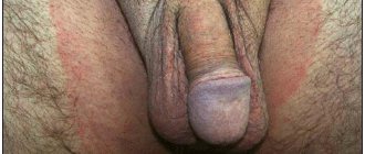
- superficial trichophytosis of the smooth/hairy scalp, smooth skin of the torso/extremities with possible involvement of vellus and coarse hair of the eyebrows, eyelashes, in men - the upper lip/chin, as well as the groin and axillary areas;
- chronic trichophytosis of smooth skin of the trunk, limbs and nails, scalp.
With zooanthroponotic trichophytosis, according to localization, it is customary to distinguish zooanthroponotic trichophytosis of the trunk/limbs, scalp, smooth facial skin, and onychomycosis. In addition, there are atypical variants of trichophytosis.
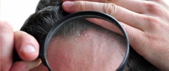
Anthroponotic superficial trichophytosis of the scalp
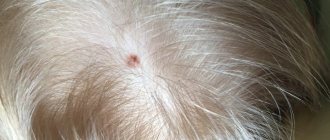
Depending on the form, small-focal and large-focal trichophytosis are distinguished. With superficial small-focal trichophytosis, flaky small lesions 1-2 cm in size with rounded/irregular outlines with indistinct blurry boundaries and covered with whitish scales are noted. Inflammatory phenomena are minor (the skin in the area of the lesions is slightly hyperemic and swollen). The lesions are located isolated, without a tendency to merge. In some cases, especially in peripheral areas, hyperemia / swelling may increase with the addition of vesicles, pustules and crusts. Characteristic is the thinning of healthy hair in the lesions, caused by their breaking off at various levels, usually at a height of 1-2 mm above the skin. Accordingly, some hair looks like gray stumps; others, when broken off at the exit from the mouth of the hair follicle, have the appearance of so-called “black dots”. Affected hair is gray, dull, loses elasticity, and partially curls/bends.
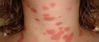
Anthroponotic superficial trichophytosis of smooth skin
Lesions are most often localized on open areas of the skin: forearms, shoulders, face, neck, and torso. They are usually presented as erythemotic spots of oval/round shape with clearly defined boundaries in the form of a hyperemic ridge with the presence of vesicles, nodules, and crusts. At the same time, the central parts of the lesions are flaky and paler. The lesion often takes the form of a ring and the lesions can merge into figures of various shapes. There is little or no itching. When the lesions are localized in the area of the chin, upper lip, eyebrows and eyelashes, the lesions are smaller, flaky with minor inflammation and the presence of short broken hair. It can occur in isolation or in combination with damage to the scalp. Hair follicles may also be affected, which is manifested by the formation of small nodules, subsequently accompanied by suppuration with the development of folliculitis.
Anthroponotic chronic trichophytosis of the scalp
The location of the pathological process is predominantly the occipital region. It manifests itself as areas of atrophy with slight whitish pityriasis-like peeling located on a lilac background. Broken hair is difficult to detect and, as a rule, there are few of them.
Anthroponotic chronic trichophytosis of smooth skin
This form is characterized by damage to the forearms and elbows, buttocks, legs, and less commonly the skin of the face and torso. It is extremely rare that mycosis spreads universally. Lesions in the form of spots without clear boundaries of a pinkish-bluish color with a flaky surface. There are no pustules/vesicles, a peripheral ridge, and the central part does not develop backwards.
When the soles/palms are affected, peeling, mild hyperemia, increased skin pattern, and sometimes thickening of the stratum corneum occur, which leads to the formation of deep grooves/cracks on the palms and soles in places of skin folds.
With trichophytosis of the soles/palms, blisters do not form. Involvement of vellus hair in the pathological process is often noted. Damage to the nail plates is often observed - a whitish-gray spot forms in the distal, proximal or lateral part of the nail, which gradually increases in size, the nails become deformed, thicken, and crumble easily. The nail plate becomes lumpy, dirty gray and dull.
Zooanthroponotic infiltrative-suppurative trichophytosis
Manifests by the appearance of one/several flaky pale pink spots of a round shape, with clear boundaries. As a rule, the peripheral ridge is formed by small vesicles and follicular papules, which subsequently shrink into crusts. Due to peripheral growth, the lesions gradually increase in size, and due to increasing infiltration and inflammation, they begin to rise above the skin level. When they merge, various types of figures are formed, the surface of which is covered with papules, vesicles, pustules and crusts. Often, vellus and long hair are involved in the process. “Stumps” of broken hair appear.
As the disease develops, inflammation in the foci located in the area of growth of the beard and mustache/scalp intensifies: hyperemia , swelling increases, hemispherical , sharply demarcated nodes with a bumpy surface of bluish-red color are formed, which are covered with multiple folliculitis, erosions, and less often ulcerations. The hair is loose, easily removed and partially falls out. Characteristic signs include significantly enlarged mouths of the hair follicles, which are filled with purulent contents, released in the form of copious drops when pressed. Gradually, the dense consistency of the nodes on the scalp softens and the lesions resemble honeycombs, and when localized in the mustache/beard area, they resemble wine berries.
When localized on smooth skin, predominantly flat plaques are formed, often extensive, with isolated papules located on their surface, gradually transforming into pustular elements. The process of suppuration contributes to the death of fungi, which are preserved mainly at the periphery of the lesions in scales. In severe cases of infiltrative-suppurative trichophytosis, regional lymph nodes may enlarge, sometimes the general condition suffers ( headaches , malaise , fever ), changes appear in the blood - leukocytosis/increased ESR. As a rule, a scar is formed as a result.
Below are photos of trichophytosis in humans.
Separately, it is necessary to dwell on such dermatomycosis as favus.
Favus of the scalp
Favus is a fairly rare, sporadically occurring low-contagious dermatophytosis in the Russian Federation, which most often affects the scalp and vellus hair, less often smooth skin and nails. The causative agent is the anthropophilic fungus Tr. schonleinii. Occurs mainly in childhood. More often, favus of the scalp occurs in a typical (scuticular) form, less often in atypical forms (squamous/impetiginous). A characteristic pathognomonic sign of the scuticular form is the so-called “scutules” (scutellums), which are dry to the touch, round saucer-shaped formations of a dense yellow consistency. At the same time, the central part of the scutula with the hair pierced from it sinks. The scutulae have a tendency to merge, which leads to the formation of continuous extensive crusts that have a moldy smell, followed by the development of persistent baldness and cicatricial atrophy of the skin under the crusts. Hair loses its elasticity and shine, becomes dull, dry, and there are no broken hairs in the area. In addition to the classic form, there are 2 more varieties of favus on the scalp - impetiginous and squamous.
The squamous form of the favus is manifested by abundant continuous peeling of a yellowish-brown color. The scutulae are often absent or very few and small in size. After removing the scales, a shiny bright red surface of the skin is visible, on which atrophic phenomena develop. The impetiginous form of favus disease is characterized by the formation of pustules at the mouths of hair follicles, which heal with the formation of dry crusts. The lesions are limited, and once the inflammatory process has resolved, cicatricial atrophy of the skin . Treatment is similar to trichophytosis .
Types of mycoses of the feet
Interdigital dermatophytosis
The most common type of foot fungus. It occurs in acute and chronic forms. In the acute form, the skin between the 3rd, 4th and 5th fingers is affected. There is softening (maceration) of the skin, the epidermis exfoliates, weeping, redness, cracks. The exfoliated epidermis is whitish in color. The adjacent areas of the foot are very quickly affected.
Rice. 5. The photo shows foot fungus. Interdigital dermatophytosis.
Deep tinea pedis
This condition is regarded as a complication of interdigital dermatophytosis. Pyogenic bacteria easily penetrate through damaged areas of the skin into the deep layers, destroying them. Without adequate treatment and with a decrease in immunity, foot fungus spreads to the entire sole and its inner part.
Rice. 6. The photo shows foot fungus. Deep dermatophytosis of the feet.
Dyshidrotic dermatophytosis of the feet
Caused by the fungus Trichophyton mentagrophytes. Rarely seen. An inflammatory reaction and multiple rashes of vesicles and blisters appear on the skin of the foot. Bubbles and vesicles resemble rashes during allergic reactions. All manifestations are associated with an allergic reaction to dermatophyte antigens. The skin of the sole, its inner part and the spaces between the toes are affected. When a bacterial infection (Staphylococcus aureus) attaches, pus appears.
Rice. 7. The photo shows foot fungus. Dermatophytes are visible - allergic rashes due to fungal infections.
Plantar dermatophytosis of the feet
Caused by the fungus Trichophyton rubrum. Initially, redness occurs with small papules along the edges, then the affected area begins to peel off and become keratinized. Covering the entire foot and its lateral surfaces, the affected skin resembles a ballet shoe.
Rice. 8. In the photo there is a fungus on the feet - plantar dermatophytosis.
Prevention
The basis of preventive measures is:
- Strict adherence to the rules of personal hygiene: do not use other people’s personal hygiene items, underwear, clothes, wash your hands thoroughly after contact with animals.
- Early (timely) identification of patients with trichophytosis with their isolation and quarantine of contact persons.
- Identification of animals with trichophytosis, carrying out deratization work.
- Carrying out current/final disinfection at infection sites (in schools, kindergartens, at home, bathhouses, hotels, hairdressers, medical institutions, dormitories, etc.).
List of sources
- Leshchenko V. M. Fungal skin diseases. In the book: Skin and venereal diseases (a guide for doctors). Ed. Yu. K. Skripkina, V. N. Mordovtseva. M., 1999. T. 1. P. 257-311.
- Kubanova A.A., Potekaev N.S., Potekaev N.N. Guide to practical mycology. – Moscow, Financial Publishing House “Business Express”, 200 p.
- Bayazitova A.A., Bayazitova A.A., Kupriyanova-Ashina F.G., Khaldeeva E.V., Ilyinskaya O.N. Primary pathogens - Pathogens of superficial mycoses // International Journal of Applied and Basic Research. – 2015. – No. 11-2. – pp. 241-248.
- Medvedeva T.V. Trichophytosis: modern ideas about the etiology, clinical picture, features of diagnosis and therapy / T.V. Medvedeva, V.B. Antonov, L.M. Leina, T.S. Bogomolova // Klin. dermatology and venereology. - 2007. - No. 4. - P. 70–74.
- Rodionov A.N. Fungal skin diseases (Guide for doctors) St. Petersburg: Peter. 1998. – 288 p.
1.General information
Dermatophytosis is a large group of infectious skin diseases caused by mold fungal cultures called dermatophytes. Translated from ancient Greek, “dermatophyte” means “skin vegetation,” which apparently reflected the ideas of ancient healers about the essence of this disease. Today, dermatophytic mycoses are a very serious problem: not presenting a direct threat to life in most cases, such infections sharply reduce its quality, causing significant physiological and psychological discomfort, and are characterized by high therapeutic resistance (resistance), chronic and persistent (persistent) course , a tendency to relapse, since immunity to dermatophytes is not developed and repeated infection with the same pathogen is possible.
It is extremely difficult to estimate the exact prevalence of dermatophytosis: apparently, not all cases are included in official medical statistics; This is especially true for mycoses of low-symptomatic or subclinical severity, in which, nevertheless, the infected person remains highly contagious.
Almost all special epidemiological studies reveal a trend towards an increase in incidence. Today, at least a fifth of humanity is infected with one or another dermatophyte fungi.
The idea of dermatophytosis as a predominantly childhood disease is erroneous: in fact, the risk of infection and the clinical picture do not depend on age.
A must read! Help with treatment and hospitalization!