Pain in the buttocks indicates the presence of pathologies, such as:
- lumbosacral radiculitis,
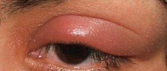
- formation of phlegmon and abscesses,
- osteomyelitis,
- furuncle
- or is considered a consequence of muscle overexertion.
Pain in the buttocks can also be caused by:
- osteochondrosis,
- coccyx cyst,
- intervertebral hernia.
If there is a disease in the lower parts of the spine, due to the location of the nerves, the pain may radiate to the legs or buttocks.
To diagnose the cause of pain, the patient should determine the nature, intensity and painful location. We recommend that you contact experienced proctologists.
The buttocks are considered symmetrical parts of the body and represent, figuratively speaking, a “layer cake”. The first, top layer is the skin. The second is the tissue of the left and right gluteal muscles, respectively. The third layer is represented by subcutaneous fat, which is located directly under the muscles and, in comparison with other parts of the body, is considered the most developed.
Pain in the butt occurs in any of the balls. A painful process in the buttocks indicates the consequences of an injury, the presence of an infectious or inflammatory process in the body, or muscle pathologies.
FIBROHISTIOCYTIC TUMORS AND TUMOR-LIKE LESIONS
Xanthoma
A rare disease, most often localized in the skin. Occurs in people with impaired lipid metabolism, usually multiple. Also localized in tendons. Presented by small nodules, partly of the xanthelasma type.
Juvenile xanthogranuloma
A small nodule in the dermis or subcutaneous tissue. Disappears spontaneously.
Fibrous histiocytoma
It is more common in middle age and is localized mainly on the lower extremities. Usually has the shape of a dense knot up to 10 cm, grows slowly. After surgical removal, recurrences are rare.
Liposarcoma - prognosis
The prognosis for liposarcoma depends primarily on the type of lesion. For example, people with well-differentiated liposarcoma can have a 5-year survival rate of up to 90-100% after appropriate treatment. Patients with liposarcoma multiforme, which is more likely to metastasize, have a much worse chance.
Another aspect to consider is the propensity for recurrence, which also varies depending on the type of tumor. Relapse of liposarcoma can occur after different times; it happens that the tumor recurs even several years after the end of treatment. This is due to the fact that lipomas grow very slowly.
ONLINE REGISTRATION at the DIANA clinic
You can sign up by calling the toll-free phone number 8-800-707-15-60 or filling out the contact form. In this case, we will contact you ourselves.
BENIGN TUMORS OF ADIPOSE TISSUE
Lipoma
One of the most common benign tumors (30-40%). It can occur anywhere there is fatty tissue. When localized in the dermis, it is usually encapsulated, in other parts of the body it is poorly demarcated. Tumors localized in the retroperitoneal space can become malignant; other localizations practically do not become malignant. Lipomas are often multiple and sometimes develop symmetrically. Their growth is not related to the general condition of the body. The tumor has the shape of a lobular node. With long-term existence in the lipoma, dystrophic changes, calcification, and ossification can develop.
There are numerous variants of mature fatty tumors, which differ from the classic lipoma both in clinical manifestations and in some morphological features.
Myelolipoma
A rare tumor, most often found in the retroperitoneum, pelvic tissue, and adrenal glands. Does not become malignant.
Subcutaneous angiolipoma
Numerous painful nodes. It occurs more often in young men on the front wall of the abdomen, on the forearm.
Spindle cell lipoma
It is observed more often in adult men (90%). The node is round in shape, dense, slowly growing, most often localized in the area of the shoulder joint and back. Recurrence and metastasis after excision have not been described, despite the fact that the tumor can infiltrate surrounding tissues.
In chondro- and osteolipomas, metaplastic areas of bone and cartilage tissue are detected.
Benign lipoblastomatosis
It is divided into nodular (kind lipoblastoma) and diffuse (kind lipoblastomatosis) forms. Boys under 7 years of age (88%) get sick more often. The tumor is localized on the lower limb, in the buttocks and on the upper limb - the shoulder girdle and hand. Lesions of the neck, mediastinum, and trunk have also been described. The tumor node is encapsulated, lobulated, spherical in shape, and can reach 14 cm. After surgical treatment, relapses are possible, sometimes repeated. Metastases have not been described.
Gebernoma (fetal lipoma)
Lipoma from lipoblasts, pseudolipoma is an extremely rare tumor, localized in places where there is brown fat (neck, axillary region, sinus, mediastinum). Presented as a lobular node, usually small in size. Does not recur and does not metastasize.
Causes of eczema on the buttocks
The prevalence and appearance of the rash on the butt, like other symptoms, largely depends on the causes of the disease.
The main reasons include:
| Contact with an irritating substance |
|
| Allergic reaction |
|
| Infection |
|
In children, the most common cause is diaper dermatitis, which is contact dermatitis by nature, but several other factors play a role in its development. These are the moist warm environment under the diaper, mechanical irritation, exposure to urine, fungal infection.
BENIGN TUMORS OF MUSCLE TISSUE
Tumors of muscle tissue are divided into smooth muscle tumors - leiomyomas, and striated muscle tumors - rhabdomyomas. Tumors are quite rare.
Leiomyoma
Mature benign tumor. Occurs at any age in both sexes. It is often multiple. The tumor can become malignant. Treatment is surgical.
Leiomyoma, developing from the muscular wall of small vessels - small, often multiple, poorly demarcated and slowly growing nodes, often with ulcerated skin, is clinically very similar to Kaposi's sarcoma.
Genital leiomyoma is formed from the muscular lining of the scrotum, labia majora, perineum, and nipples of the mammary gland. May be multiple. Cellular polymorphism is often observed in the tumor. Hormone dependent. Treatment is surgical.
Angioleiomyoma from the trailing arteries
Clinically, a sharply painful tumor that can change size under external influences or emotions. The sizes are usually small, more often found in older people, on the limbs, near the joints. It is characterized by slow growth and a benign course.
Rhabdomyoma
A rare mature benign tumor based on striated muscle tissue. Affects the heart and soft tissues. It is a moderately dense node with clear boundaries, encapsulated. Metastases of rhabdomyoma have not been described. Relapses are extremely rare. Microscopically, 3 subtypes are distinguished - myxoid, fetal cell and adult. Rhabdomyoma of the female genitalia is also distinguished. The adult type mainly recurs.
Liposarcoma - diagnosis
Patients with different types of tumors under the skin often wonder how to recognize liposarcoma? To make a diagnosis, just one examination is not enough, since the tumor is very similar to a harmless lipoma. Therefore, first of all, in order to distinguish the quality of the tumor, the dermatologist prescribes magnetic resonance imaging.
Magnetic resonance imaging
This study allows, among other things, to suspect liposarcoma, however, the diagnosis requires clarification using a histopathological examination that reveals cancer cells. First of all, histopathology allows us to accurately determine whether the change described in the studies is liposarcoma or lipoma. In addition, microscopic tests allow you to diagnose a specific type of tumor, identifying, for example, liposarcoma, myxoids.
Knowing the type of change is important to determine the patient's prognosis for cure. Material for histopathological examination is obtained by biopsy.
BENIGN TUMORS OF BLOOD AND LYMPHATIC VESSELS
These lesions include various processes, a significant number of them are considered in dermatology. Some of them relate to malformations of the vascular system of a tumor-like nature, some to true tumors.
Capillary angioma
True neoplasm with proliferation of endothelial cells.
Benign hemangioendothelioma
Congenital pathology, occurs in newborns and infants, more often in girls, with localization in the head area.
Capillary hemangioma
After lipoma, the most common tumor of soft tissues, often multiple, reaches its maximum size by 6 months of age; with multiple lesions, localization in internal organs is possible
Cavernous hemangioma
A formation consisting of bizarre sinusoid-type cavities of various sizes. Localized in the skin, muscles, internal organs. Has a benign course.
Senile hemangioma
A true tumor is characterized by proliferation of capillaries followed by their cavernization with secondary changes.
Hemangioma
A mature benign tumor of vascular origin, common. It most often affects middle-aged people and is localized on the mucous membrane of the nose, lips, skin of the face, extremities, and in the mammary gland. It is a clearly demarcated grayish-pink node 2-3 cm in size. The tumor can often become malignant and develop into angiosarcoma.
Arterial angioma
A conglomerate of malformed vessels, no signs of a tumor.
Glomangioma (glomus tumor, Barre-Masson tumor)
It occurs as an isolated tumor or as multiple disseminated familial glomusangioma. The tumor is benign and occurs in older people, in the hands and feet, most often in the nail bed area. May affect the skin of the lower leg, thigh, face, and torso. In isolated observations, it was observed in the kidneys, vagina, and bones. When localized in the skin, the tumor is sharply painful. Does not recur and does not metastasize.
Hemangiopericytoma
It is rare and can occur at any age. Localized in the skin, less often in the thickness of soft tissues. It looks like a delimited dense red node. The tumor can become malignant - giving relapses and metastases, it is considered a potentially malignant process. Malignancy in up to 20% of cases has been described in adults. The process in children is benign.
Lymphangioma
It is observed more often in children as a malformation of the lymphatic vessels, but can occur at any age. Most often localized on the neck, oral mucosa.
Diagnostic methods
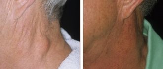
In most cases, a physical examination is sufficient to make a diagnosis if there is a lump on the skin. It is performed by a therapist. The patient then receives a referral to visit other doctors (if necessary) and diagnostic tests.
A dermatologist examines neoplasms similar to warts and papillomas. Abscesses and benign tumors are examined by a surgeon, and malignant ones by an oncologist.
Seal diagnostic methods:
- general and detailed blood test;
- Ultrasound;
- siascopy - high-quality visual analysis using light waves;
- dermatoscopy - study of the morphological structures of the skin;
- screening tests - displaying skin condition in 3D resolution.
High-quality diagnosis is impossible without detailed laboratory, biochemical, histological, cytological, microscopic and x-ray studies.
BENIGN TUMORS AND TUMOR-LIKE DISEASES OF SYNOVIAL TISSUE (JOINTS)
Benign synovioma without giant cells
The existence of benign synoviomas is debated. Most authors are inclined to believe that all synoviomas are malignant, regardless of the degree of maturity. The tumor mainly affects the knee joint, in the form of small dense nodes. Treatment is surgical, but patients should be observed for 5-9 years. The disease can cause relapses and metastases.
Benign giant cell synovioma (nodular tenosynovitis)
Pseudotumor process occurs quite often. In 15% the process occurs in the area of the synovial membrane of the joints, in 80% in the tendon sheaths, in 5% in the mucous bursae. It is a nodular formation, most often localized on the fingers, less often on the feet, and even more rarely in the area of large joints. Favorite localization is the interphalangeal joints. It is more common in women 30-60 years old. If it persists for a long time, it can cause atrophy of surrounding tissues, including bones. The process often recurs, most of the relapses are associated with incomplete removal. Does not give metastases.
Pigmented villonodular synovitis
It is located inside the shell of the joints, most often in the area of the knee, elbow and shoulder joints. Occurs in middle age. The etiology is not clear.
Characteristic symptoms
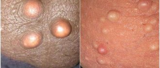
The symptoms of a lump on the gluteus medius muscle depend on what caused its appearance. Many benign formations may not manifest themselves for years or may increase in size but not cause physical discomfort. Features of different types of seals:
- A fatty tissue, an intermuscular lipoma, is a painless nodule, a lump under the skin on the buttock. It has a soft structure and deforms when enlarged.
- Subcutaneous lipoma is a mobile, round-shaped tumor. The maximum size in diameter is 1-2 cm.
- Erythema nodosum is a nodular formation in the form of a hemisphere. It is distinguished by its red color due to the inflammatory process. When pressed, the lump in the gluteal muscle hurts, accompanied by fever, chills, nausea, and drowsiness.
- Atheroma has a pronounced clinical picture. The lump itself is small, soft, painless. The presence of secretion inside causes an unpleasant odor. With inflammation, redness of the skin at the site of the tumor, suppuration, and pain are observed.
- Fibroma is a medium-sized growth, painless. Sometimes causes itching and sensitivity to touch. When it is injured, bleeding occurs.
- Gluteal fibroid is a slow-growing, mobile tumor. It has a densely elastic consistency and is painless. By the time of discovery it often reaches large sizes.
- A cyst is a soft and painless spherical formation. When infected, it becomes red and painful. Swelling, suppuration, and breakthrough of atheromatous masses may occur.
- Cancer - a malignant formation has pronounced borders and is red in color. Accompanied by intense itching, ulcerative lesions, and an increase in size of the lump. It almost always has a foul odor and causes pain when touched. At the same time, general malaise, intoxication, loss of appetite, and fever are observed.
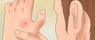
If the lump appears as a result of an injection, there is a post-injection infiltrate inside it. The formation hurts and itches. Rashes and redness may appear around it. However, in many cases the problem goes away on its own.
If the lump does not resolve within a few days, consult a doctor. Special ointments (Troxevasin, Lyoton, Triumel), an iodine mesh or a compress of aloe juice or cabbage leaf can help the skin return to normal.
BENIGN TUMORS OF PERIPHERAL NERVES
Traumatic or amputation neuroma
Arises as a result of post-traumatic hyperregeneration of the nerve. It is a small painful node.
Neurofibroma
A single, slowly growing benign tumor of the mesenchymal sheath of the nerve trunk of any location, but most often develops on the sciatic nerve and intercostal nerves. Occurs in people of any age. Clinically defined as a small, densely elastic consistency with a smooth surface of the tumor node, upon palpation of which the pain radiates along the nerve. Some tumors can reach large sizes. Tumor growth can occur both to the periphery of the nerve and in the thickness of the nerve trunk, which is revealed during its morphological examination.
Treatment is surgical. The prognosis is good. A special disease is multiple neurofibromatosis (Recklinghausen's disease), which belongs to the group of dysplastic processes. Cases of malignancy of one of the multiple neurofibromas in this disease have been described.
Liposarcoma - treatment
Surgery plays a fundamental role in the treatment of liposarcoma. Patients who develop a tumor, for example, on the hip, often fear having to amputate the entire limb. Unfortunately, this risk exists, but in general, amputations are only carried out when this is the final possible solution. If possible, conservative procedures are considered first.
The most important thing in the treatment of these changes is their complete removal with a supply of healthy tissue.
In addition to surgery, radiation therapy and chemotherapy are used. They are especially useful when a patient's existing tumor has invaded important structures, such as major blood vessels, and the tumor cannot be completely removed. In this case, radiation therapy is effective. Then, when the patient develops multiple metastases, chemotherapy becomes the main treatment option.
Chemotherapy for the treatment of liposarcoma
What could be bumps in the anus?
Most often, a lump near the anus is a hemorrhoid, swollen and inflamed veins of the rectum and perianal area. Provoking factors for the appearance of hemorrhoids are:
- constant constipation;
- sedentary lifestyle;
- hereditary predisposition to hemorrhoidal pathology;
- excessive use of laxatives;
- lifting weights;
- prolonged static sitting or standing;
- overweight;
- smoking and alcoholism;
- pregnancy.


