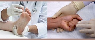
This article is not a call for self-medication, please consult your doctor!
At the same time, the author of the article is convinced that we should know about all scientifically based approaches to treatment, their advantages and disadvantages. Only then, instead of simple executors of the doctor’s will, we will become his allies on the path to health.
Nail psoriasis is a specific form of psoriasis that affects the fingernails and/or toenails. Doctors call this type of disease psoriatic onychodystrophy (from the Greek onychos - nail, dys - disorder, trophe - nutrition).
From this article you will learn about the causes of the development of nail psoriasis, its symptoms, which do not always clearly indicate the correct diagnosis, as well as dangerous misconceptions regarding this form of the disease.
Note. The article contains many photographs that may frighten an unprepared reader. Therefore, we specifically reduced their sizes for viewing on a computer monitor. If necessary, click on the photo to view the details.
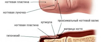
Where do nails grow from?
To understand the problem of nail psoriasis, it is important to understand how the so-called nail apparatus works.
The nail performs two functions: working and aesthetic. Firstly, the nail protects the fingertips from damage, increases accuracy and sensitivity when working with small objects, can be a weapon of attack or defense, and finally, we itch with the help of nails. Secondly, the aesthetic, or cosmetic, function of nails is also important, especially for women.
Nails are formed from the outer layer of skin - the epidermis. The nail apparatus includes:
- nail plate - the nail itself,
- matrix - it produces the nail plate, the nail socket, or lunula, is the only visible part of the matrix, this is a white moon-shaped area at the base of the nail plate,
Diagram of the nail apparatus
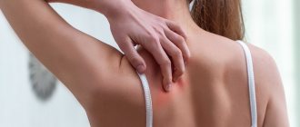
Complications of urticaria
It is important to know what hives look like and to be able to provide first aid correctly.
This will help prevent the development of severe complications that can lead to death. Often urticaria is accompanied by Quincke's edema, which is also called angioedema. Its development in the larynx area is especially dangerous, as it can compress the trachea and impair breathing.
Another serious complication is anaphylactic shock. This is a life-threatening immediate allergic reaction that occurs when the human body is hypersensitive to a particular allergen. Usually develops upon repeated contact with the allergen and requires immediate medical attention.
Causes and mechanism of development of nail psoriasis
In its course - with periodic exacerbations and remissions - psoriasis on the nails resembles the vulgar form of the disease.
It is believed that nail psoriasis develops for the same reasons and in the same pattern as typical psoriatic rashes. These reasons include external and internal factors.
The main internal factor is genetic predisposition. External causes are numerous and include, for example, injury, poor diet, intoxicants (alcohol and tobacco), infections and certain medications.
The standard mechanism for the development of nail psoriasis under the influence of these causes can be briefly described as follows.
- Triggers, such as trauma, activate immune cells.
- Activated immune cells migrate to the nail matrix or nail bed area.
- Immune inflammation develops in these areas.
- The division of skin cells accelerates sharply, and their maturation is disrupted.
- Characteristic symptoms of psoriasis appear on the nails.
Also, the cause of nail psoriasis can be considered as a result of the body’s inability to adapt to unfavorable environmental conditions. According to this point of view, the main cause of psoriasis is an environment that is evolutionarily unusual for humans.
As a result, this evolutionary approach considers poor nutrition, lack of sun and clean water, excess toxins, lack of normal physical activity, sleep disturbances and chronic stress as the direct causes of the disease.
Nail psoriasis and psoriatic arthritis are linked
The connection between nail damage and psoriatic arthritis has long been known.
Based on observations, scientists have found that psoriatic arthritis is accompanied by nail damage in nine out of ten cases.
But the mechanism of this connection has not been fully studied. However, the authors of several studies, for example from the Institute of Molecular Medicine in Leeds (UK), tried to explain this connection beyond the concept of immune inflammation.
In their opinion, the fact is that the finger joint is located next to the nail and is anatomically connected to it.
Therefore, microtraumas and the Koebner phenomenon, which cause primary inflammation of the joint - psoriatic arthritis - also cause secondary pathological changes in the nearest nail.
This is why psoriatic arthritis is accompanied by nail lesions so often.
Psoriasis with damage to the nails and inflammation of the joints (arthritis) of the toes
Thus, symptoms of nail psoriasis often indicate the presence of psoriatic arthritis.
Let's now look at the main myths accompanying this disease and why they are dangerous.
Myth 1: Nail psoriasis is rare
Not really. Apparently, with psoriasis, the nails suffer very often.
According to various sources, nail psoriasis occurs in the range from 6% to 82% of cases of vulgar psoriasis. Such a large scatter in the assessment of the prevalence of this pathology is explained by problems in its recording. Medical statistics record that patients with the vulgar form turn to doctors first, and attention is paid to nails secondarily. In scientific studies, cases of nail psoriasis are also usually studied only in addition to the main object of interest - psoriasis with skin lesions.
However, a number of publications say that
up to 80-90% of patients with psoriasis vulgaris reported recurrent nail damage.
And also that nail psoriasis occurs in 90% of patients with psoriatic arthritis and psoriasis of the scalp.
It should be noted that adults usually suffer from this form of the disease.
According to various sources, in children, nails are affected in approximately 7-37% of cases of psoriasis. Unfortunately, often the manifestations of psoriasis on a child’s nails are not given due importance. Parents or doctors believe that this is a variant of the norm or a consequence of injury, or simply do not notice due to the mild severity of the symptoms.
Onychodystrophy as a sign of other diseases
A much more serious problem than isolated onychodystrophies are those that arise as a result of the development of other disorders in the functioning of the body. In such situations, patients require comprehensive treatment aimed at eliminating existing diseases and improving nail nutrition.
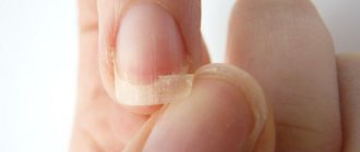
Onychorrhexis
Onychorrhexis is onychodystrophy, accompanied by longitudinal separation of the nail plate in any part of it, including at the base of the nail. Such changes may signal the development of chronic dermatosis, most often caused by frequent contact with specific chemicals.
Scleronychia
Scleronychia is a clear manifestation of endocrine diseases, i.e. hormonal imbalances as a result of excessive or, conversely, insufficient function of one or another endocrine gland. This type of onychodystrophy is characterized by a change in the color of the nail to yellow-brown, but unlike hyperpigmentation, this is accompanied by its thickening, loss of elasticity and transparency. Subsequently, the nail may separate in places from the nail bed, as with onycholysis. Sometimes the lunula disappears.
Trachyonychia
Trachyonychia is an uncommon type of onychodystrophy, characteristic of immunodeficiency states. It is characterized by paleness of the nail plate, loss of natural shine and the formation of small scales on it. The lunula has also disappeared.
Thimble-shaped nails
This form of the disease is characterized by the formation of small depressed dots on the surface of the nail, as if from a needle. This can happen when:
- psoriasis;
- lichen planus;
- exfoliative dermatitis;
- alopecia areata.
Myth 2: It’s easy to recognize nail psoriasis by its symptoms.
Actually not always. The fact is that
The nail can react to various diseases with only a limited number of symptoms. Therefore, the manifestations of various diseases on the nails may look the same.
Of course, nail psoriasis can be suspected if the patient has severe symptoms of vulgar psoriasis. However, the damage to the nails may be minor compared to the manifestations of the disease on the skin, and the doctor can easily ignore them.
Usually, the more active the psoriasis is on the skin, the more severe the damage to the nails.
The first to be affected are the fingernails.
And it is also important to know that in 5% of cases, nails may be the only initial manifestation of psoriasis. That is, the classic manifestations of psoriasis on the skin may be completely absent.
What nail psoriasis looks like depends on where the pathological changes originate - in the matrix or the nail bed.
The source of symptoms - matrix or bed - is important to consider when choosing treatment. Therefore, it is necessary to define it correctly.
Manifestation of psoriasis symptoms on the nails
Symptoms that originate in the nail matrix are:
- thimble symptom,
- white spots and dots (leukonychia),
- red dots on the hole,
- crumbling nails.
Although the cause of these symptoms is at the matrix level, as the nail grows, pathological changes appear on the nail plate.
Symptoms that are caused by the nail bed are:
- nail detachment (onycholysis),
- longitudinal hemorrhages,
- subungual hyperkeratosis,
- oil stain symptom.
Next, we will dwell in detail on each symptom separately. And let's start with the manifestations that originate in the matrix.
Thimble symptom
The thimble symptom manifests itself on the surface of the nail plate as holes or pits that are similar to the indentations of a thimble.
Such defects occur mainly on the fingernails, but they rarely appear on the toes. As the nail grows, the pits move from the nail fold to the edge of the nail plate.
The pits in nail psoriasis are usually deep, large and randomly located. They arise due to the desquamation from the surface of the nail of loose accumulations of cells in which division and keratinization are impaired.
Thimble symptom - multiple depressions on the surface of the nail plate
The more severe the psoriasis, the more often the thimble symptom occurs.
However, it should be taken into account that in addition to psoriasis, pits on the nails are also characteristic of focal alopecia (baldness), eczema, dermatitis, and can also occur, for example, with a fungal infection.
Counting the total number of pits on all nails will help make the correct diagnosis.
- Less than 20 is not typical for psoriasis,
- from 20 to 60 - psoriasis can be suspected,
- more than 60 - confirm the diagnosis of psoriasis.
White spots (leukonychia)
Leukonychia is a symptom that appears as white spots or dots on the nails.
Psoriatic leukonychia appears as smooth white dots
With leukonychia (from the Greek leukós - white and onychos - nail), in contrast to the superficial pits with the thimble symptom, cells with impaired division and keratinization are located in the thickness of the nail plate. The surface of the nail remains smooth. And the white color of the spots occurs due to the reflection of light from clusters of loosely located cells.
However, some studies suggest that leukonychia is so common in healthy people that it is not a hallmark symptom of psoriasis. For example, the cause of leukonychia can be an injury during a manicure.
Crumbling nails
When superficial pits (thimble symptom) and deep zones of leukonychia (white spots) merge, the nails begin to crumble.
Nails crumble when areas of leukonychia and pits merge
Typically, nail crumbling occurs with long-standing nail psoriasis.
And the more intense the inflammation of the nail matrix, the more the nail plate is destroyed. In severe cases, the nail may completely break down and fall out.
Red dots on the nail hole
Apparently, red dots in the area of the hole and its general redness occur due to increased blood flow to the vessels under the nail.
Also, red dots on the hole are formed due to a disruption in the structure of the nail plate itself: it becomes more transparent and thinner. And because of this, firstly, the vessels become better visible, and secondly, the thin nail plate puts less pressure on the vessels beneath it, and they become more filled with blood.
Red dots in the area of the nail hole (lunule)
Thinning of the nail plate can also cause the entire nail bed to become red.
Nail detachment (onycholysis)
Now let's look at the symptoms that originate from the nail bed.
Onycholysis is the separation of the nail plate from the bed due to the accumulation of cells under the nail with impaired division and keratinization.
Onycholysis extends from the edge of the nail to the nail fold
Onycholysis itself (from the Greek onychos - nail and λύσις - division) is not necessarily a sign of psoriasis and can develop, for example, as a result of a nail injury.
Initially, the loss of contact between the nail and the bed occurs in the hyponychium zone - along the outer edge of the nail plate. Then onycholysis spreads towards the nail fold in the form of a semicircular line. The peeling area turns white due to the accumulation of air under the nail.
A reddish border (scientifically called erythema) along the edge of onycholysis, which is usually visible on the fingers, is characteristic of psoriasis and helps to make the correct diagnosis.
Onycholysis - separation of the nail from the nail bed; a reddish border (erythema) is visible along the edge of onycholysis.
With prolonged onycholysis, the nail bed loses its properties and the growing new nail will most likely not be able to attach to it normally. Therefore, even with complete renewal of the nail plate, onycholysis often persists.
Due to the fact that onycholysis facilitates the penetration of bacteria and fungi, an infection may occur. This sometimes leads to a change in the color of the nail. For example, a greenish color can occur when the bacteria Pseudomonas aeruginosa (Pseudomonas aeruginosa) and others are attached.
Dark green coloration of the nail due to infection of the onycholysis area with Pseudomonas aeruginosa (Pseudomonas aeruginosa)
Longitudinal subungual hemorrhages
Longitudinal subungual hemorrhages occur in the nail bed and appear as dark red lines 1-3 mm long.
Increased blood flow and swelling in the area of inflammation of the nail bed lead to rupture of capillaries, which manifests itself in the form of such hemorrhages.
Longitudinal subungual hemorrhages at the edge of the nail
Due to the nature of the blood supply, most hemorrhages occur closer to the free edge of the nail - in the hyponychium zone.
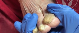
Subungual hyperkeratosis
Subungual hyperkeratosis is a collection of keratinized cells under the outer part of the nail plate.
Subungual hyperkeratosis on the thumb
In psoriasis, subungual hyperkeratosis (from the Greek hyper - excessively and keras - horn) is usually silvery-white in color, but can also be yellow. And when an infection occurs, it can become, for example, greenish or brown.
The more the nail is raised above the nail bed, the higher the activity of the pathological process.
On the fingers, subungual hyperkeratosis usually appears as loose layers under the nail plate. On the feet, these masses are tightly fused to the thickened nail.
Severe subungual hyperkeratosis and psoriatic plaques on the toes
Also, psoriasis with damage to the toenails is characterized by a combination of subungual hyperkeratosis with onycholysis (separation of the nail).
Symptom of an oil stain
The symptom of an oil stain appears under the nail plate in areas of yellow-red (salmon) color.
They appear on the nail bed closer to the nail fold and move toward the edge of the nail as it grows.
Symptom of an oil stain - salmon-colored areas near the area of nail plate separation (onycholysis)
The cause of this symptom is inflammation of the nail bed with dilation of capillaries and accumulation of cells involved in inflammation, as well as cells with impaired division and keratinization.
Oil stains can come in different shapes and sizes. They can occur both in the center of the nail and on the edge, near the area of onycholysis.
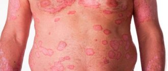
Symptoms of pityriasis
Clinical manifestations of pityriasis rosea are caused by exposure to infectious pathogens and the development of allergic reactions. The skin disease is manifested by the following symptoms:
- General weakness, enlarged lymph nodes, elevated body temperature.
- The formation on the body of small pinkish and mauve spots that have a symmetrical shape and appear along Langer’s lines. Rashes appear on the back, limbs, chest, neck, groin and other parts of the body.
- The appearance on the skin of 2-3 bright red maternal plaques (their diameter is 4 cm), dotted with scales. After a week, small pink rashes form from these large spots.
- Dropout spots spread throughout the body and increase in size (their diameter is 1-2 cm), can peel off, and resemble medallions in shape.
- Severe skin itching.
- Increased irritability.
With proper therapy, the symptoms of pityriasis disappear after 5-8 weeks, and the patient fully recovers. Longer therapy is required if the rash has dense nodules, blisters or papules. In exceptional cases, pityriasis rosea turns into eczema, purulent inflammation of the skin, folliculitis, streptococcal infections, etc. develop. The development of complications is facilitated by the patient’s excessive sweating, a tendency to allergies, constant friction of the skin and improper treatment.
Myth 3: Nail psoriasis is only a cosmetic problem
Actually this is not true. Although more than 90% of patients do complain about the unsightly appearance of psoriasis nails, it is not just a cosmetic problem.
According to various studies, nail psoriasis significantly reduces the quality of life of patients:
- 52% of patients also complain of pain,
- 59% - to problems in daily activities,
- 56% - due to difficulties at home and
- 48% - due to difficulties at work.
Nail psoriasis dramatically reduces the quality of life of patients
Therefore, it is very important to make a correct diagnosis and begin treatment as early as possible, since improving the condition of the nails significantly improves the quality of life of patients with psoriasis.
Can it be cured?
At the current stage of development, medicine is not yet able to cope with genetic diseases, so modern treatment of ichthyosis is aimed at reducing clinical manifestations and alleviating the patient’s condition. For this purpose they prescribe:
- vitamin complexes, iron-containing compounds, immunoglobulin;
- blood plasma transfusions;
- hormone therapy (in difficult cases);
- baths with the addition of various drugs, emollient ointments and creams;
- physiotherapy, spa treatment according to indications.
The choice of treatment methods depends on the degree of skin damage and the characteristics of the disease.
Myth 4: Nail psoriasis is not dangerous
In reality this is not the case. Talking above about the causes of this form of the disease, we have already written that
Nail psoriasis is an important symptom of psoriatic arthritis.
It is important to keep in mind that external manifestations of arthritis may be completely absent. In this case, we can talk not only about the fact that the joints of the fingers and toes are affected, but also the joints of the spine and pelvic bones can be involved.
You can check your joints for arthritis using ultrasound (US) or magnetic resonance imaging (MRI).
This severe form, called arthritis mutilans, occurs in 5% of cases of arthritis in psoriasis
Even if there are no obvious symptoms of arthritis, but there are manifestations of nail psoriasis, it is very important to make sure that all joints are in order.
And then regularly monitor the condition of the joints. Otherwise, psoriatic arthritis can be easily missed! A late diagnosis will lead to late treatment and ultimately to irreversible joint damage and disability.
Therefore, if the doctor did not order insurance tests, citing the absence of visible signs of arthritis, you need to go to the clinic yourself and undergo, for example, an ultrasound scan on a paid basis.
Pityriasis rosea in pregnant women
Pityriasis occurs more often in women than in men. It is especially dangerous when the skin disease occurs in pregnant women. If you notice any rashes, it is important to immediately visit a dermatologist and undergo treatment. It is unacceptable to risk the baby’s health and expect the plaques to disappear on their own. If the disease is not treated, then bacterial infections appear, which are much more difficult to deal with.
If a pregnant woman has not been diagnosed with pityriasis rosea, it is nevertheless important to adhere to the following recommendations:
- clothing made from cotton and linen is preferable to synthetic and woolen fabrics
- limiting heavy physical activity
- For hygienic purposes, use only warm water
- timely moisturizing of damaged skin areas
How to diagnose nail psoriasis
It is important to be able to recognize the many symptoms of nail psoriasis that we described above, as they help to establish the correct diagnosis. But since nail changes characteristic of psoriasis can also occur in other diseases, it can be difficult to immediately make a correct diagnosis.
Onycholysis and leukonychia developed as complications after manicure. Reminds me of psoriasis symptoms
In this case, the presence of several symptoms simultaneously on different nails can help in diagnosis.
Important signs of psoriasis on the nails are:
- thimble symptom: more than 20 pits on all fingernails indicate the possibility of psoriasis, and more than 60 pits confirm the diagnosis of psoriasis,
- detachment of the nail (onycholysis) with a reddish border along the edge,
- oil (salmon) stains on the nail bed.
Difficulties in diagnosing nail psoriasis based on a single symptom
It is especially difficult to diagnose nail psoriasis if it only shows one symptom.
For example, if it manifests itself only as onycholysis on the hands or only as subungual hyperkeratosis on the hands and/or feet.
The only method for making a reliable diagnosis for isolated onycholysis (detachment of the nail) is probably to examine the hyponychium using a special microscope - a dermatoscope.
For this purpose, a videodermatoscope with high magnification is used. Please note that a manual dermatoscope does not provide the necessary magnification. What you need is a videodermatoscope with a magnification of at least 40 times. Then dilated capillary loops characteristic of psoriasis become visible.
Dermatoscopy with 40x magnification: typical dilated capillaries of the hyponychium confirm psoriasis
With isolated subungual hyperkeratosis, the likelihood of psoriasis is high if the accumulation of scales under the nail is whitish-silver in color, as well as if all nails on the fingers or toes are affected.
Psoriasis or nail fungus?
Approximately 30% of patients with psoriasis of the nails also have a fungal infection - scientifically called onychomycosis.
Externally, hyperkeratosis and onycholysis (separation of the nail) in psoriasis may resemble manifestations of a fungal infection. Therefore, it can be difficult to carry out differential diagnosis, that is, to identify the true cause of changes in the nail plate.
Moreover, both psoriasis and fungus can affect the same nails at the same time. This most often occurs on the toes and is primarily characteristic of older patients.
Also, with a fungal infection, one or both nails on the big toes often suffer. With psoriasis, as a rule, several nails are affected at once.
Onycholysis and subungual hyperkeratosis in onychomycosis
The following symptoms speak in favor of psoriasis:
- oil stains and/or thimble symptom on fingernails,
- signs of psoriasis on the scalp and/or large folds of skin,
- periodic remissions and exacerbations of nail damage.
In favor of onychomycosis they say:
- longitudinal stripes on the affected nail,
- detection of fungi when examining a scraping from the affected nail treated with potassium hydroxide under a microscope (KOH test),
- positive culture for fungus.
In general, based on external manifestations alone, it is impossible to completely exclude fungal nail infections in patients with psoriasis.
You also need to remember that a fungal infection can cause Koebner phenomenon on the nail and surrounding skin, which will cause symptoms of psoriasis. So anyway
It is useful to contact a mycologist and conduct research on fungi and, if they are found, begin antifungal therapy.
Diagnostics
If signs of onychodystrophy occur, you should contact a dermatologist who will interview and examine the patient, including using a dermatoscope. It is a compact device that provides a multiple magnification of the surface under consideration.
If necessary, the doctor will additionally prescribe a blood test, nail microscopy and bacterial culture. These tests are mainly required to diagnose infectious complications of onychodystrophy.
Important conclusions and what to do
Let us summarize important information about nail psoriasis and its symptoms.
Diagnostic features
- Nail psoriasis is very common but often goes undiagnosed.
- Manifestations of nail psoriasis can be minor, so even specialists often do not pay attention to it.
- In 5% of cases, nail damage may be the only symptom of incipient psoriasis.
- The manifestations of different diseases on the nails may look the same, which further complicates diagnosis.
The main manifestations of nail psoriasis:
- a symptom of a thimble - pits on the nail,
- white dots,
- crumbling nails,
- red dots in the hole area,
- nail detachment,
- longitudinal subungual hemorrhages,
- subungual hyperkeratosis - loose accumulations under the nail,
- oil stain symptom.
Psoriasis and fungus
- Nail psoriasis is often accompanied by a fungal infection.
- To definitely exclude it, you need to contact a mycologist and conduct additional research.
Nail psoriasis and psoriatic arthritis
- Nail psoriasis is a common accompaniment of psoriatic arthritis.
- It is important to detect pathological changes in joints as early as possible in order to begin treatment on time and avoid irreversible complications and disability.
- Even if there are no external symptoms of arthritis, but nail psoriasis is detected, it is necessary to undergo an examination of the joints using ultrasound or MRI.
Read all about the treatment of nail psoriasis: why medicine only helps temporarily and what can be done to get rid of the disease forever.
- If this article was useful to you, please share it with those who may also benefit from it. And in the comments to the article you can share your experience and thereby help other readers. Thanks a lot! We appreciate your attention!
Appendix: list of references
Some of the scientific articles used:
- Rich P et al. Baseline nail disease in patients with moderate to severe psoriasis and response to treatment with infliximab during 1 year. J Am Acad Dermatol. 2008 Feb;58(2):224-31.
- Zaias N. Embryology of the human nail. Arch Dermatol. 1963 Jan;87:37-53.
- McGonagle D et al. The pathogenesis of psoriatic arthritis and associated nail disease: not autoimmune after all? Curr Opin Rheumatol. 2009 Jul;21(4):340-7.
- Cannavò SP et al. Treatment of psoriatic nails with topical cyclosporin: a prospective, randomized placebo-controlled study. Dermatology. 2003;206(2):153-6.
- Singh SK. Finger nail pitting in psoriasis and its relation with different variables. Indian J Dermatol. 2013 Jul;58(4):310-2. doi:10.4103/0019-5154.113955.
- van der Velden HM et al. Fingernail psoriasis reconsidered: a case-control study. J Am Acad Dermatol. 2013 Aug;69(2):245-52.
- Baran R. The burden of nail psoriasis: an introduction. Dermatology. 2010;221 Suppl 1:1-5.
- Gelfand JM et al. Prevalence and treatment of psoriasis in the United Kingdom: a population-based study. Arch Dermatol. 2005 Dec;141(12):1537-41.
- Gupta AK et al. A higher prevalence of onychomycosis in psoriatics compared with non-psoriatics: a multicentre study. Br J Dermatol. 1997 May;136(5):786-9.