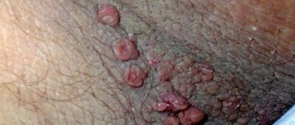
A pressing problem in modern gynecology that many girls and young women face is papillomas on the labia. In these intimate areas on the mucous membrane from some types of HPV, the so-called appear. "genital warts". If a wart on the genital organs is detected or suspected, each patient should know how the disease manifests itself, what treatment should be carried out and what tests should be taken for diagnosis.
These formations are very common. Warts on the labia minora and majora in girls are mainly represented by genital warts, the causative agents of which are types 6 and 11 of the virus. These types of HPV are of low oncogenic risk, that is, the risk of their degeneration into a malignant tumor is low. Children, teenage girls and girls with virgin status cannot be excluded from possible risk groups for the presence of these formations, because sexual is not the only way of transmitting the virus (see more details).
Causes and risk factors for vulvar cancer
Currently, scientists know two mechanisms for the occurrence of vulvar cancer. In young women (especially smokers), tumors associated with infection with the human papillomavirus are most common. Probably, by penetrating a cell, the pathogen can cause changes in genes that lead to the production of abnormal proteins and, as a result, uncontrolled cell proliferation.
Women over 55 years of age most often do not have HPV infection, but they do have a mutation in the p53 gene, which plays an important role in preventing cancer.
In addition to human papillomavirus infection, other risk factors for vulvar cancer are known:
- Age . Women under 50 account for less than 20% of cases, and women over 70 account for more than 50%.
- Smoking . Traditionally, exposure to tobacco smoke has been associated with an increased risk of lung cancer. But in fact, smoking increases the risk of many cancers.
- HIV infection . The immunodeficiency virus weakens the body's defenses and increases the risk of HPV infection.
- Vulvar intraepithelial neoplasia . It is a precancerous disease. Abnormal cells appear in the vulva area, initially they are in the superficial layers of the skin, but over time they can transform into cancer.
- Diagnosed with cervical cancer . There is an increased risk of vulvar cancer.
Prevention
To avoid HPV infection and the formation of papillomas in the vagina, women should adhere to preventive measures and recommendations.
- maintaining intimate hygiene;
- maintaining a healthy lifestyle;
- no abuse of tobacco and alcohol products;
- regular examinations by a gynecologist;
- use of barrier methods of contraception during sexual intercourse;
- increasing immunity (regular consumption of vitamin complexes, minerals and trace elements);
- maintaining a proper balanced diet.
Classification of vulvar cancer
The most common type of vulvar cancer is keratinizing squamous cell carcinoma . As a rule, it develops in older women (over 55 years old). Less common are warty and basaloid (usually in young women), superficial squamous cell carcinomas.
In 8% of cases, malignant tumors of the vulva are represented by adenocarcinomas , which originate from glandular cells. Typically, they develop in the Bartholin glands . These glands are located in the thickness of the labia majora; they moisturize the vagina during sexual intercourse. Tumors of the Bartholin glands are often mistaken for cysts. Sometimes adenocarcinomas develop from cells of the sweat glands, which are located in the area of the external female genitalia.
, basal cell carcinomas can be found in the vulva area .
In 6% of cases, melanomas are found in the vulva area - malignant tumors that originate from cells that produce the pigment melanin. This localization is not entirely typical for melanoma; more often it occurs in open areas of the skin exposed to sunlight.
Classification of vulvar cancer by stages:
- Stage 1 : at stage 1, a tumor that grows within the vulva and does not affect neighboring organs or lymph nodes.
- Stage 2 : In stage 2, the tumor has grown into adjacent structures, such as the urethra, vagina, and anus.
- Stage 3 : Stage 3 is a tumor in which cancer cells have spread to the lymph nodes.
- Stage 4 : with stage 4 vulvar cancer, the damage to the lymph nodes is more pronounced compared to stage 3, tumor growth is observed in the upper part of the urethra, vagina, bladder, rectum, pelvic bones, and the presence of distant metastases.
The stage of the tumor determines treatment approaches and further prognosis.
Find out the exact cost of treatment
Ideal shape and color of the labia: you will need a mirror
Ideal shape and color of the labia
If you look closely at the private parts in the mirror, you will see that the labia are not exactly the same. Just like the face, breasts and even legs are not completely symmetrical. The labia also differ from each other, and sometimes very significantly.
A 2005 study found that about half of women have longer inner labia than outer labia. Of course, this is not beautiful, but this structure of the labia cannot be called a critical defect. Although this upsets many women, especially if the labia minora hang down significantly, and also unevenly.
Unfortunately, such a problem cannot be masked by any means. Therefore, gynecologists offer to “remove the excess” using labiaplasty.
Labiaplasty
As for color, everything is simpler here. Although the labia majora are made of normal tissue and covered by normal skin, they may be slightly different in color from the rest of the body. For example, they may be slightly purple, pink or brown. This is all completely normal. And just as the inside of the lips is a different color, the labia minora are a different color from the larger lips.
In this case, you need to worry only when the labia become red, itchy or painful.
Wearing uncomfortable underwear or pants that are too tight can cause abrasions. So if you have this problem, choose comfortable underwear and switch to skirts for at least a few days until the irritation goes away.
Symptoms of vulvar cancer
Precancerous changes and early stages of vulvar cancer may have no symptoms. When the tumor grows deeper and cancer cells begin to spread to the lymph nodes, almost all women experience some symptoms:
- An area of skin in the vulva that differs from the surrounding healthy tissue: it may be lighter or darker, reddish or pink.
- A lump, plaque, wart, or ulcer appears on the skin.
- Itching, burning, and pain occur.
- Many women are concerned about bleeding from the vagina outside of menstruation.
Most often, these symptoms are not caused by cancer. But there is always a small chance that it is cancer.
Adenocarcinoma, which develops from the Bartholin gland, appears as a mass in the labia that can be felt. A cyst manifests itself with similar symptoms.
Clinical signs
The disease usually develops gradually. In the initial stages, a compaction nodule can be detected on the labia. There may be several such nodules. The patient usually complains of pain or severe itching in the groin area. Next, clinical signs increase in the following sequence:
- Inflammatory nodules quickly increase in size. The affected area swells, turns red, and the local temperature rises.
- A purple, pear-shaped formation forms on the labia. The nodes are tightly fused to the skin. At this stage of the disease, the patient’s general condition suffers - the temperature rises, malaise, weakness, and headache develop.
- The maturation stage is accompanied by softening of the nodes and a breakthrough of purulent discharge. The pus may contain blood. The patient's general condition improves after the pus breaks through.
- After healing of some nodes of suppuration, others may develop in the neighborhood. Thus, the process can become chronic.
If you notice the first signs of the disease, you should not postpone your visit to the doctor. An experienced surgeon at our clinic will prescribe the necessary examinations and provide effective treatment.
Useful information about visiting a surgeon at the clinic:
- How to prepare for a surgeon's appointment
- What diseases does the surgeon treat?
- Calling a surgeon to your home
- Surgical care in the clinic
- Surgical care at home
- What symptoms should you contact a surgeon for?
- Treatment of surgical diseases
- Treatment of intestinal pathologies
- Treatment of skin surgical pathologies
- Treatment of bedsores and necrosis
- Treatment of parasitic diseases
- Treatment of inflammatory processes of soft tissues
- Treatment of diseases of the musculoskeletal system
- Diagnosis of surgical diseases
Diagnosis of vulvar cancer
The main method for diagnosing vulvar cancer is biopsy. Examining a tissue sample under a microscope helps to accurately distinguish benign from malignant tumors. In order to understand where to take tissue from, the doctor examines the vulva using a colposcope and treats the skin with a solution of acetic acid or toluidine blue: as a result, pathological foci become more visible.
According to indications (if the tumor is large and there is a suspicion that it has spread beyond the vulva), other diagnostic methods are prescribed:
- Endoscopic examination of the bladder and rectum.
- CT and MRI help to detect foci of tumor growth in the lymph nodes and various organs.
- A chest x-ray is performed to look for metastases in the chest.
- PET scanning is an effective method for searching for distant metastases.
Swelling of the penis and testicles.
Swelling of the genital organs can be caused by both completely harmless factors (passionate sex, tight underwear, allergies, poor hygiene) and serious reasons. Therefore, if the swelling does not subside within a day or additional symptoms appear (itching, burning, redness, discharge, rash, inability to open the foreskin), you should consult a specialist.
Why does the penis and/or testicles swell?
Various reasons can cause swelling of the penis and testicles:
Balanitis, posthitis, balanoposthitis.
Balanitis (inflammatory disease of the head of the penis) and posthitis (inflammatory process of the foreskin) are usually inseparable, which is why they are combined under the common term “balanoposthitis.” In a man, erosions form on the head of the penis and in the area of the foreskin, pain, swelling, and hardening of the inguinal lymph nodes are possible.
Cavernite.
Pathological processes in the cavernous bodies cause swelling of the shaft of the penis.
Phimosis.
Due to the narrowing of the foreskin, the head of the penis is not fully released, which leads to inflammation and swelling.
Paraphimosis.
Pinching the head of the penis by the foreskin causes redness and swelling of the organ, and pain when urinating. In the absence of timely medical care, tissue necrosis is possible.
Thrombophlebitis.
As a result of inflammation of the dorsal vein, the penis swells and the temperature rises sharply. Physical activity and movement cause pain.
Allergy.
Itching and swelling of the penis can be caused by synthetic underwear, poor-quality lubricants, condoms, and long-term use of antibiotics.
Microtraumas.
The slightest bruise can provoke swelling of the penis and the appearance of a hematoma, which is explained by the increased sensitivity of the organ. Sometimes the cause of microtrauma can be a strong impact on the penis during sexual intercourse or masturbation, or wearing too tight underwear.
Infections.
Sexually transmitted diseases (trichomoniasis, chlamydia, gonorrhea, syphilis) often cause swelling of the genital organs. With syphilis, a growing compaction also becomes noticeable, and the general condition worsens. Swelling of the penis is also possible with systemic infections (tuberculosis).
Tumor.
The cause of swelling can be a malignant neoplasm, but swelling of the entire trunk is extremely rare.
Surgical treatment of vulvar cancer
Surgery is the main treatment for vulvar cancer. During the operation, the doctor tries to completely remove the tumor tissue, while maintaining the aesthetic appearance of the genital organs, normal evacuation of urine and stool (this is especially important when the tumor is close to the urethra and anus). Depending on the stage, one of the following surgical options is used:
- Local excision of the tumor. Possibly in the early stages. The surgeon excises the tumor and approximately 1.3 cm of surrounding tissue and subcutaneous fat. The removed tissue is sent to a laboratory for microscopy. No tumor cells should be found at the edges of the incision (negative resection margin) - this will indicate that the tumor has been completely removed, and most likely there are no more cancer cells left in the body.
- Vulvectomy . The surgeon removes all or most of the vulva. There are different options for vulvectomy. In some cases, after surgery, it is possible to maintain the normal appearance of the genital organs; in some women, it is subsequently necessary to resort to reconstructive plastic surgery.
- Evisceration (exenteration) of the pelvis is the most complex and traumatic operation; it is performed if the cancer has spread to the pelvic organs. The bladder, lower part of the colon and rectum, and the uterus along with the cervix and vagina are removed. The volume of intervention depends on where the tumor has managed to grow.
- Removal of lymph nodes . Previously, it was always carried out - just in case, since the surgeon could not know for sure whether the tumor cells had spread to the nearest lymph nodes. Currently, it is possible to conduct a sentinel biopsy - a study of the lymph node, which is the first on the path of lymph outflow from the tumor. If there are no cancer cells in it, then it makes no sense to remove the remaining lymph nodes.
Sometimes, if there is a high risk of relapse after surgery, surgical treatment is combined with a course of radiation therapy and chemotherapy.
Write to an oncologist
Radiation therapy for vulvar cancer
Radiation therapy is used for vulvar cancer in the following cases:
- Before surgery in combination with chemotherapy. This helps to reduce the tumor and convert inoperable cancer into operable one.
- After surgery to prevent relapse.
- Radiation therapy can be used as the only treatment for inguinal and pelvic lymph nodes.
- It is used alone or in combination with chemotherapy to treat women who are contraindicated for surgery.
Causes of synechiae
- Genitourinary infections - vulvovaginitis, vulvitis, etc. Infection is possible in the maternity hospital or at home, when swimming in stagnant natural bodies of water.
- Violation of intimate hygiene rules. Both a lack of hygiene and its excess are harmful.
- Allergy in a child. A reaction can occur to food (from the diet of a girl or a nursing mother), components of care products, washing powder, etc.
- Difficulties during pregnancy - intrauterine infections, severe toxicosis. If a mother had difficulties during pregnancy, her daughter increases the risk of developing synechiae.
- Genetics. The tendency to fusion of soft tissues can be transmitted from mother to daughters.
- Wearing tight synthetic underwear, which creates all the conditions for inflammation.
- Reduced estrogen levels. This is normal before puberty, but in some girls it causes synechiae.
- Dysbacteriosis. Pathogenic intestinal flora can provoke adhesions. Source: Z.K. Batyrova, E.V. Uvarova, L.S. Namazova-Baranova, N.Kh. Latypova, A.E. Donnikov Fusion of the labia minora in girls during early childhood: tactics of a pediatric gynecologist // Issues of modern pediatrics, 2012, vol. 11, no. 2, pp. 118-121
Prognosis for vulvar cancer
How long do people live with vulvar cancer?
Even if treatment is successful and remission occurs, the woman remains at risk of cancer recurrence in the future. Therefore, all patients require long-term observation, over several years. Prognosis is assessed by 5-year survival rate, an indicator that shows the percentage of women surviving 5 years after diagnosis. This indicator depends on the stage of the cancer and how much it has spread beyond the vulva:
- For local cancer that is located within the vulva (corresponding to stages I and II), the 5-year survival rate is 86%.
- For regional cancer, when tumor cells have spread to the lymph nodes and neighboring organs (corresponding to stages III and IVA) - 54%.
- For advanced cancer, when there are distant metastases (corresponding to stage IVB) - 16%.
Can vulvar cancer be prevented?
The main measures to prevent vulvar cancer are to prevent infection with the human papillomavirus. It is necessary to limit the number of sexual partners (ideally there should be one), use condoms. The vaccine effectively protects against dangerous strains of HPV.
Since vulvar cancer is more common among women who smoke, quitting the bad habit is an effective preventive measure.
Even if a woman leads an absolutely “correct” lifestyle, this does not guarantee that she will not get sick. Therefore, early diagnosis is important and you need to regularly visit a gynecologist.
Prices for treatment of vulvar cancer at Euroonco
The cost of treatment for vulvar cancer depends on the stage of the tumor, the extent of surgery, and whether it needs to be supplemented with chemotherapy, radiation therapy, and other types of treatment. At Euroonko you can get care at the level of leading Western oncology centers, but at a lower price.
Appointment with an oncologist-gynecologist - 6,900 rubles. Photodynamic therapy - from 170 rubles (calculated depending on the patient’s weight).
Book a consultation 24 hours a day
+7+7+78