According to official international statistics on the prevalence of fungal infections
For many years in a row, they have been ranked second among all skin diseases. They not only reduce the quality of life and cause discomfort, but also provoke allergic reactions, chronic inflammatory processes and many other pathologies.
Due to the peculiarities of their microbiological nature, fungal diseases can be actively transmitted from a carrier to a healthy person, and in addition to the skin, they can affect the nail plates, hair, internal organs and cause various pathogenic changes in them. Some fungi are prone to relapses, have a fairly long incubation period and very similar external manifestations. At the same time, some types of mycoses are treated only with oral medications, others - with cutaneous forms, so it is strictly forbidden to self-medicate and contact specialists at the first symptoms.
When are tests for fungal infections prescribed?
External signs can only make a primary diagnosis of a fungal disease. Therefore, in case of redness of the skin, focal lesions of the smooth and scalp, as well as itching in the foot or skin folds, changes in the shape and color of the nail plate, dermatologists prescribe laboratory tests. This allows you to correctly diagnose the nosological form, and, therefore, select the most effective treatment package, take the necessary measures to localize the infection and minimize possible side effects.
Immunological and biological studies
Immunological research methods are used to identify specific changes in the body and serological diagnosis of fungal diseases. To detect specific antibodies in serum samples, the following serological reactions are carried out: agglutination, precipitation, complement fixation, immunofluorescence with the corresponding antigens. The allergic condition of the patient's body is detected using allergic skin tests. Allergens are applied to scarified skin according to Pirquet or by rubbing into the skin according to Moreau, intradermally according to Mantoux, or by injection into the skin. With the help of these tests, allergic reactions of both immediate and delayed types are detected, which makes it possible to assess the state of humoral and cellular immunity. To identify specific sensitization of lymphocytes, reactions of basophil degranulation, agglomeration and alteration, blast transformation test, suppression of macrophage migration, etc. are used. Comparison of the results of serological and allergic reactions turns out to be useful both for diagnosis and for prognosis of the course of mycoses. Biological method. Used for laboratory diagnosis of deep and especially dangerous mycoses. It is based on the infection of animals with pathological material from a patient or a culture of the fungus being studied. Carried out in special laboratories.
Types of fungal diseases
Microscopic pathogenic fungi in medical practice have a common name - mycoses (Greek mycosis). Today, more than 100 types of parasitic and pathogenic microfungi are classified; we will highlight the main infections that affect adults and children.
Dermatomycoses
Common fungal diseases that affect the skin, nails and hair. The source of infection can be a person or an animal. They manifest themselves in various symptoms; we will indicate only the most common diseases in our geographical area:
- Rubromycosis
is a disease caused by the anthropophilic fungus Trichophyton rubrum. It is characterized by a variety of clinical manifestations and localization of lesions on any part of the body; it can affect smooth skin, hair follicles and nails; - mycosis of the foot
(athlete's foot), which also affects the interdigital folds. Very similar to candidiasis, and sometimes polymycosis infection occurs; - favus
is a rare form accompanied by severe baldness of the head. Can be transmitted through combs, underwear and shaving and haircutting tools; - microsporia
– trichomycosis, the causative agent of which is the microsporum fungus. On smooth skin it appears as red spots of a clear shape with a peripheral ridge, and on the scalp – as small flaky lesions; - trichophytosis
(synonymous with ringworm). Externally manifested by pink-red focal skin lesions on any part of the body; - epidermophytosis of skin folds.
Accompanied by itching, redness and peeling.
Keratomycosis
Mycoses are predominantly of the stratum corneum. Among them, the most famous are piedra, erythrasma and pityriasis versicolor. Superficial mycoses are often ignored by many ordinary people, because if lichen versicolor can be primarily identified by yellowish-brown spots covered with pityriasis scales, then erythrasma is often perceived as age-related darkening of the skin. This is due to the fact that the disease progresses slowly and is localized on the inner thighs, in the groin folds and under the mammary glands in women. Faint darkening of the skin affected by erythrasma is covered with small pityriasis-like scales, and although the disease causes almost no itching, it sharply reduces the protective properties of the skin and spoils the appearance of the infected person.
Candidiasis
Diseases caused by yeast-like fungi of the genus Candidosis, which includes more than 150 species. They are considered the most dangerous and most common, as they are found throughout the world and can be transmitted from people, birds and pets. Moreover, in addition to skin manifestations, candidiasis can:
- affect mucous membranes;
- penetrate deeply into tissues and organs;
- cause septic diseases and allergic changes in the body.
Candidiasis can manifest itself as a localized and widespread rash on the hands and feet, lesions of the nail folds and scalp, in the form of stomatitis, cheilitis and gingivitis. With internal infection, they cause vulvovaginitis, urethritis, pleuropneumonia, endocritis, meningitis and other diseases.
Visceral and systemic mycoses
Fungal infections of internal organs caused by infection with taxonomic microfungi and accompanied by severe damage to the skin, visceral (internal) organs, subcutaneous tissue, nervous system and even the musculoskeletal system. This group includes more than two dozen fungi, among which there are pathogenic and conditionally pathogenic. The most common are actinomycosis, aspergillosis, histoplasmosis, coccidiosis, sporotrichosis, chromycosis and others. As a rule, deep mycoses are transmitted indirectly and are difficult to diagnose. Some pathogens demonstrate amazing survivability and resistance to drug therapy, high pathogenicity and significant contagiousness and can lead to disability and death. Most systemic mycoses act as opportunistic infections in patients with hepatitis and AIDS.
Fungal infections are damage to the skin, nails, mucous membranes, and internal organs caused by pathogenic fungi.
Fungal microorganisms are widespread in the environment, some of them are constantly present in the human body (for example, candida is part of the normal microflora of the oral cavity and intestines).
There are infections that are caused by pathogenic fungi that live only in certain areas (endemic mycoses). In such cases, the main routes of infection into the human body are:
- aerogenic – when inhaling spores of pathogenic fungi; the first manifestation of infection may be pneumonia;
- contact – the development of skin lesions as a result of direct contact of the skin with soil containing pathogenic fungi.
With a decrease in the body's defenses and immunodeficiency states (for example, in patients with HIV), opportunistic fungal infections can develop. They occur with increased growth and reproduction of fungi that are constantly present in the body.
The most common group of mycoses are superficial mycoses of the skin and nails. The causative agents of these infections can be transmitted both from person to person and as a result of the spread of pathogens to the skin from the environment. However, the disease does not develop in all cases, since susceptibility to fungal infections may vary from person to person.
Fungi multiply intensively in warm and humid conditions, which causes their frequent localization on the skin of the toes, feet, skin folds, and nails.
Antifungal medications are available to treat infections caused by fungi. Depending on the type of infection and the severity of the disease, these drugs can be used in the form of injections, tablets, ointments, creams, and solutions for topical use.
Synonyms Russian
Mycoses.
English synonyms
Fungal Infections, Mycoses.
Symptoms
Fungal infections of the skin and mucous membranes can manifest themselves with the following symptoms:
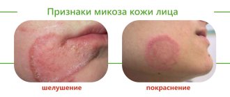
- redness of the skin;
- itching, burning;
- skin rashes;
- weeping on the affected areas of the skin (separation of serous fluid through the smallest defects of the epidermis);
- small blisters on the skin that can burst and form crusts;
- peeling of the skin;
- painful sensations in the area of damage to the skin, mucous membranes;
- plaque on the mucous membranes (for example, white cheesy plaque in the mouth, vagina with candidiasis);
- hair loss in the affected areas.
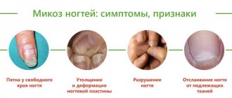
Symptoms of fungal nail infection:
- thickening of nails;
- fragility of nail plates;
- distortion of the shape of nails;
- loss of shine, change in nail color (for example, darkening or yellowing);
- painful sensations in the nail area.
In primary and endemic mycoses, the clinical picture depends on the prevailing involvement of certain organs in the infectious process. For example, with coccidioidomycosis, inhalation of fungal spores can affect the lungs. As a result, they develop:
- cough;
- increased body temperature;
- dyspnea;
- chest pain.
General information about the disease
Fungal infections are diseases of the skin, nails, mucous membranes, and internal organs caused by various types of fungi.
Some types of fungi (for example, candida) are constantly present in the human body without causing harm to health.
Under favorable conditions, these fungi begin to actively multiply, which leads to the development of a fungal infection. Factors contributing to the development of fungal diseases include:
- warm and humid environmental conditions;
- failure to comply with personal hygiene rules;
- increased sweating;
- tight clothing, shoes that do not allow air to pass through well;
- taking antibiotics - they can destroy not only pathogenic, but also beneficial bacteria that form normal microflora, this is fraught with increased growth and reproduction of fungi;
- decreased efficiency of the immune system: the immune system resists pathogens of various diseases; its work may be disrupted when taking certain drugs (for example, glucocorticoids), endocrine diseases (for example, diabetes), or damage to the immune system itself (as with HIV).
The most common forms of fungal infection are local lesions of the skin, mucous membranes, and nails. This group of diseases includes:
- Candidiasis. Candida is a yeast-like fungus. They are permanent inhabitants of the skin and mucous membranes. With a decrease in local and systemic defenses of the body, candida can cause local and generalized (damage to the entire body involving internal organs) fungal infection. Systemic candidiasis most often develops in people with immunodeficiency (for example, in patients with HIV). Cutaneous candidiasis most often develops in areas such as the armpits, gluteal folds, under the mammary glands, and in the spaces between the fingers. In these places, favorable conditions are created for the existence of candida: increased temperature and humidity.
The disease manifests itself in the form of areas of redness of the skin, rashes (small blisters filled with liquid). In the presence of skin diaper rash, erosions (damage to the surface layer of the skin) sometimes occur. These manifestations may be accompanied by burning and itching.
When the periungual skin ridges are affected, swelling and redness of the skin around the nail develops. The spread of infection under the nail plate can cause gradual thickening and discoloration of the nail plates. Onycholysis (detachment) and even nail loss are less common.
- Candidiasis of the mucous membranes. With candidiasis, white plaques and areas of redness of the mucous membrane form in the oral cavity and oropharynx. Painful cracks appear in the corners of the mouth.
Damage to the mucous membranes of the genital organs (usually the vaginal mucosa in women) is accompanied by the formation of a white cheesy coating and discharge. At the same time, a burning and itching sensation is felt in the vagina.
- Dermatophytosis. Dermatophytosis is caused by molds that use the keratin (protein) of the skin, hair, and nails to survive. The infection can be transmitted from people, animals, through contact with infected socks, shoes, or when visiting swimming pools or baths. It is manifested by hair loss in limited areas, peeling, redness of the skin, and itching. When the nail plates are damaged, the nails thicken, their shape and color change, they can become brittle and crumble.
- Pityriasis versicolor. This fungus causes white spots to appear, which become especially visible after sunbathing. There are usually no other symptoms.
The causative agent of pityriasis versicolor is part of the normal microflora of the skin. The disease can manifest itself under certain conditions, for example when the immune system is weakened, during pregnancy.
There are mycoses that are predominantly distributed in certain areas (endemic mycoses). One of them is coccidioidomycosis, the highest incidence of which is noted in the southwestern United States and northern Mexico. The infection can be transmitted by inhaling spores of pathogenic fungi or by skin contact with soil. The disease is characterized by damage to the lungs and skin.
In people with immunodeficiency, fungi that do not cause any symptoms in a healthy person can lead to systemic mycoses. Such infections are called opportunistic.
The causative agents of opportunistic systemic mycoses can be candida, aspergillus and other fungi. The clinical picture consists of damage to the skin and lungs with possible further spread to internal organs: brain, kidneys, liver, gastrointestinal tract, heart valves.
These forms often develop against the background of severe damage to the immune system and significantly reduced body resistance, so the prognosis in many cases is unfavorable.
Who is at risk?
- Sweating intensely.
- Individuals who have a weakened immune system (for example, patients taking glucocorticoid medications)
- Suffering from diabetes.
- Working in damp, warm conditions.
- Those who walk barefoot in public places (gyms, swimming pools).
- Wearing clothes, socks, shoes that have poor ventilation ability.
- Neglecting the rules of personal hygiene, using other people's towels, shoes, and bed linen.
- Have completed a course of antibacterial therapy.
- Located in areas where the incidence of a certain type of fungal infection is endemic.
Diagnostics
Diagnosis of fungal infections consists of identifying characteristic clinical signs of the disease, conducting laboratory tests to identify the type of fungal infection that causes the fungal infection, which is important for further treatment.
Laboratory research
- Determination of the genetic material (DNA) of the causative agent of fungal infection in material (urogenital smear, biopsy, bronchoalveolar lavage (wash water), blood) by polymerase chain reaction (PCR).
This method is the most specific and informative for diagnosing many types of fungal infections. Its essence is to detect fungal DNA in material samples, which highly accurately confirms the presence of a pathogen in the material under study.
In the blood, pathogens of fungal infections are found in generalized forms of fungal infection, which can develop against the background of immunodeficiency states.
- Determination of IgG class antibodies to pathogens of fungal infections. In response to the penetration of infectious agents into the human body, the immune system produces antibodies, or immunoglobulins. They come in different classes (eg, IgA, IgG, IgM).
IgG is synthesized some time after the pathogen enters the body and persists for a long time. In this regard, an increase in this indicator may indicate the presence of an acute or past disease. A marked increase in IgG levels can occur with systemic mycoses.
- Sowing on nutrient media with determination of sensitivity to antifungal drugs.
The method consists of inoculating the test material (for example, urogenital smear material) on special nutrient media that promote the growth of fungi. This analysis allows not only to identify the causative agent of a fungal infection, but also to select the most effective antifungal drugs. This is of great importance for further treatment of the disease.
- Analysis of vaginal microbiocenosis using polymerase chain reaction. Many forms of fungal infections develop against the background of disruption of the normal composition of the microflora. Using this analysis, it is possible to determine with high accuracy and in the shortest possible time the quantitative and qualitative species composition of the main representatives of the vaginal microflora and select the optimal therapy - the most effective and with the minimum number of side effects.
Depending on the severity and prevalence of the fungal infection, it may be necessary to conduct clinical tests of blood, urine, cerebrospinal fluid, and determine the biochemical parameters of the body’s activity (for example, liver tests, indicators of nitrogen metabolism, water-electrolyte composition of the blood, etc.).
Other studies
- Radiography. Using chest x-ray, you can assess the extent of damage to lung tissue due to fungal infections. A fairly simple but highly informative study.
- Ultrasound examination of internal organs (ultrasound). The principle of the method is based on the action of ultrasound. Allows you to visualize internal organs and identify pathological space-occupying formations (for example, abscesses) that can occur when internal organs are affected by a fungal infection.
- Computed tomography (CT). CT allows you to obtain highly informative layer-by-layer images, which is of great importance in the diagnosis of damage to internal organs, including fungal infections.
Treatment
Treatment for fungal infections involves the use of antifungal medications. Depending on the severity and type of infection, medications may be prescribed in the form of injections, tablets, ointments, creams, and solutions for topical use. The duration of the course of therapy is determined by the attending physician.
Prevention
To prevent fungal infections, the following principles should be observed:
- wear fresh underwear, change socks daily;
- do not wear wet clothing (eg swimsuits, sportswear);
- observe the rules of personal hygiene, do not use other people’s towels, clothes, bed linen, shoes;
- do not wear shoes that are too narrow;
- wipe your hands and feet dry after bathing;
- do not walk barefoot in gyms and swimming pools;
- Use antibiotics only as prescribed by your doctor.
Recommended tests
- Candida albicans, DNA [real-time PCR]
- Candida albicans, IgG, titer
- Culture of Candida spp./yeast-like fungi with selection of antimycotic drugs
- Analysis of vaginal microbiocenosis. 8 indicators, quantitative DNA [real-time PCR] (urogenital smear)
- Aspergillus fumigatus, DNA [PCR]
- Aspergillus fumigatus, IgG
Literature
- Dan L. Longo, Dennis L. Kasper, J. Larry Jameson, Anthony S. Fauci, Harrison's principles of internal medicine (18th ed.). New York: McGraw-Hill Medical Publishing Division, 2011. Chapter 198. Diagnosis and Treatment of Fungal Infections.
Methods for diagnosing fungal infections
The Northwestern Center for Evidence-Based Medicine offers traditional and innovative research methods. We carry out the following analyses:
Microbiological methods
Microscopy
– the most accessible and simplest diagnostic method. The study is aimed at confirming infection with superficial mycosis, for which biological material is taken from the patient, which is potentially considered infected: scraping of a pathologically changed nail or skin, eyelash, hair. Refers to qualitative analyzes and allows only to establish or refute the fact of infection. The study takes several days: the resulting material is processed (stained) in a certain way and submitted for microscopic examination to identify the elements of the fungus (spores, hyphae). Microscopy allows you to quickly confirm mycosis, but the type of pathogen and its quantitative concentration are established only for yeast-like and mold species. Therefore, it is often supplemented with cultural research (bacterial culture). The results of microbiological studies must be interpreted by the treating specialist.
Enzyme-linked immunosorbent assay (ELISA)
Linked immunosorbent assay
– a modern and highly reliable method for identifying fungi in a patient’s venous blood. It is a qualitative and quantitative method and can be used as a primary diagnosis and act as a confirmatory analysis of superficial and visceral mycoses.
The method is based on the detection and identification of immunoglobulin protein to a specific pathogen. Antibodies and antigens provide reliable information about infection with aspargillosis, candidiasis, cryptococci and dimorphic microfungi. The test result is interpreted as “positive” (there is an infection) or “negative” (there is no fungal infection). In some cases, the study may give a questionable result; as a rule, this happens if the patient has recently taken antibacterial drugs.
The study takes from 1 to 5 days. If it is necessary to identify the dynamics of the disease, ELISA is carried out every 14 days.
PCR
A highly accurate research method based on the polymerase chain reaction and taking no more than 3 days. It can be used to register any fungal pathogens, but has one drawback - targeted research. This means that the laboratory must obtain information about the specific species of microfungus whose spores and hyphae need to be identified. Blood, sputum, prostate secretion or urine can be provided for analysis, but in the last three options it is necessary to ensure maximum purity of the biological material. The most effective and appropriate in the complex diagnosis of systemic and visceral forms of fungal diseases. The study provides qualitative and quantitative results that are interpreted only by the attending physician.
Serological method
Classic studies in which IgG prepitins, enolase, proteinase and mannoprotein antigens are more often detected. The study is variable and can be based on the agglutination reaction, titration and RSC. They allow you to obtain information only about the fact of carriage of a mycotic infection or indicate a previously suffered fungal disease. The analysis may be based on blood serum testing. With extensive serodiagnosis, microfungus can be detected in other physiological fluids of the patient.
The results are interpreted by the treating specialist. In this case, the serological method is often used as a control study for subsequent adjustment of therapy and determining the effectiveness of treatment.
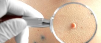
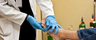
What tests are done for nail fungus?
The main analysis is a scraping for fungus. This procedure is completely painless and is performed by a mycologist. The scraping is obtained using scissors, a scalpel or a dissecting needle. The choice of instrument depends on the area of the nail from which the material for examination is taken.
The material obtained by scraping is transferred to glass and examined using a microscope for the presence of fungal spores. If spores are detected, the next step is to inoculate the material to determine the type of fungus. After the results of scraping and culture are known, the mycologist prescribes individual treatment for the patient. Please note that on average it takes approximately three weeks from the scraping to the culture results.
Risk groups and prevention of fungal infections
Fungal pathogens are found in minimal quantities on the skin of any person. But uncontrolled use of medications, especially hormones and antibiotics, can provoke their active growth and subsequent lesions. It should also be taken into account that deep mycotic infections can enter the body through open wounds. Compliance with sanitary rules for treating any injuries associated with a violation of the skin minimizes the risk of disease with systemic and visceral microfungi.
High humidity and constant above-zero temperatures are an ideal environment for the life and reproduction of microscopic fungi. Accordingly, the risk group a priori includes employees and visitors of swimming pools, fitness clubs, bath complexes, spa salons, as well as workers in laundries and catering establishments.
People with weakened immune systems, a depressed nervous system, a tendency to allergies, critical weight loss and metabolic disorders are also prone to fungal infections. Therefore, the most effective prevention is strengthening the immune system, impeccable adherence to personal hygiene rules and systematic examinations by a therapist and dermatologist.
Cultural examination
The material is inoculated on standard Sabouraud medium, often with added antibiotics. In the diagnosis of dermatophyte infections, it is customary to add cycloheximide to Sabouraud’s medium, which suppresses the growth of contaminant fungi coming from the air. Ready-made commercial media with added antibiotics and cycloheximide are available. It should be remembered that many non-dermatophyte molds and some Candida species do not grow on medium with cycloheximide, so it is recommended to inoculate on Sabouraud's medium with cycloheximide and on medium without it. Identification of species is usually carried out by microscopic examination of the grown culture or by subculture on selective media (Fig. 2-15). Rice. 2. Culture of the fungus T. rubrum isolated from affected nails. Obtained on Sabouraud medium (left) and corn agar (right) Fig. 3. Culture of the fungus T. mentagrophytes var. interdigitale isolated from affected nails. Obtained on Sabouraud medium. Rice. 4. Culture of the fungus Candida albicans. Obtained on Sabouraud medium. Rice. 5. Culture of the fungus Torulopsis glabrata isolated from affected nails. Obtained on Sabouraud medium. Rice. 6. Culture of the fungus Ulocladium sp., isolated from affected nails. Rice. 7. Micromorphology of Acremonium sp. isolated from affected nails. Rice. 8. Micromorphology of Fusarium sp., isolated from affected nails. Rice. 9. Micromorphology of Scopulariopsis sp. isolated from affected nails. Rice. 10. Micromorphology of Candida albicans isolated from affected nails. Rice. 11. Micromorphology of Altemaria sp. isolated from affected nails. Rice. 12. Micromorphology of Aspergillus sp. isolated from affected nails. Rice. 13. Micromorphology of Ulocladium sp isolated from affected nails. Rice. 14. Micromorphology of Chaetomium sp. isolated from affected nails. Fig. 15. Panel of nutritional compounds for the identification of dermatophytes (on the left - T rubrum culture, on the right - T mentagrophytes var. mterdigitale). From left to right: Sabouraud's medium, Baxter's medium, Christensen's medium, corn agar It should be noted that some molds, including dermatophytes, grow slowly in culture, within 2-3 weeks. Even if all the rules for collecting material are observed, with good equipment of the laboratory and highly qualified personnel, the number of positive results of cultural research is very small. According to foreign literature, the percentage of positive studies does not exceed 50. The percentage of positive results in the best domestic laboratories barely reaches 30. Thus, in 2 out of every 3 cases of onychomycosis, its etiology cannot be established.
Price
The coordinated work of all employees and structural divisions of the center, combined with the rational organization of laboratory and functional studies, allows us to create favorable competitive prices for all types of services. At the same time, the Northwestern Center for Evidence-Based Medicine has installed ultra-modern equipment and uses a unique coding system for biological material, which makes it possible to accurately identify pathogens of fungal diseases and conduct all research promptly and strictly confidentially.
Where can I get tested for nail fungus?
We invite you to get tested for nail fungus in Moscow by a mycologist at our clinic. We guarantee confidentiality to all patients who contact us. Our mycologist will take tests and prescribe treatment based on their results. To get rid of fungus, we use unique German equipment - the PACT Med device.
PACT Med destroys the fungus using light waves. This method of fighting fungus is called photodynamic antimicrobial therapy. The uniqueness of this technique lies in the fact that light waves successfully affect any type of fungus, while classical treatment does not always give excellent results and involves taking drugs that are harmful to human health.
Photodynamic antimicrobial therapy is painless and has no contraindications. It does not require additional use of antifungal agents, so it is prescribed even to pregnant women and children.
Do you want to make an appointment with a mycologist for testing?
Sign up by phone. (free call within Russia) or leave a request on the website.
Preparation and collection of biomaterial for analysis
The patient should prepare in advance for the upcoming manipulation: a few days before the date of the study, hygiene procedures should not be carried out; It is prohibited to use cosmetics and apply them to the places from which the biomaterial will be taken; for a sample near the nail plates, you cannot first apply coloring varnishes and cosmetics to them; you should stop using certain medications (it is advisable to consult a dermatologist about the use of medications).
Biomaterial from the affected area of the epidermis is taken with a sterile scalpel or special instruments. It is necessary to separate the damaged skin near the inflammatory and affected area, since in this place the concentration of microorganisms will be maximum; to scrape the area between the fingers, it is necessary to take the stratum corneum.
After taking a scraping, a special solution is added to the biomaterial to create an alkaline environment and destroy the keratin. A day later, the sediment in a container or test tube is examined using various methods.
Best materials of the month
- Coronaviruses: SARS-CoV-2 (COVID-19)
- Antibiotics for the prevention and treatment of COVID-19: how effective are they?
- The most common "office" diseases
- Does vodka kill coronavirus?
- How to stay alive on our roads?