Lipoma - what is it?
Lipoma is a benign fatty tumor. Lipoma can be limited capsularly from other tissue, or it can be diffusely present in tissues.
This tumor occurs in females aged from thirty to fifty years (middle age).
A tumor forms in areas of the body that are rich in adipose tissue. This can be: subcutaneous tissue, skin, adipose tissue near the kidneys, fat behind the peritoneum, muscle areas of adipose tissue, intestines, mammary gland, as well as the meninges, lungs, myocardium, bones and nerve trunks. Often the latter are formed from embryonic tissue if the tissue falls into the wrong area during formation and development.
By structure
Lipomas according to these characteristics are divided into:
- Classic (there is only fatty tissue inside);
- Angiolipomas (have vessels inside);
- Hibernomas (in the fiber there are formations similar to the formations of hibernating animals);
- Myelolipomas (hematopoietic and adipose tissue are located together);
- Myxolipomas (contain mucous tissue elements inside);
- Myolipomas (muscle fibers are found together with fatty tissue);
- Fibrolipomas (there is connective and fatty tissue inside).
To sum up all of the above, we can say
- Lipoma and atheroma are benign formations of different natures - lipoma simply consists of altered adipose tissue, and atheroma is made of a sebaceous gland with a capsule filled with secretion - sebaceous atheromatous masses.
- Conservative, incl. Treatment of lipoma with folk remedies, as well as treatment of atheroma, is absolutely ineffective and often harmful.
- A small (2-3 cm) lipoma can not be operated on, but observed. In case of growth, as well as any discomfort, surgery to remove the lipoma is indicated.
- Removal of atheroma is always desirable, because they tend to increase in size and fester.
- If you find a subcutaneous formation in yourself, you need to consult a doctor, because... under the guise of a lipoma or atheroma, other formations can develop - dermatosarcomas, liposarcomas, hygromas, lymphadenitis, etc.
Dr. Elshansky I.V. has been involved in the diagnosis and surgical treatment of benign formations of the skin and subcutaneous tissue for many years.
By appearance
Based on how the tumor looks, lipomas can be divided into:
- Ring-shaped (location – neck, similar to a necklace);
- Tree-like (located inside the joints);
- Capsule-shaped (with a dense shell and clear edges);
- Diffuse (without capsule, clear boundaries);
- Ossified or petritic (when calcium deposits form inside);
- Pedicled (with a pedicle where the vessel and nerve are located);
- Dense;
- Soft.
The reasons may be as follows:
- Neutrophic;
- Lack of nutrition to the nerves in a certain area.
Lipoma contains fat cells of different sizes. Between the simple fat cells there are rudimentary ones, which affect the growth of the former.
Lipomas grow slowly. With frequent trauma, there is a risk of lipoma malignancy.
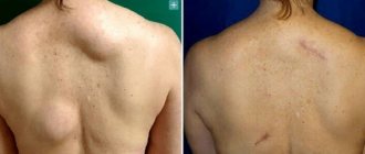
When localized superficially, the lipoma looks like a painless, mobile formation ranging in size from 0.5 to several centimeters. Lipoma can be either dense or dough-like. It does not hurt when touched, although it can compress nervous and vascular tissues. If it is located in a vulnerable place, then pus and inflammatory processes may appear from friction.
Most often, this tumor grows on the head, face, neck and waist, back, chest, but can also occur on the legs and abdomen.
The patient will complain of breathing problems if the lipoma is on the neck.
Typically complaints are cosmetic and aesthetic in nature.
During examination, a specialist doctor determines a lipoma by its location (if it is located in a place where adipose tissue accumulates), soft or compacted consistency, mobility, painlessness on palpation, tissue retraction when the skin is stretched.
Next, the puncture is taken for study.
They may prescribe ultrasound, x-rays, electroradiography of soft tissues, contrast x-rays, magnetic resonance and computed tomography.
The doctor must distinguish a lipoma from an atheroma.
You should not treat the problem yourself. After consultation with a surgeon, surgical treatment is prescribed, if required.
Prostate adenoma
Lipoma, or wen
Jaundice
22918 December 17
IMPORTANT!
The information in this section cannot be used for self-diagnosis and self-treatment.
In case of pain or other exacerbation of the disease, diagnostic tests should be prescribed only by the attending physician. To make a diagnosis and properly prescribe treatment, you should contact your doctor. Lipoma: causes, symptoms, diagnosis and treatment methods.
Definition
Lipoma is a common tumor consisting of fat cells (adipocytes) and is usually delimited from surrounding tissue by a thin connective tissue capsule. Lipomas can form anywhere where fat cells are present, usually located subcutaneously, but are sometimes found on internal organs (for example, the stomach, esophagus or intestines, bronchi, heart or muscles).
Lipomas are benign neoplasms, do not have a tendency to become malignant and do not cause discomfort to patients. The exception is when lipomas are poorly located (for example, in the joint area) or grow quickly, which happens extremely rarely.
Lipomas are the most common mesenchymal tumors (mesenchymal tumors are soft tissue tumors and specific bone tumors).
The incidence is about 2.1 per 1000 people per year, with men diagnosed slightly more often than women. Causes of lipomas
The exact causes of lipomas are unknown. Some researchers agree that genetic abnormalities play a role in their formation. Another theory suggests a connection between the occurrence of lipoma and previous trauma. Risk factors for the development of lipoma may include obesity, alcohol abuse, liver disease, impaired glucose tolerance, and hyperlipidemia. The appearance of a lipoma may be a consequence of another disease, for example, familial multiple lipomatosis.
Classification of the disease
Lipomas can be simple or multiple (multiple lipomatosis). The latter include Dercum's disease, benign symmetrical lipomatosis (Madelung's disease), familial lipomatosis, and congenital infiltrating lipomatosis. Multiple lipomatoses account for approximately 5-10% of all detected cases of lipomas.
Depending on the components of which tissues are involved in the pathological process, fibrolipomas (with connective tissue elements), myolipomas (contain muscle fibers), angiolipomas (include blood vessels), myxolipomas (contain mucous tissue), myelolipomas (contain hematopoietic tissues).
Lipoma symptoms
Lipomas in the subcutaneous fatty tissue are soft and mobile to the touch, not fused with the surrounding tissues. Lipomas are characterized by slow growth and their size usually ranges from 1 to 10 cm. Larger lipomas are called “giant”. The growths are usually painless unless they involve joints, nerves, or blood vessels. The skin over the lipoma is not changed.
In the gastrointestinal tract, lipomas are submucosal fatty tumors. They are asymptomatic, but can cause ulceration and bleeding. Esophageal lipomas can make it difficult to swallow food and liquids and cause belching, vomiting, and reflux.
Lipomas of the small intestine are diagnosed, as a rule, in older people, are most often located in the ileum and are dangerous due to blockage (obstruction) of the intestinal lumen.
In addition, intestinal lipomas cause pain, obstructive (obstructive) jaundice, and intussusception (the insertion of one part of the intestine into another).
It is extremely rare that lipomas form in the heart: subendocardially - under the inner lining of the heart (endocardium) or intramurally - inside the muscle layer (myocardium). Typically, cardiac lipomas are not covered by a capsule and appear as a yellow mass protruding into the cavity of the heart. Cardiac lipomas can cause chest pain, arrhythmias, and shortness of breath.
Lipomas formed in the bronchi or lung parenchyma lead to respiratory dysfunction.
Diagnosis of lipoma Diagnosis
requires a clinical examination by a doctor and an ultrasound examination of soft tissues.
Reasons for appearance and localization features
Causes of compaction on the thighs and thighs:
- Violation of carbohydrate, fat, and mineral metabolism negatively affects the synthesis of enzymatic and hormonal substances.
- Benign tumors from adipose tissue occur when the endocrine system does not function properly.
- Poor ecology, food, water are factors that provoke the proliferation of atypical cells and the occurrence of formations of any nature.
- Hypo-vitaminosis negatively affects the functioning of the sebaceous glands.
- Smoking and alcohol provoke toxic poisoning of the body. A person with a weak immune system and chronic diseases is easily susceptible to the negative influence of the external environment.
- Hereditary predisposition.
- Fat spots appear on the legs due to excessive physical activity.
- Harmful working conditions provoke a pathological process.
- Physical inactivity, metabolic syndrome.
- The predominance of fatty foods in the diet guarantees metabolic disorders, hypercholesterolemia and the development of the disease.
A benign tumor can be located in different layers of the skin and penetrate deep inside.
Types of formations by localization:
- Subcutaneous wen.
- The perineural reaches the nerve fibers.
- Tendons arise in the tendons of the legs.
- Intermuscular muscles grow between the thigh muscles.
- The diffuse compactions look like a cluster-shaped group of adipocytes. They are located diffusely and do not have a capsule.
Subcutaneous lipomas can grow and reach the thigh muscles and tendons. The condition is accompanied by severe discomfort and pain during movement.
What does diagnostics provide?
After the diagnosis, if the diagnosis is confirmed, further treatment is carried out by the surgeon, who decides whether surgical intervention is necessary or not. The fact is that a disease such as lipoma can be life-threatening, because in old formations you can see a well-developed vascular system.
It is also worth considering that lipoma is very similar to liposarcoma; it is far from safe for human life. Therefore, as a rule, after the tumor is removed, it is sent for examination to the laboratory to rule out the presence of malignant cells.
Diagnostic methods in surgery:
- Doppler in surgery
- Colonoscopy
- Angiography
- CT scan
- Gastroscopy
- MRI
- Abdominal ultrasound
- X-ray
- Endoscopy
Prices:
| Code | Name of service | Prices |
| 1 | Initial appointment | 1200 |
| 2 | Repeated appointment | 900 |
| 3 | Calling a surgeon to your home | 3500 |
| 4 | Abdominal ultrasound | 2200 |
| 5 | Ultrasound of veins and vessels | 2400 |
| 6 | Doppler 2-3 trimester | 1200 |
| 7 | Rectoscopy | 1500 |
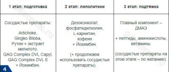
Methods for safe lipoma removal
They get rid of formations using surgical and hardware methods, use medication, and use folk remedies. Each method is characterized by advantages and disadvantages. Conservative, alternative therapies help to avoid violation of the integrity of the skin, common postoperative complications - wound suppuration, severe bleeding. The duration of the conservative course depends on the diameter and shape. Ointments and tinctures do not remove the capsule of the wen located on the thigh. There is a risk of relapse.
The operation helps get rid of a benign tumor by eliminating the source of the problem.
Need advice from an experienced doctor?
Get a doctor's consultation online. Ask your question right now.
Ask a free question
Minimally invasive techniques reduce the likelihood of complications. Disadvantage of the hardware method of treatment: high cost. Laser or radio wave removal does not guarantee the absence of recurrence of the disease.
The choice of treatment is determined depending on the results of additional studies. For differential diagnosis of lipomas, ultrasound, MRI, puncture biopsy, and histological analysis are performed.
Effective solutions:
- The surgery is performed under local anesthesia. Depending on the location of the seal - on the left, right thigh, back or front, a comfortable position for the patient is selected. The surgeon excises the benign lesion with a scalpel, then the sample is sent for histological examination to determine the etiology of the condition.
- The puncture-aspiration technique allows you to remove pathological contents using special equipment; the puncture is made with a needle. The capsule remains, which guarantees the development of a relapse.
- Laser destruction is considered a safe, low-traumatic way to combat subcutaneous formations. Under the action of the laser, adipocytes and connective tissue capsules are destroyed. There are no scars left after the procedure.
- Radio wave surgery. The seal on the thigh is destroyed by high frequency waves. A slight redness remains, which disappears after 1-2 days.
Clinical types of lipomas
- Perineural. Develops near nerve endings and is most painful and difficult to remove.
- Lumbosacral. It occurs predominantly in the spinal canal (usually underdeveloped), less often near the vertebrae.
- Intermuscular. They affect muscles by growing through them. After removal they often appear again.
- Myolipoma. It affects soft tissues and almost never develops under the skin, being located directly on it.
- Angiolipoma. It appears in the form of a dense subcutaneous node, typical for men.
- Adenolipoma. In many ways it is similar to atheroma, affecting not only fatty tissue, but also the sweat glands.
- Articular. It affects the synovium of the joint, as well as the vagina and tendons.
Atheroma
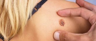
This is a deep cyst of the sebaceous gland, located in the thickness of the skin.
The excretory ducts of the sebaceous glands can open directly on the surface of the skin. When the excretory ducts of the sebaceous glands are blocked, the secretion they secrete accumulates inside. An atheroma is formed - a retention cyst of the sebaceous gland. This formation never occurs on the palms and soles. It can appear on any part of the body where there are sebaceous glands.
The inside of the cyst cavity is surrounded by a capsule. A sebaceous cyst contains sebum, dead skin cells, and cholesterol.
As a rule, treatment of atheroma comes down to its surgical excision along with the capsule - this is a guarantee that there will be no relapse.
Symptoms of soft tissue lipoma
Soft tissue lipoma of the thigh is not accompanied by specific symptoms. Manifestations of the pathological process when reaching large sizes:
- The seal protrudes above the surface of the skin. On palpation, a soft consistency, rounded shape, and small lobules are felt. There is no pain, itching. The skin around the affected area may become stretched.
- A benign tumor easily moves to the side and is not fused to the skin.
- Inflamed growths are manifested by hyperemia, pain, itching, and a local increase in temperature.
- The color does not differ from the surrounding fabrics. Upon closer inspection, you can notice yellowness.
- The location of the wen on the inner side of the thigh leads to frequent injuries when moving.
Rehabilitation after removal
The duration of the recovery period directly depends on the number and size of the operated lipomas. It usually lasts several days and consists of protecting the postoperative wound from the penetration of pathogenic microorganisms into it. Therefore, the doctor will recommend that the patient not expose the site of the removed lipoma to sunlight and water for a week, and will also give recommendations on how to care for it.
Have you discovered a suspicious subcutaneous bulge in yourself or your loved ones? Experienced modern medical specialists are always ready to correctly diagnose the tumor and promptly offer you the most appropriate method to get rid of it, avoiding relapses and complications!
Services
Vaccination against tick-borne encephalitis
Kinesiology
Cardiologist appointment
ALL SERVICES
Useful articles
Let's say no to hair loss!
Luxurious smoothness of your skin
Primary sources of lipoma development
The appearance of wen is associated with various pathologies, sometimes they arise as an independent pathology. Provoking factors for the appearance of lipoma are presented:
- heredity - if the parents have the disease, the probability of a neoplasm occurring is 75%;
- disorders of embryonic development - tumors arise against the background of dystopia of embryonic lipid cells;
- individual diseases - Cowden, Madelung, Dercum;
- endocrine disruptions – with hypofunction of the pituitary gland, pancreas, thyroid gland, altered estrogen levels, malignant neoplasms of the upper respiratory tract;
- sedentary lifestyle - congestion provokes blockage of glands and fat deposits;
- dysfunction of tissue trophism - due to problems with the nutrition of nerve cells, free lipids begin to accumulate next to them;
- injuries - when local metabolism is disrupted due to damage;
- with age - a slowdown in metabolic processes and a decrease in protective mechanisms after 40 years becomes a source of wen.
The development of lipoma is influenced by bad habits and an incorrectly selected diet. Also, pathology is formed due to diseases of the digestive organs, genitourinary tract, liver, and hormonal imbalance.