From this article you will learn:
- what is the nasolacrimal/tear trough,
- filling the nasolacrimal trough – reviews, photos,
- choice of fillers, possible complications.
Age-related changes in the area around the eyes include: hyperpigmentation, swelling, circles under the eyes, dynamic and static wrinkles, drooping eyebrows, decreased elasticity of the skin of the eyelids, the appearance of “bags under the eyes” (hernial protrusion of fatty tissue), as well as the appearance of furrows. The latter include the nasolacrimal, eyelid, nasozygomatic, and nasobuccal grooves. In this article we will focus in detail only on the correction of the nasolacrimal groove with fillers based on hyaluronic acid.
But it must be said right away that filling the nasolacrimal trough with fillers will be contraindicated for patients with: 1) fatty hernias of the lower eyelids, 2) with excess skin of the lower eyelids, 3) severe elastosis, 4) with allergies to filler components, 5) with infectious process in the eye area, 6) if the patient has previously had silicone, polyacrylamide gel or fillers of unknown origin implanted in this area.
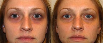
Nasolacrimal trough: photo
This article is written primarily for patients, and is intended to focus their attention on the need to very carefully select a cosmetologist who will perform this procedure. Reviews for correction of the nasolacrimal trough can be very negative. Due to the individual anatomy of this area, it is not always possible to achieve a good result in 1 procedure, and 2-3 procedures may be required. And in some patients, anatomical features may not allow achieving a good result at all.
According to statistics, complications after correction of the nasolacrimal groove develop in 50% of cases, and in most cases are associated with downward displacement of the filler. The latter will lead to the formation of visually clearly visible “sausages” just below the tear troughs and will require longidase injections to dissolve the filler. Less commonly, contour plastic surgery of the nasolacrimal groove can also result in vascular complications, for example, necrosis of the skin of the infraorbital region or irreversible visual impairment. Such complications arise as a result of occlusion or embolism of the angular artery located under the orbicularis oculi muscle (i.e., exactly where the filler is supposed to be removed).
How often does swelling occur?
Correction of the nasolacrimal trough very rarely causes complications, but much depends on the qualifications of the doctor, the correct selection of the drug, and compliance with recommendations during the rehabilitation period. Edema is not a complication - it always appears to one degree or another, but can be more or less pronounced. How long the swelling will last after correction of the nasolacrimal trough depends on the individual characteristics of the body, the drug used, and the number of punctures.
The main techniques used to correct the nasolacrimal groove area:
- Spot.
- Fan.
- Retrograde linear.
- Using a cannula.
The most gentle are injections using cannulas and the fan-retrograde technique. After injections, the rejuvenation effect is noticeable, but it can be finally assessed after 7-14 days. The result is smoothing, filling in depressions, visual rejuvenation of the face, elimination of dark circles under the eyes. Normally, swelling after correction of the nasolacrimal trough goes away on its own within a few days (up to two weeks). If the reaction persists longer, contact a cosmetologist - he will find out the possible causes of this phenomenon and give recommendations on how to relieve swelling. If the gel is administered excessively, hyaluronidase injections are performed.
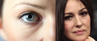
Why is she becoming so visible?
The main reason for the bluish color of the furrow is that the vessels and orbicularis oculi muscle are visible through the skin. Triggers for the formation of this deficiency are age-related or genetic “weight loss” of adipose tissue under the eyes. Because of this, the relief of this area changes, unnecessary shadows are formed, which visually gives the eyes a tired look.
Most often, a pronounced furrow becomes noticeable after 35-40 years.
How dangerous is swelling?
Swelling always occurs after correction of the nasolacrimal trough; normally, this condition is not dangerous. The cause of swelling is minor tissue trauma, which always occurs when the skin is punctured. If you look at the photo of the swelling after correction of the nasolacrimal trough, you can see that it is small and does not interfere with the aesthetics of the face. Using decorative cosmetics to hide imperfections is strongly discouraged. If necessary (for example, you don’t want to appear in front of colleagues after a beauty procedure), take a short vacation.
Correction of the nasolacrimal groove before and immediately after the procedure. Doctor - Syomova A.M.
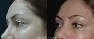
Correction of the nasolacrimal groove before and immediately after the procedure. Doctor - Golubeva M.S.
If minor swelling occurs after correction of the nasolacrimal trough, you do not need to do anything - just wait a few days. You may also be concerned about redness (it goes away in a few hours), small bruises at the injection sites (they also go away on their own). The recovery period takes no more than 1-2 weeks. Remember that effective non-surgical correction of the nasolacrimal groove without injections is almost impossible. There are a number of hardware techniques that can improve the condition of the skin in this area, but it is contour plastic surgery that allows you to fill the missing volume.
In the practical aspect of the modern concept of facial rejuvenation, morphometric and aesthetic analysis serves as the most important tool. What parameters of such an assessment should you pay special attention to? Carrying out correction in the upper and middle third of the face – which zones and points are dominant?
Rejuvenation and harmonization of the face require its correction as a single, holistic object with precise definition of the main zones - this is the modern general concept. However, doctors often deviate from this principle, concentrating their efforts on solving individual, sometimes rather insignificant, problems. What is this connected with? The reasons may be different: misunderstanding, ignorance or ignorance of the basics of aesthetic and morphometric analysis, lack of necessary professional skills for correcting certain areas of the face, lack of mastery of certain methods, and sometimes following the patient’s lead. As a result, the cosmetologist is limited to correcting a single aesthetic defect, and the patient cannot even imagine what full-scale effect of facial rejuvenation is possible in his case.
The wishes (and even demands) of our patients, as a rule, do not cover the picture as a whole, but are concentrated on its details, which, in their opinion, spoil the whole thing: “It’s because of these wrinkles around my mouth that I look older, just remove them " In fact, performing some kind of procedure to eliminate one or another aesthetic imperfection, usually considered a sign of facial aging, has nothing to do with the real concept of rejuvenation. For example, correction of nasolabial folds has become a kind of social dominant among other anti-aging procedures. It is surprising that patients who do not have this very conditional defect at all come to cosmetologists to get rid of it. But nasolabial wrinkles when smiling and other facial movements appear even on the face of a baby. In accordance with the general concept of facial rejuvenation, we should not talk about isolated correction of individual aesthetic defects, but about the priority of zones in the complex correction of the entire face.
Morphometry and aesthetic analysis in the applied aspect
To emotionally assess a face along the “young-old” axis, no special analytical thinking is needed; involutional changes can be easily diagnosed by any specialist in aesthetic medicine. And morphological disturbances - what is inharmonious and what is wrong - are also simply determined. However, it is important not only to note them - the professional must explain exactly how and why the violations occurred and be able to eliminate them.
To identify aesthetic and morphological defects of the face, it is enough to determine its height and width as a whole, its upper, middle and lower thirds and the proportional relationship of these parameters and, based on the assessment, understand what violates the correct proportions.
If the face is characterized by a sharp “geometry” rather than smooth transitions from one zone to another, this is also clearly visible (“a smooth transition” here is perhaps the main characteristic). You just need a careful appraising look to notice that there has been a narrowing of the forehead in its lower or upper part, that it is not high enough, there are depressions on it, the temple is deepened, there is no smooth transition from the forehead area to the side of the cheekbone, there is practically no subcutaneous fat layer in the area of the temporal suture or the bony lateral edge of the orbit (this area is skeletonized), there is no gradual (soft) transition from the eyelid area to the anterior convexity of the cheekbone, under the eyes there are depressions that absorb light, the cheekbone has a “mountain-valley” relief, but should be a single smooth one a convexity from the surface of which light is uniformly reflected, etc.
These are the elementary foundations of morphometry and artistic analysis, which at the diagnostic stage allow a modern specialist to determine on the face the point of focus of attention of others (let’s call it the central focus), and, what is extremely important, the dominant zones, evaluate their gradation and joint aesthetic significance and in accordance With this, we can direct the patient’s wishes in the right direction. Only then do real opportunities arise to obtain optimal visual results of facial rejuvenation.
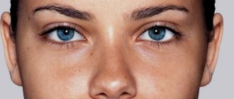
On the face, the center of attraction is, of course, the eyes. Beautiful eyes - almond-shaped, with white sclera, bright irises, black eyelashes and regular canthus. (Correct is when the lateral corner of the eye is higher than the medial one. This position of the canthus ensures a higher position of the lacrimal glands compared to the lacrimal sac and ducts and regulates the physiological outflow of tears under the influence of gravity.)
The eyes, as the central focus of the face, require aesthetic perfection from the areas surrounding them - the lower orbital, zygomatic, temporal and frontal. These zones dominate the perception of the face. Their morphological disorders and involutional changes distract from the beauty and expressiveness of the look, “turning the arrows” towards themselves; they are the ones who primarily age the face.
We will consider aesthetic correction of zones using fillers and their anatomical justification in this material.
Correction of infraorbital grooves and anterior zygomatic zone with gel
Anatomically, the face is divided into two sections - anterior and lateral, the border between which is defined as a line drawn along the medial edge of the masticatory muscle.
The anterior section of the face is more mobile, all facial muscles are concentrated in it, it is responsible for facial expressions, articulation, and food grasping occurs here. In most of this section, the fifth layer of tissue is represented by the mucosa.
The lateral section, on the contrary, is more static. It contains the chewing muscles: the temporal and masseter muscles; SMAS is represented by the superficial fascia - parotid-masseteric and external temporal.
According to this division of the face in the zygomatic zone, the anterior and lateral sections are also distinguished.
The infraorbital grooves and the anterior zygomatic zone are priority targets for correction. It was from applying uniform light to this area that classic Photoshop and other image-enhancing programs grew. The purpose of the correction is to eliminate the nasolacrimal, nasobuccal and palpebromalar grooves, mask the minimal palpebral and malar hernial sacs, restore the anterior convexity of the cheekbones, and create a smooth transition from the eyelid area to the cheekbone.
The nasobuccal (midbuccal) groove is an area of depression separating the superficial infraorbital compartments (malar hernial sac) and the underlying superficial nasolabial compartment.
To correct the nasobuccal groove with fillers, the drug can be safely administered with a 25G/40 mm cannula. The entry point for the cannula is determined under the zygomatic ligament, but above the septa of the angular vein - on the convexity of the cheekbone along a line roughly drawn through the lateral corner of the eye.
Hyaluronic gels with high viscoelastic properties are preferred, providing long-term results of volumetric correction. If facial tissues rise strongly when smiling and protrude into its anterior plane, then in this case dynamic gels, specially adapted to the natural biomechanics of tissues, elastic under stretching and squeezing, are more suitable.
The injection is performed slowly, supraperiostally, under SMAS.
Deformation of the nasolacrimal groove as an aesthetic defect is expressed in the formation of a depression zone, limited above by the periosteal attachment of the orbicularis oculi muscle, and below by the septa of the angular vein. In this area, the muscle fibers are dense, with good blood supply, there is no surface fat above them, so the muscle is visible through the thin skin tissue.
Correction of the nasolacrimal groove: the cannula is inserted through the same access point as when working with the nasobuccal groove, at an angle of 45 degrees until it touches the periosteum, towards the medial canthus, and then it is positioned parallel to the periosteum.
It is important to take into account that in the vestibule of the orbit the level of the periosteum changes, and in accordance with this it is necessary to change the course of the cannula. The use of a needle in this area is dangerous, since the angular vein, the superior branch of the infraorbital artery, the angular artery (in 30% of patients) and its duplex (in 15% of cases) are localized here.
To ensure maximum aesthetic results when correcting the nasolacrimal groove, it is necessary to smoothly connect the end of the nasolacrimal groove and the beginning of the nasobuccal groove.
The formation of the palpebromalar groove is due to the fact that the outer cheek (zygomatic fat) does not reach the level of the bony edge of the orbit, which causes a difference in tissue level. This zone is conditionally dangerous, so needle work is acceptable here. The needle pierces all tissues slightly at an angle to the periosteum and touches it lightly. The drug is administered as a bolus, in a volume of no more than 0.1 ml per injection. In the presence of palpebral and malar hernial sacs, insertion should be ensured exactly between them (between the bony edge of the orbit and the ligament of the orbicularis oculi muscle) in order to mask their bulging.
This zone is characterized by a low level of tissue hyaluronidase, so here the long-term effect is provided by low-viscosity and low-elastic, well-distributed drugs that do not exert high pressure on the orbicularis oculi muscle.
To create the anterior convexity of the cheekbone as a whole, fillers are injected primarily into the deep medial buccal compartment, easily identified on any face. Its upper border is the zygomatic ligament, the lower is the septa of the angular vein, the lateral border is determined by a line conventionally drawn along the minor zygomatic ligament. Selective increase in the anterior convexity of the cheekbones visually enhances the deepening of the infraorbital grooves, therefore both zones are corrected step by step sequentially and in one procedure.
To reduce pain and ensure patient comfort during the procedure, it is better to choose lidocaine-containing drugs. They are first inserted into the deep medial buccal compartment, and then the nasolacrimal groove is corrected. This ensures maximum anesthesia: lidocaine has time to accumulate in the premaxillary space, where the exit point of the infraorbital branch of the trigeminal nerve is located, which innervates this entire area of the face, and in particular the area of the infraorbital grooves. It is better to first administer lidocaine-containing drugs antegradely, in small portions. Next, the specialist can use any injection technique, since anesthesia has already been achieved.
Gel injections into the anterior zygomatic area are performed sequentially on both sides of the face until optimal correction parameters are achieved.
For successful aesthetic correction, it is necessary to determine the dominant zones and their subordination at the diagnostic stage, and base all cosmetic interventions on aesthetic and morphometric analysis.
Gel correction of the lateral cheekbone
Many patients consider insufficient convexity of the lateral part of the cheekbone to be a significant aesthetic defect.
Anatomically, this section of the cheekbone is partly formed by its body and partly by its temporal process, or the so-called arch. Its superficial fatty compartments are separated by rather weak, vertically oriented connective tissue septa. In this regard, superficial introduction of hyaluronic gels can lead to their displacement into the underlying buccal area. To prevent this displacement and create a sculptural convexity of the lateral cheekbone, the main part of the drug must be injected into the lateral portion of the deep inferior orbital fat (SOOF): its upper border is the suspensory ligament of the orbicularis oculi muscle (ORL), the lower - the zygomatic ligament (ZL), the “roof” "- orbicularis oculi muscle (OOM), "bottom" - periosteum of the zygomatic bone.
The introduction of gels into this anatomical formation allows you to obtain long-term correction results and create a strong lateral vector of the cheekbone.
We insert the cannula from the same point as for correcting the nasobuccal groove and direct it laterally into the above compartment. Further, to ensure a smooth transition from the zygomatic zone to the cheek, correction can be supplemented with superficial administration of the drug.
It should be taken into account that an increase in the convexity of the lateral part of the cheekbone can lead to a visual deepening of the overlying temporal and underlying buccal zones, so at the final stage of correction it is sometimes necessary to ensure smooth transitions between them.
Contour correction of the temporal region
The objectives of correction in the temporal region are to eliminate depressions, mask signs of skeletonization and ensure a visual expansion of the boundaries of the forehead and a smooth transition from the frontal zone to the temple.
The upper border of the temporal zone is the superior temporal septum - a vertically oriented connective tissue running from the temporal suture to the surface of the skin (to simplify understanding, it is better to consider it the temporal suture itself). In front, the temporal region is limited by the lateral bony edge of the orbit (connected by the frontal process of the zygomatic and the zygomatic process of the frontal bones).
The lower border of the temporal zone is represented by the upper edge of the zygomatic arch.
The anatomical posterior border is also the temporal suture, but for contour correction, the aesthetic border of this zone is relevant - the hairline.
Layer anatomy demonstrates 5 traditional layers:
1) skin, 2) subcutaneous fat, 3) superficial temporal fascia, 4) weak areolar vascular tissue (analogous to deep fat), 5) deep temporal fascia.
In this area, it is most easy, safe and effective to inject filler between the superficial and deep temporal fascia using a 25G/40mm cannula from the entry point located in the forehead. We mark the puncture point with the trocar on the frontal bone 1 cm above the eyebrow, above the temporal suture. We insert the cannula supraperiostally, perpendicular to the surface of the forehead, until it touches the periosteum. Then we unfold it parallel to the periosteum, pierce the superior temporal septum and automatically move to the surface of the deep temporal fascia (the cannula slides along the fascia).
To avoid discomfort in the patient while determining the injection level, the puncture site with the trocar should be anesthetized with lidocaine and adrenaline. Here it is also recommended to first use antegrade injection of lidocaine-containing filler, and then, after the onset of anesthesia, the usual retrograde one.
Depending on the severity of the depression, low- or medium-viscoelastic preparations are preferred, ensuring good integration with this delicate area.
Usually, 1 ml of the drug is enough to correct one temporal zone.
It must be remembered that eliminating the depression in the temporal zone can lead to the visual effect of increasing the depressions in the frontal zone, so if necessary, the drug must also be injected into it. The goals and objectives of correction are considered completed if all signs of skeletonization can be masked.
If in other zones the integration of the gel takes an average of 4 weeks, then in the temporal zone it may take longer, during which the surface of the zone looks uneven due to poor distribution of the gel under thin tissues.
Forehead correction
Elimination of depressions and masking of signs of skeletonization in the temporal zone often make morphological and involutional changes in the adjacent frontal zone more noticeable.
Here are the neurovascular bundles: supratrochlear (projected on the perpendicular passing through the medial edge of the iris) and supraorbital (on the midpupillary perpendicular), which emerge from the corresponding supraorbital recesses on the upper edge of the orbit and cross the forehead from bottom to top. It is easy to assume that the septa of these particular neurovascular bundles divide the frontal fat into compartments.
In addition, the forehead correction area is separated from the temporal by the superior temporal septa, which is the lateral border of the temporal compartments.
To create a smooth transition between the interconnected temporal and frontal zones, visually widen the forehead, eliminate depressions and avoid embolism, it is better to administer gels here using large-diameter cannulas.
We choose the entry point for the cannula arbitrarily, next to the depression zone, insert the cannula perpendicular to the surface of the forehead and up to the periosteum, then turn it around and move it parallel to it.
I would like to emphasize that needle insertion is more dangerous. When it is perpendicular to the periosteum, the gel is distributed only within one compartment, so longitudinal administration through the cannula is more preferable.
The forehead area is characterized by significant pain during injection, so it is necessary to use lidocaine-containing preparations.
To obtain the necessary correction parameters, as a rule, gels of medium and low viscosity and elasticity are sufficient.
So, we looked at methods of administering stabilized hyaluronic acid preparations for aesthetic correction of the upper and middle third of the face. In conclusion, I will once again emphasize the most important points.
A person should be assessed as a whole, as a set of structural units - zones. Correcting them in isolation is impractical and incorrect, since improvements achieved in one zone can cause visual deterioration in others adjacent to it. For successful aesthetic correction, it is necessary to determine the dominant zones and their subordination at the diagnostic stage, and base all cosmetic interventions on aesthetic and morphometric analysis.
And we remember the main thing in the new image being created - the smoothness of lines and transitions.
Margarita Egorova, dermatologist, cosmetologist, medical director, Moscow
Is it possible to eliminate swelling on your own?
It is impossible to completely avoid the appearance of swelling, since it is a normal reaction to tissue injury and the introduction of hyaluronic acid under the skin (the element attracts water). To minimize the risk of complications, take into account contraindications for correction of the nasolacrimal trench and follow the doctor’s recommendations during the rehabilitation period:
- In the first 2 weeks after the procedure, saunas, swimming pools, steam baths, and hot baths are contraindicated for you. Staying in the sun for a long time or visiting solariums is also not recommended.
- You need to take care of your skin carefully and only use those cosmetics that your doctor has approved for use. Give up decorative products for a while (do not mask swelling and bruises - this may prolong rehabilitation).
- Touch your skin less with your hands to avoid infection at the injection sites.
- Use sunscreens with a high SPF factor.
- Put off all cosmetic procedures for a while - they cannot be done immediately after filler injections.
- Ask your doctor what absorbable creams and decongestant gels can be applied topically to quickly relieve swelling and other side effects.
- Any self-massage in the area of contouring is contraindicated.
All recommendations are clear and easy to follow. There is no need to make sacrifices - just choose the right method of rejuvenation, a proven clinic, a competent doctor, a suitable drug and limit yourself a little for 1-2 weeks after the procedure. Now you know what to do if swelling occurs after correction of the nasolacrimal trough.
GMTClinic is a business class clinic. We employ only professional cosmetologists who use proven quality products in their work. All this allows us to guarantee maximum effectiveness of procedures and the absence of side effects. Appointments are by appointment only - choose, book an appointment and take a step towards your ideal beauty.
Methods for correcting the nasolacrimal trough
The epidermis in the periorbital zone is extremely thin, so only highly professional specialists can work with such a delicate area.
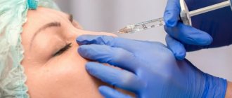
The nasolacrimal groove is removed using fillers. A dermal filler introduced into the area of the nasolacrimal groove smoothes out the relief, fills the intercellular space, and gives the skin a healthy tint.
Thanks to components that stimulate the production of collagen, elastin and hyaluronic acid, powerful skin rejuvenation occurs and the water balance of cells is normalized.
Fillers strengthen the connection between the skin and muscle fibers, increasing muscle tone, thereby eliminating gaps at the junction of muscles and subcutaneous tissue. This helps smooth out the relief. As a result, the face looks tightened, fresh and rested.
Gutsenko Liliya Anatolevna
Cosmetologist
Line of drugs STYLAGE
Stylage fillers in Moscow are available in several variations. This gives the specialist the opportunity to select the most suitable type and dosage to correct the required area. The line includes the following drugs:
- Stylage S. Quite liquid filler. Used to get rid of small and medium wrinkles. Can be used to correct the area under the eyes, periorbital area, lips. The result lasts for up to 9 months.
- Stylage M. A denser gel, suitable for the correction of deep wrinkles. It is used to eliminate nasolabial folds, marionette lines, lip and nose correction, and hand rejuvenation. The effect lasts for up to 1 year.
- Stylage L. High density gel. It is used to correct nasolabial folds, shape the chin, eliminate skin creases, and rejuvenate the hands. Gives effect for up to 1 year.
- Style XL. The densest filler in the series. Suitable for volumetric facial correction and contour restoration. The result lasts up to 1 year.
- Stylage Special Lips. The drug is intended specifically for the lip area. It allows you to adjust the volume, improve the contour, get rid of asymmetry, and level out age-related perioral wrinkles. The result lasts for 6-9 months.
- Stylage Hydro and Hydro Max. These fillers are intended for biorevitalization. Use on the neck, arms, and décolleté. They include unstabilized hyaluronic acid, which promotes tissue rejuvenation. The effect lasts up to 4 months.
The price list shows the cost of injections with Stilage drugs. The required number of sessions is determined by a specialist.
The filler injection procedure lasts about 15-40 minutes. The cosmetologist first determines the correction zone, the number of injections, and the injection technique. Thanks to lidocaine, injections are painless. The final result will become noticeable in about 1-2 days, after the final restoration of the skin. Stilage preparations have proven themselves well in cosmetology and are in great demand both among specialists and patients.
How does the procedure work?
The first stage of the procedure is examination and consultation. After this, an anesthetic gel is applied to the face, and as soon as it takes effect, the doctor begins the injections.
The filler is injected using a thin, flexible cannula, a type of blunt-tipped needle. Since the skin in this area is very thin and prone to stretching, the doctor is required to have exquisite precision and masterful technique. Gels are used in a minimal dosage - if necessary, the result can be adjusted after a couple of weeks.
Important! Entrust contouring only to an experienced and qualified cosmetologist. The main components of success are the correct administration technique, accurate dose calculation and a high-quality certified drug.
Prevention
It is not always possible to prevent the appearance of a nasolacrimal trough. For example, if this defect is due to genetics and appears at an early age, preventive measures will be useless. In other cases it is recommended:
- Do not get carried away with salty, spicy, smoked foods, as well as simple carbohydrates - especially before bed.
- Try to maintain a stable weight, avoid extreme diets and fasting, and do not overeat.
- Drink enough fluid - mainly in the first half of the day.
- Use only products specifically designed for this area to care for the skin around the eyes.
- Monitor the health of the heart and kidneys, treat diseases that cause swelling.
Why clients choose us
- The Expert Cosmetology Clinic has valued its impeccable reputation since 2009!
- Our doctors have developed proprietary programs to effectively solve even complex problems.
- Specialists will never perform a procedure if it is not indicated for you.
- We attach great importance to your safety and compliance with sanitary standards. Checko systems control the microclimate in the clinic premises.
- We carefully select our staff; the procedure is performed only by cosmetologists.
- We use only original drugs and equipment to ensure that the procedure is as safe and accurate as possible.
- We have our own parking, no need to waste time looking for a space on neighboring streets.
- Thousands of grateful clients confidently recommend us to their family and friends because they TRUST US!
Call the Telos Beauty Prof clinic by phone or
Contraindications
The use of Stylage fillers is safe and has a minimum of contraindications:
- pregnancy and feeding;
- individual sensitivity to components;
- intolerance to anesthetics;
- use of anticoagulants;
- bleeding disorders;
- oncological diseases;
- exacerbation of chronic diseases;
- skin inflammation.
Before the session, you need to inform the cosmetologist about all known contraindications - this will help prevent possible side effects.