Medical Internet conferences
Dry skin syndrome. Modern view of the problem
Sazonova S.A.
Scientific supervisor: candidate of medical sciences, associate professor Morrison A.V.
Federal State Budgetary Educational Institution of Higher Education Saratov State Medical University named after. V.I.Razumovsky Ministry of Health of the Russian Federation
Definition. Dry skin is a common skin condition in which there is a decrease in the function of the sebaceous and sweat glands, and, as a result, a decrease in moisture in both the dermis and epidermis. Dry skin syndrome can be the result of both exogenous factors (domestic and environmental conditions) and the manifestation of endogenous disorders (genetic, hormonal and immune). Although much of the literature refers to dry skin as “xerosis of the skin,” I believe that this term is most appropriate to describe dry skin as a mild form of ichthyosis vulgaris in the presence of a genetic predisposition.
Clinical manifestations. Dry skin is characterized by pathological peeling, the presence of small cracks, thickening of the skin and its roughening (lichenification), decreased turgor and elasticity due to a lack of lipid content. Subjectively, patients may be bothered by: mild itching, a feeling of tightness and burning. With long-term itching, if it is accompanied by scratching and rubbing, dry skin can thicken and become pigmented. Often, dry skin is a prerequisite for the development of various inflammatory skin diseases such as atopic dermatitis, pyoderma, eczema, etc.
The classification according to the ethology and pathogenesis of dry skin syndrome can be divided into three main groups: • acquired; • associated with age-related changes; • constitutional.
First group
- these are patients with acquired dry skin. Firstly, these are people whose “dry” skin is caused by the direct influence of harmful environmental factors on the surface layers of the skin. These factors include: improper daily skin care (substances contained in soaps, shower gels and shampoos disrupt the functioning of the sebaceous glands) or adverse effects of climatic conditions (insolation: UVA and UVB rays lose the ability of collagen and elastic fibers to bind water), as well as contacts of chemically aggressive substances with the skin (when swimming in a pool with chlorinated water, the protective substances that ensure the normal condition of the epidermis are destroyed and washed away under the influence of chlorine).
Secondly, it should be noted that the clinical picture of some malignant diseases (lymphomagranulomatosis), infectious lesions (HIV/AIDS, viral hepatitis), mental disorders (psychogenic anorexia), endocrine pathology (diabetes mellitus), and renal failure may be accompanied by acquired dry skin. Dry skin syndrome is also a consequence of side effects from the use of certain pharmaceuticals. For example, externally applied retinoids, benzoyl peroxide, azelaic acid lead to severe dryness of the treated skin areas, and the use of vitamins (especially nicotinic acid), statins and diuretics leads to generalized dryness of the skin. [2] In addition, peeling, hyperemia and thinning of the skin can be the result of repeated aggressive cosmetic procedures: peeling, laser resurfacing, various types of masks (especially clay-based). Second group
(due to age-related changes): the condition of the skin is significantly influenced by physiological changes that occur in the body during certain age periods. During life, there are several peaks that are characterized by high dryness of the skin:
The first period begins on the 1st–2nd day of the child’s life, which is explained by the removal of caseous lubricant and a decrease in skin edema (physiological catarrh). Increased dryness of the skin can continue during the first 2–4 weeks of life and is observed in every third child.
The second period is for children aged 2 to 8 years, because they experience a period of minimal activity of sex hormones (estrogens, testosterone, progesterone, cortisol) and reduced production of sebum by the sebaceous glands.
The third age period is senile age. 75% of people over 70 years old have dry skin, which leads to the formation of microcracks. Dry skin is explained by involutive dystrophic processes occurring in the skin and a decrease in the level of sex hormones, which are responsible for stimulating the sebaceous glands.
Third group
(constitutional): dry skin is caused by genetic mutations that lead to structural and functional disorders in the superficial layers of the epidermis. These changes in the skin, as a rule, accompany patients from the moment of birth until old age, and the influence of harmful environmental factors and poor lifestyle can aggravate the severity of dry skin.
Mutations of the FLG gene, which is responsible for the synthesis of the filaggrin protein (from the English filaggrin - filament-aggregating protein) - a protein that promotes the aggregation of filaments, are well studied. Filaggrin deficiency leads to transepidermal water loss and disruption of the epidermal barrier. In addition, fillagrin differentiates to form amino acids (NMF - natural moisturizing factor), which can retain water in the epidermis.
Physiological conditions.
During pregnancy, many organs undergo changes, the skin is no exception. On average, 89% of women experience a change in hormonal levels during gestation, which is manifested by a decrease in the formation of the female hormone estrogen, which entails a decrease in the secretion of the sebaceous glands, so the skin is not sufficiently moisturized. The concentration of progesterone also decreases, resulting in a decrease in skin elasticity and flaking. The period of pregnancy is characterized by the possible development of hypothyroidism, which is characterized by brittle hair, nails and dry skin.
Conclusion. Dry skin syndrome can be caused by genetic mutations, associated with age-related physiological changes, and also acquired under the influence of harmful environmental factors and be a manifestation of hormonal and immune disorders. in organism.
Kinds
Depending on the damaged chromosome, there are 8 subtypes of the disease: A, B, C, D, E, FG and Young's pigmented xerodermoid. The first 7 types of xeroderma pigmentosum have a similar clinical course and are determined only after molecular genetic analysis. Young's xerodermoid pigmentosa has a more favorable prognosis - the symptoms of the disease appear later, and the pathological process itself is mild.
An independent clinical form of the disease is De Sanctis-Cacchione syndrome. It is considered the most aggressive type of xeroderma pigmentosum and is characterized by pronounced changes in the central nervous system. The syndrome can develop with any chromosomal variant of the disease, but most often occurs with subtype A.
Treatment methods
There is no etiopathogenetic treatment for xeroderma pigmentosum. In essence, any methods of therapy are symptomatic and are aimed at eliminating the existing manifestations of the disease. Comprehensive treatment of the disease includes:
- vitamin therapy courses;
- antimalarial drugs - the effectiveness of drugs from this group in eliminating the inflammatory process and sensitization of the body has been noted;
- aromatic retinoids;
- cytostatic antitumor drugs.
Patients are also advised to adjust their lifestyle, limit exposure to the sun as much as possible, and before going outside, take care to protect their skin from sun rays.
All benign skin elements are subject to mandatory removal and histological examination. When a neoplasm transforms into a malignant one, treatment tactics are determined based on the type of tumor and the stage of the oncological process.
Forecast
The quality and length of life depend on several factors, primarily on the timing of seeking medical help, making the correct diagnosis and early initiation of comprehensive treatment. With timely diagnosis of the disease and constant dynamic monitoring by a dermatologist or oncodermatologist, the prognosis is relatively favorable.
With warty growths on the skin, about 35% of patients do not survive to adulthood and die at the age of 13-15 years. This is due to the high tendency to malignancy and rapid metastasis of the tumor to internal organs.
De Sanctis-Cacchione syndrome has a poor prognosis. The aggressive course of the disease leads to death from oncopathology in childhood in 70% of patients.
Book a consultation 24 hours a day
+7+7+78
General symptoms
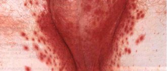
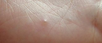
Dry skin has specific symptoms and can be recognized without even touching the body. Typically, the skin looks irritated and tight. It shows inflamed, slightly reddish areas. Pores are practically invisible behind the exfoliating layer.
Below is a list of signs that accompany the occurrence of xeroderma:
- The skin quickly loses moisture; conventional moisturizing creams do not help normalize its balance. It becomes rough and wrinkled.
- After water procedures, a feeling of tightness appears, which disappears only after thoroughly moisturizing it.
- If left untreated, the skin begins to itch and peel.
- In place of the fallen scales, redness forms, then thin cracks, which become deeper over time, begin to bleed and hurt.
You need to get rid of the above symptoms immediately. At the first and second stages of development, people with xeroderma can be treated at home, following the recommendations of the attending physician.
In case of severe and large-scale skin damage, patients are advised to hospitalize, which, as a rule, begins with diagnosing the patient’s current condition.
Classification
Taking into account the reasons contributing to the occurrence of xeroderma, this form of pathology is divided into 3 types. So, xeroderma happens:
- Acquired - appears as a result of exposure to external adverse factors on the skin. A striking example is long-term treatment with external or systemic glucocorticoids (Betamethasone, Prednisolone).
- Senile is the result of the natural aging process. The skin is characterized by increased dryness, wrinkles and pigmentation.
- Constitutional (atopic) - its development is associated with hereditary predisposition, i.e. If in the family one of the relatives previously suffered from dry skin, then this form of pathology was inherited.
Note! Most often, symptoms of xeroderma can be seen on the skin of the hands, which are daily exposed to adverse factors, in particular contact with household chemicals.
A practicing dermatovenerologist will tell you more about the symptoms and forms of xeroderma, which is also called ordinary or vulgar ichthyosis, in the following video:
Stages of disease development
There are 4 stages of development of xeroderma:
- It is practically asymptomatic. The person does not notice any structural changes in the skin (there is no wrinkling or itching), slight dryness is felt only on the feet.
- Over time, the skin begins to peel , thickens, becomes rough and hard. The patient constantly feels tightness of the skin. Begins to notice that from time to time itching and redness appear in certain areas of the body.
- Excessive exfoliation is observed. Painful cracks appear on the skin. Inflammation, itching and redness occur. In deep wounds, pus forms.
- This stage is characterized by a strong change in the structure of the affected skin. Trophic ulcers form (heal for a long time). The disease affects not only the protective layer of the skin, but also the dermis, which contains elements (elastin, collagen) responsible for the firmness and elasticity of the skin.
For reference: Symptoms of the first and second stages of xeroderma can be removed with the help of a deeply moisturizing cream, for example, such as “Losterin”. The third and fourth stages of pathology development require specialized treatment, including the use of medicinal antimicrobial and restorative drugs (see list below).
Folk remedies
Below are several recipes that will help you cope with the initial stages of xeroderma.
In addition, we recommend using these recipes during the cold off-season, when skin health especially needs support.
Bath "Cleopatra"
Ingredients:
- Fresh warm milk - 4 tbsp.
- Natural liquid honey - 0.5 tbsp.
- Warm olive oil - 5 tbsp.
How to prepare: Mix all ingredients. Pour the mixture into a bathtub with water at a comfortable temperature.
How to use: Lie in the healing liquid for 15-20 minutes. Dry your body with a soft towel without rubbing. Apply a cream with a restorative and moisturizing effect, for example, La Roche Posay, which is often recommended by dermatologists.
Result: You will be able to notice an improvement in skin condition after the first procedure. When you take such a bath every day, you will forget about the problem with xeroderma forever.
Hot compress for face
Ingredients:
- Mint petals.
- Calendula flowers.
- Linden blossom.
How to prepare: Take equal parts of the ingredients, pour boiling water over them, cover the container and wrap it up. Let the mixture sit for 10 - 15 minutes.
How to use: Soak a soft towel in the herbal infusion, cooled to a comfortable temperature. Apply to your face for 1.5 - 2 minutes. Repeat the procedure 5 - 6 times. Lubricate your skin with a nourishing cream; the Riche Desalterante cosmetic product has proven itself well in this regard. (Clean 2 times a week).
Result: The compress expands and cleanses pores, removes peeling.
Diagnostics
A detailed history taking is of great importance in making the correct diagnosis. The collected data (onset of the disease in early childhood, seasonality of symptoms, high photosensitivity, consanguineous marriages) suggest xeroderma pigmentosum. The diagnosis is confirmed after cytological and histological examination of the affected areas of the skin.
Additional examinations include examination by a dentist, otorhinolaryngologist, ophthalmologist and neurologist to exclude damage to the central nervous system, oral apparatus, visual and hearing organs. For De Sanctis-Cacchione syndrome, an X-ray examination or computed tomography of the skull bones, and magnetic resonance imaging of the brain are also performed. In this case, small sizes of the sella turcica, a decrease in the volume of the cranium and brain, and underdevelopment of the cerebellum and pituitary gland are detected.
Complications
The main complication of the disease is the appearance of skin and visceral (internal) malignant tumors. Multiple warty neoplasms have the highest risk of oncogenic transformation. The incidence of malignant tumors also correlates with the severity of skin pathological processes. With the early onset of the disease and its aggressive course, oncological tumors appear in adolescence (12-14 years) and quickly metastasize to internal organs.
Among malignant tumors, squamous or basal cell carcinoma most often develops, and melanoma of the skin or eye is the least common. Cases of more than 50 different types of oncology have been described: fibrosarcomas, angiosarcomas, histiocytes and others.
The disease can also lead to severe deformities of the face (mouth, nose, ears). The result is social maladjustment of the child and psychological problems.