from February 4, 2021
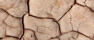
Keratosis or hyperkeratosis is a non-inflammatory form of dermatosis, which is accompanied by excessive keratinization of the skin.
For healthy skin of any person, the process of keratinization is a normal physiological state. The process of keratinization provides one of the most basic functions of the skin - protective.
MECHANISM OF KERAINIZATION IN NORMAL AND IN HYPERKERATOSIS
Normally, as a result of their vital activity, skin cells gradually move from the lower layer to the upper, while gradually accumulating keratin - a protein that makes them more durable and resistant to external influences. The top layer of skin is nothing more than completely keratinized cells or horny scales, which are peeled off and replaced with new ones during human life. Such cells lack viability, but at the same time, thanks to keratin, they provide protection to living cells located underneath them from negative environmental factors.
With the development of hyperkeratosis as a pathological condition of the skin
- on the one hand, there is an active accelerated division of epidermal cells that produce keratin;
- on the other hand, there is a delay in the process of normal physiological exfoliation of horny scales from the surface layer of the skin.
Prevention
There are no special methods for preventing keratosis pilaris. To reduce the risk of its occurrence, the following are relevant:
- relief of skin irritation;
- increasing the body's immune forces.
If you take precautions, treatment of keratosis if it occurs will be more successful.
The method of preventing actinic keratosis is to avoid exposure to direct sunlight. The risk of developing seborrheic keratosis can be reduced if you take proper care of your skin throughout your life - thanks to this, the aging process of the skin will occur later.
General recommendations for the development of any type of keratosis are to follow a healthy lifestyle and avoid prolonged exposure to UV rays on the skin.
On our website Dobrobut.com you can get more detailed information about how keratosis occurs, what it is, and how to treat it.
TYPES OF KERATOSIS
Most often, the phenomena of hyperkeratosis are localized on the legs (heels, feet, knees) and arms (palms, elbows), less often - on the scalp, face in the area of the nose, cheeks, forehead. Basically, this pathology is formed in areas of irritation by any external mechanical or environmental factors. However, hormonal imbalances, heredity and improper skin care can also provoke hyperkeratosis.
The most common types of manifestations of hyperkeratosis are:
- Actinic (solar) keratosis on the face and body (keratomas). It develops due to excessive exposure of the skin to ultraviolet rays and photodamage. Appears as flat, rough yellow or brown spots;
- Palmar and plantar keratoderma - calluses, corns on the soles of the feet and palms, heel cracks. Formed in areas exposed to prolonged friction or pressure, during infectious and fungal diseases;
- Follicular or pilaris keratosis (“goose bumps”). Dermatosis occurs as a result of both accelerated keratinization and disruption of the physiological desquamation of horny scales. Against this background, the normal secretion of sebum is disrupted and local inflammation of the hair follicles on the body occurs. It looks like a rash of small multiple nodules of bright pink and gray color on the face, shoulders, legs, buttocks;
- Seborrheic keratosis is the most common form of hyperkeratosis. It is a hyperpigmented “plaque” with clear boundaries from light to dark brown, covered with keratinized skin. It can be either single or multiple.
- Actinic keratosis - develops in old age in the form of beige-brown spots and is localized on the face, shoulders, back, and back of the hands.
Introduction
In recent years, the incidence of skin diseases associated with ultraviolet radiation has increased sharply.
These diseases primarily include actinic keratosis (AK), also known as solar keratosis, which is the most common precancerous skin growth [1, 2]. The first mentions of ACT date back to the end of the 19th century, but already to the beginning of the 20th century. started talking about the malignant potential of this disease [5]. Moderately expressed AK occurs frequently with a predominance of 15.4 and 34% of men and 5.9 and 18.2% of women at 40 and 70 years of age, respectively. The high incidence of AK is primarily associated with the cumulative effect of exposure to the sun or irradiation in a solarium, i.e. It is not at all necessary to get sunburn; it is enough to receive the usual dose of solar radiation to exposed skin for a number of years for the average person. Due to the predominant damage to the skin of the face, neck, and back of the hands, patients develop cosmetic, personal, psychological and social problems.
The main risk factors are old age, light skin tone, frequent sun exposure throughout life and long-term immunosuppression [6, 8, 9]. It is important to note that although most cases of AK do not develop into invasive squamous cell carcinoma of the skin, the majority of invasive squamous cell carcinomas develop at the site of AK [10].
AK diagnostics
Currently, many methods are used to diagnose AK, including histological examination, cytological examination of the skin, ultrasound of the skin, and dermoscopic examination. AK is usually diagnosed clinically and confirmed by cytology and dermoscopic or ultrasound examinations. When the clinical diagnosis is doubtful or in cases of clinical signs indicating malignant degeneration of AK, histological examination is necessary.
Microscopically, AK is characterized by proliferation of intraepidermal keratinocyte atypia (large pleomorphic and hyperchromic nuclei) with loss of polarity and mitotic parameters. These cells are similar to keratinocytes in invasive squamous cell skin cancer, which subsequently leads to the degeneration of a number of AK lesions [3]. The incidence of invasive squamous cell carcinoma in patients with AK, according to various sources, is 5–20% within 10–25 years, and the rate of transformation of AK into squamous cell carcinoma is 0.1–0.24% within 1 year. It was found that 8.4–100% of patients with invasive squamous cell carcinoma on open skin areas had a history of AK [3].
Clinical picture
Clinically, AK manifests itself in the form of single or multiple erythematous, slightly infiltrated spots of small sizes with round or oval outlines, covered with tightly fitting gray or yellowish-brown scales, after removal of which a papillary, sometimes eroded surface is revealed.
Treatment methods for AK
Taking into account the above facts, the issue of treatment of AK is relevant. In accordance with Russian, American and European clinical guidelines, today there are several methods of treating AK. The choice of treatment method is determined by the number of affected areas, their size, duration of the disease and localization of the lesion, and also depends on concomitant diseases, desired cosmetic results and the wishes of the patient. Treatment methods for AK include surgical excision, curettage and electrocoagulation, laser destruction, photodynamic therapy, application of ointment with 5-fluorouracil, cryodestruction. The importance of curing AK explains such a wide range of methods used. At the same time, each of the methods used has a number of disadvantages.
Surgical treatment is used extremely rarely and only in cases where the AK is a dense plaque with the possibility of deeper invasion. The choice of this technique is usually determined by the presence of signs indicating a high likelihood of developing squamous cell carcinoma. After excision of the affected area, a cosmetic suture is applied, however, the likelihood of cosmetic defects with this method is very high.
Curettage is a type of surgical intervention to remove a tumor using curettage. This method is considered very outdated and is rarely used for the treatment of AK due to its lack of effectiveness, as well as the frequent occurrence of cosmetic skin defects after its implementation, the duration of skin restoration and frequent infection of the area where it is performed due to insufficient patient compliance with the doctor’s recommendations for care the corresponding zone.
Electrocoagulation is the destruction or destruction of a formation by cauterization with high-frequency electric current. The effectiveness of this method for the treatment of AK has not been confirmed by clinical studies, and the likelihood of scarring is high.
Dermabrasion is used to treat multiple AKs. Diamond cutters or wire brushes are used with rotation speeds ranging from 800 to 33 thousand rpm; the inconvenience of the method is that the patient must stay in the hospital for at least a week.
Three types of drug treatment for AK are widely known: applications of ointment with 5-fluorouracil - the action of the drug is aimed at disrupting DNA synthesis and inhibiting the division of pathological cells; the use of imiquimod - the action of the drug is aimed at stimulating the synthesis of interferon: a substance that destroys cancer and precancerous cells. Treatment with this drug lasts about 4 months; gel with diclofenac and hyaluronic acid is prescribed for highly sensitive skin to other types of medications. The active component in this case is diclofenac, its action is aimed at preventing the appearance of squamous cell carcinomas.
These treatment methods do not take into account the proliferative capacity of cells, which determines their ability to invade, which leads to relapse of the disease. Cryodestruction is a method based on the local effect of low temperatures on the affected tissues, however, the lesion is not always limited to the affected cells and healthy tissues are often affected. The changes occurring in the cells are associated with the transformation that the water in the cell undergoes: when cooled, it turns into ice directly in the tissues, respectively, the tissue cells are compressed by the ice. As ice crystals form, they rotate around the center of crystallization and literally “cut” cellular and intracellular membranes. During freezing, blood circulation stops, the delivery of oxygen and nutrients stops, all biochemical reactions stop and the cells die. In addition, freezing causes a jump in osmotic pressure that the cells cannot tolerate. Exposure to cold leads to irreversible tissue destruction - necrosis. However, unlike laser therapy, this method does not make it possible to determine the required depth of tissue damage, as a result of which healthy tissues suffer, which increases the regeneration period of the skin.
Treatment with laser destruction (CO2 laser) of AK occurs by grinding the upper layers of the skin, is used only in the initial stages of the disease, has a number of contraindications associated with the presence of concomitant diseases (diabetes mellitus, autoimmune diseases, psoriasis, etc.), old age [11–24]. In addition, cosmetic problems that arise after the use of these treatment methods significantly aggravate socio-psychological and interpersonal relationships for most patients, often causing anxiety and a decrease in quality of life.
Photodynamic therapy solves the issue of adverse cosmetic consequences [25] and more than others meets the requirements of the doctor and the patient, however, the economic costs of providing medical organizations with laser units and photosensitizers limit its widespread use. In the last 20 years, special attention has been drawn to the possibility of using hyperthermia, in particular laser-induced thermotherapy (LITT), as an independent organ-preserving therapeutic technology for some solid human tumors. The LITT method is based on local heating of the tumor to 43–45°C using a laser whose wavelength ranges from 800 to 1064 nm.
As a result of the effect of laser radiation on tissue, selective heating of atypical cells occurs, associated with an altered structure of the vascular bed in the area of the neoplasm, which leads to the formation of microthrombosis in the affected area and, as a consequence, to hypoxia of atypical cells, which in turn triggers the processes of necrosis and apoptosis. It is known that LITT allows one to achieve a consistently high cytodestructive effect on actively proliferating cells with minimal damage to surrounding healthy tissues. There are reports in the literature about the effectiveness of LITT for basal cell carcinoma, Kaposi's sarcoma, and it is emphasized that the method, unlike surgical excision, is not invasive, is not toxic compared to chemotherapy, and, unlike radiation therapy, does not involve ionizing radiation and provides better cosmetic effect [26, 27]. It is important to note that there are no data on studies examining LITT for AK in the available literature.
The prevalence of the disease, the threat of transformation into a malignant process, the patient’s awareness of cosmetic unacceptability, the adverse effect on the psycho-emotional sphere and social adaptation determine the relevance of this problem and the need to develop new effective means and treatment regimens [5, 26, 28, 29].
Clinical case
Patient K. born in 1945, retired. She was admitted in January 2022 to the Department of Dermatovenereology and Dermato-Oncology of the State Budgetary Institution of Healthcare of the Moscow Region MONIKI named after. M.F. Vladimirsky with complaints of a spot on the skin of his right cheek. She considers herself sick since June 2016, when, for no apparent reason, she noted the appearance of a red spot measuring 0.3x0.2 cm on the skin of her right cheek, which gradually increased in size. During the spring-summer period, I did not use sunscreen during my life. On examination: on the skin of the right cheek there is a bright pink, rounded plaque with clear boundaries and a diameter of 0.7×0.6 cm, the surface of which is covered with scales and crust (Fig. 1). General condition is satisfactory.
Cytological examination results: changes in the dermis are represented by keratinocyte dysplasia and hyperkeratosis.
Ultrasound of AC formation revealed an increase in the thickness of the epidermis in the form of a strip of increased echogenicity and a hypoechoic zone, occupying a third of the dermis, corresponding to solar elastosis. Based on complaints, anamnesis, clinical picture of the pathological process, and examination data, a diagnosis of “actinic keratosis, erythematous form” was established.
The patient underwent LITT; a semiconductor laser device LAMI was used as a source of laser radiation (registration number 29/10020203/5212-03 [05/20/2003], code - OKP 944420, class - IIA, wavelength - 1064 nm, radiation power at the end of the fiber – 2.5 W).
A flexible quartz monofilament light guide with a diameter of 0.06 cm was used to supply light. To measure the laser radiation power at the light guide output, a DI-6A power meter was used. An assessment of the temperature field and the temperature achieved when exposed to laser radiation was carried out before the start of the study using the CEDIP IR camera.
Previously, intradermal infiltrative anesthesia of the tumor area was performed with 0.5 ml of a 2% lidocaine solution. After 5 minutes, the end end of the light guide was brought to the surface of the tumor by 0.2 cm without direct contact with the surface of the skin. The exposure lasted for 10 minutes. Directly during and immediately after the LITT procedure, signs of hyperemia and slight swelling of the surrounding tissues were noted in the area of the lesion. The treatment was well tolerated. No changes in general condition were observed. After 3 days, slight peeling of the skin was visualized at the site of lesion destruction; there were no subjective sensations.
The effectiveness of LITT was determined at the end of the therapy session, 24 hours later, on the 3rd day after treatment, as well as after 3, 6 and 12 months.
The treatment effectiveness was assessed according to the following criteria:
- regression of the lesion - the absence of a visible and palpable lesion with confirmation of the absence of atypical cells by cytological or histological studies;
- partial regression - a decrease in size by more than 50% or a visible absence of the lesion, but when atypical cells are detected in the cytological or biopsy material;
- no effect – reduction in the size of the lesion by less than 50%, the condition is unchanged and an increase in the size of the lesion.
When assessing the cosmetic effect, the presence or absence of a scar, atrophy, skin thickening, pigmentation or hypopigmentation, and hyperemia was taken into account.
3 months after treatment, an area of congestive hyperemia of a violet-pink color formed at the site of a pre-existing AC lesion; a pseudo-mesh with foci of hyperpigmentation, branching structures forming a rounded lesion with clear boundaries and “ragged” edges was visualized dermoscopically in the field of view, with ultrasound of the area of the previously existing formations in the epidermal zone there was no band of increased echogenicity and a hypoechoic zone in the dermis.
After 6 months, an area of pink hyperemia clinically remained in the area of the treated AC lesion. After 12 months, slight hypopigmentation remained clinically in the area of the pre-existing AK lesion (Fig. 2). Cytologically, there was no keratinocyte dysplasia or hyperkeratosis.
The cosmetic result was assessed as good (an area of slight hypopigmentation remained).
In order to prevent the appearance of new lesions of AK, the patient was recommended to avoid sun exposure and use sunscreens with the maximum protection factor when going out in the sun.
Conclusion
The prevalence of the disease, the threat of transformation into a malignant process, the patient’s awareness of cosmetic unacceptability, the adverse effect on the psycho-emotional sphere and social adaptation determine the relevance of this problem and the need to develop new effective means and treatment regimens.
We believe that the LITT method is interesting for further study in relation to the treatment of AK.
REASONS FOR THE DEVELOPMENT OF KERATOSIS
Factors causing the development of hyperkeratosis are divided into external and internal.
External factors that provoke hyperkeratosis include chronic inflammatory processes on the skin, infections, ultraviolet radiation, exposure to aggressive chemicals, as well as strong pressure, for example, tight shoes, a tight hat, hairpins, headbands, improper skin care and incorrectly selected cosmetics, which causes dry skin, as well as a lack of vitamins A, C, D, etc.
If the pathology has formed without prior irritation or injury, then skin hyperkeratosis is probably caused by internal factors. In this case, it may be hereditary diseases, dry skin due to metabolic disorders, liver disease, gall bladder, thyroid disease, diabetes mellitus, congenital pathologies. In addition, manifestations of hyperkeratosis occur and accompany some chronic skin diseases - eczema, psoriasis, lichen planus, seborrhea, erythroderma, keratoderma.
Etiology and risk factors
In addition to regular sun exposure, the development of keratosis can be triggered by frequent visits to the solarium. Risk factors contributing to its development include:
- average age of patients (40 years or more);
- living in sunny regions;
- excessive love of tanning;
- pale skin, red, blond hair or eyes;
- presence of freckles;
- history of cancer and other skin diseases.
Those who frequently get sunburned are also susceptible to the disease. A weakened immune system due to chemotherapy and certain medications (such as immunosuppressants) also plays a big role.
HOW TO TREAT HYPERKERATOSIS
Symptoms of hyperkeratosis usually do not cause pain and are considered a cosmetic defect. The exception is calluses and corns on the feet, which cause discomfort when walking. In this case, you should immediately seek qualified help, especially if the manifestations of hyperkeratosis cause pain, discomfort, there are symptoms of infection (redness, swelling, accumulation of pus) or if you have diabetes.
Hyperkeratosis will not go away on its own; in any case, it is necessary to start treatment.
The method of treating hyperkeratosis is chosen by the doctor, taking into account the cause, location, prevalence and shape of the thickenings, as well as compensation for the underlying chronic disease as a result of which the pathological process was formed.
To treat hyperkeratosis, the following are most often used:
- ointments, creams with a softening effect; creams rich in fat;
- taking acitretin;
- Daivonex ointment (calcipotriol);
- medical hardware manicure (if the process is localized on the palms, feet, arms and legs);
- creams with urea;
- peeling, including chemical and laser peeling (face and body);
- radiosurgical removal (keratomas);
- surgical cutting (calluses, corns).
You cannot remove calluses, corns or warts on your own, as there is a risk of infection and other complications.
Diagnostics
A dermatologist will help you determine what type of tumor it is, as well as select treatment methods. Differential diagnosis is very important - keratoses resemble other skin diseases. For this purpose, the following is carried out: - Visual inspection. — Laboratory studies - histology is carried out during surgical treatment and if there is a possibility of keratoma degeneration. — Instrumental diagnostics – dermatoscopy (examination under magnification), biopsy (sampling of material), ultrasound examination.
Reducing the risk of precancerous diseases and skin cancers
Solar radiation is the most common cause of precancerous diseases and skin cancers. Exposure to external irritants (chemical and physical factors), radiation treatment and genetic predisposition also play an important role. In case of skin changes, the appearance of new growing formations and rashes, changes in moles, as well as wounds that cannot be treated, the right decision would be to visit a doctor.
The information contained in this publication should not be used as a substitute for medical care or consultation with a physician.
author of the publication Mikhail Sergeevich Betekhtin, PhD, dermatovenerologist, oncologist.
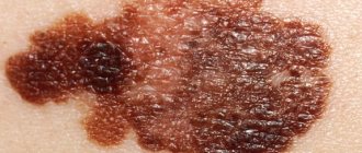
Dysplastic nevus (atypical mole)
An atypical mole is not cancer, but it can become one. It can be found on areas of the skin exposed to the sun. The size of a mole ranges from 5-6 mm to several centimeters, the shape of a mole can be different, the edges are often uneven, the surface is smooth or uneven. The color of such a mole can be any color or even a combination of several colors (pink, brown, black). If a mole has become asymmetrical, changed color or become multi-colored, changed boundaries, and increased by more than 6 mm, there is a reason to consult a doctor to rule out melanoma (a malignant skin tumor).