Correct histological diagnosis has a decisive role in the treatment of melanoma and the prognosis of the disease.
Histology helps determine:
Type of melanoma
Melanoma stage
Classification of melanoma
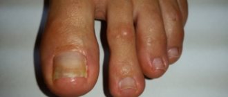
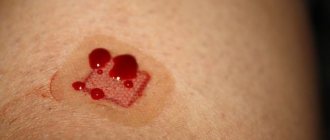
Moles are small, flesh-colored to dark brown spots. In shape, they can be in the form of papules or nodules, consisting of a cluster of pigment cells. Their main medical significance (other than cosmetic) is their resemblance to melanoma. Pigmented lesions are assessed by a set of characteristics (appearance, borders, color, pruritus, bleeding, etc.) that help exclude atypical nevi or melanoma.
Removing a mole is a relatively simple procedure. It is performed for aesthetic purposes or to eliminate pathological changes in the skin that may turn out to be malignant. The only way to know for sure whether a suspicious lesion will require further treatment depends on pathological examination of the tissue. Analysis of moles and other suspicious areas of skin, sending tissue material for histology makes it possible to answer these questions.
What does the histology of a mole show?
This is a more accurate tissue diagnostic method than cytology. It is carried out in a special laboratory, after pre-treatment, staining of a tissue sample, using a powerful microscope. Intradermal nevi, atypical moles, and non-pigmented skin formations can be differentiated by many distinctive features during histology.
Melanoma histology
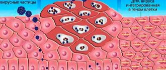
Microscopic diagnosis of pigmented skin lesions is based on identifying and determining the properties of malignant melanocytes. Aggregation (clustering) of atypical pigment cells can be observed in atypical dysplastic nevi. They are sometimes difficult to distinguish from superficial spread of malignant melanoma. Rare forms such as desmoplastic melanoma are morphologically similar to benign skin changes, which can cause diagnostic errors.
All cases of histological testing of tumors require experience and high professional skill.
When conducting histology of malignant melanoma of the skin, the pathologist has to evaluate many factors:
- type of melanoma;
- tumor thickness;
- presence of ulceration;
- infiltration;
- degree of vascular invasion;
- fields of surgical resection (tumor edges);
- mitotic rate or Clark levels.
In addition, the doctor comments on the possible invasion of lymphocytes, the presence of necrosis, cell regression and the level of neurotropism (tumor growth relative to the nerves).
Based on the results of histology of skin cancer, the type of tumor is determined in accordance with the characteristics of cellular morphology.
Tumor thickness is the most important factor in histological evaluation. It is measured in micrometers (μm) and represents the longest diameter from the top of the tumor to the lowest (deepest) epidermal tumor cell.
The pathologist, examining the removed pathological tissue, must make sure that the entire tumor has been resected and sent for examination, and determine the type and stage of melanoma growth. Pathological sections should include fragments with expected deep infiltration.
Any suspicion of skin cancer must be accompanied by histology. Histological information is important for treatment selection and further evaluation of patients with melanoma. Satellites present in the dermis must also be included in the preparation. Assessment of tumor thickness may be uncertain or impossible if there have been regressive changes in the tumor.
The pathology report of malignant tissue should include the following factors:
- histological type (invasive or in-situ);
- tumor thickness during germination (Breslow) in tenths of mm;
- ulceration (present or not);
- other factors (vascular infiltration, lymphocyte infiltration);
- marginal resection margins.
For resected lymph nodes, the following data are decisive for diagnosis and treatment:
- number of metastatic nodes;
- total number of resected nodes;
- perinodal tumor growth;
- properties of the tumor in the area of the resected area.
Histological examination of malignant melanomas of the mucous membranes reveals a variable microscopic picture. Lymphocyte infiltration is less pronounced compared to skin lesions. About 1/3 of melanomas in the nose and mouth are amelanotic. This type of tumor is difficult to differentiate from other types of mucosal cancer. Melanoma immunohistochemistry is useful in this regard.
Preventing and reducing the risk of melanomas
The postulates in the prevention of skin melanoma are: screening, limiting damaging factors, removing altered pigment formations.
Screening includes non-invasive diagnostic methods - dermatoscopy or epiluminescence microscopy annually, and for risk groups at least 2 times a year. Eluminescent microscopy is a non-invasive method for studying skin formations in a special immersion environment that can save a digital image, enter it into a database and track changes by comparing it with existing and subsequent images.
Primary prevention is to avoid the damaging effects of ultraviolet radiation, chemicals and other external factors. Secondary prevention is the removal of all moles suspicious for malignancy.
Tips that can prevent the development of melanoma are quite simple and effective. Will help reduce the risk of mole degeneration:
- wearing dark, thick clothing;
- eating vitamin D;
- removal of altered or injured moles.
The widespread use of screening and preventive measures will have a beneficial effect on the timely diagnosis and treatment of this dangerous disease.
Information for foreign patients
Foreign patients wishing to receive qualified oncological care in Israel can count on correspondence consultation with a specialist at the pre-hospital stage. In addition, if necessary, a histological examination of the material can be carried out.
In Israeli clinics, a sentinel lymph node biopsy procedure is used, according to which lymph nodes with possible metastatic lesions are eliminated.
In Russia and the CIS countries, the procedure of sentinel lymph node biopsy has not yet become widespread.
The use of this technique is most relevant if histological examination reveals an increased mitotic index and ulceration in the area of primary melanoma.
The type of melanoma, its classification and determination of the stage of tumor growth are the main characteristics of histological examination.
In specialized cancer centers such as the Melanoma Unit, histological testing of a tissue sample is carried out for more than 20 parameters. In addition to structural features, the pathologist takes into account many additional characteristics, including the properties of cell nuclei and enzyme activity.
Patient Andrey M., 31 years old, diagnosed with melanoma of the right shoulder
The conclusion of Russian pathologists speaks of increased mitotic activity (from 2 to 5 mitoses in the field of view), melanoma is described as a tumor of the 3rd level of invasive growth according to Clark with a thickness of 2.5 mm. Israeli oncologists established level 4 melanoma according to Clark with a tumor thickness of 4.25 mm. The mitotic activity rate was 20/mm2.
Examples of differences in the histological reports of Russian and Israeli pathologists.
The study of tumor material involves determining its following properties:
- Tumor thickness
- Ulceration
- Clark level
- Histological type
- Cell type
- Primary localization
- Signs of regression
- Number of mitoses
- Lymphocytic infiltration
- Vertical growth stage
- Invasion of blood vessels
- Invasion into the lymphocytic zone
- Ploidy
- S phase of the cell cycle
- DR1 gene expression
- DNA Index
- Heat shock protein expression
- Positive staining for HLD-DR
- P53 protein mutation
- Cell adhesion factor expression
- Protease expression
- Migration marker molecule
- Angiogenesis factor
- Oncogene expression
- Presence of an estrogen receptor
- Cytokine, growth factor
Our clinic offers correspondence consultations and remote examination of histological samples. This greatly facilitates diagnosis and increases the efficiency of medical care.
Histology of the removed mole
The process of malignancy can affect small moles, and when performing digital dermatoscopy, external changes in the nevus may be absent. In this regard, an erroneous treatment tactic may be undertaken to radically remove the mole using a laser, cryodestruction or electrocoagulation. In this case, it is impossible to submit the mole for histology, since the nevus tissue is burned out during the procedure. In this case, a tactical error (performing an operation without histology) can have very serious consequences.
Histology of a mole after its removal is an indispensable condition of modern oncology. Based on the results of nevus histology, one can judge the effectiveness and radicality of the chosen method. Surgical protocols for melanomas and questionable moles categorically exclude the use of techniques in which nevus histology is impossible.
“Acute” surgical excision of any suspicious area of changed skin using a scalpel, followed by examination of the resected material, is the gold standard of modern oncological surgery. Unfortunately, in Russia and the CIS countries there are no strict protocols and “imperative” instructions prohibiting the use of auxiliary techniques (laser, electrocoagulation, radioknife, etc.) “at the discretion” of the surgeon, as a result of which the likelihood of error increases, and histology of the removed mole is carried out not 100% of the time.
The only “bad” sign my mole has is uneven coloring—is it dangerous?
If this is the case, most likely we are talking about a dysplastic (atypical) nevus. These nevi are called that way because they often have uneven coloring or an uneven edge. Upon examination, they may resemble melanoma and it can be difficult for a person not associated with oncology to distinguish one from the other.
Immunohistochemistry of melanoma
An effective diagnostic method that allows one to determine the properties of a tumor, as well as its sensitivity to certain types of antitumor drugs, is immunohistochemistry. More than half of cutaneous melanomas have mutations in the BRAF and NRAS genes. Thanks to immunohistochemical analysis, it is possible to identify specific mutations in the nucleus of tumor cells, which makes it possible to assess the degree of effectiveness of certain target drugs and prescribe the most accurate therapy.
The method is also effective for the morphological diagnosis of anonymous metastatic neoplasms.
Specialists at the Israeli Oncology Center have extensive experience in histological diagnosis. The diagnosis of melanoma in Russia and the CIS countries differs from the diagnostic protocols used in Israel. The lack of clinical experience and the lack of high-tech equipment in most medical institutions in the CIS leads to cases of discrepancies in diagnoses and stages in the expert opinions of specialists.
In Israel, if doubts arise about the morphology and stage of the tumor, other specialists are brought in for examination (second opinion). This approach allows us to reduce the likelihood of subjective error in diagnosis to zero.
Advantages of histological diagnosis in Israel
- Diagnostics are carried out by highly qualified specialists with extensive clinical experience.
- Testing is carried out promptly, within 1–3 days.
- During histological evaluation of the material, additional criteria are applied that are not used in medical institutions of the CIS countries.
- Remote examination of material samples is possible.
- Pathological laboratories in Israel have the necessary certificates for conducting any tissue research, incl. genetic.
Melanoma is easily curable in the initial stages and is actually curable even at the stage of micrometastasis - a timely and correct diagnosis is crucial.
Sources
- https://www.cancernetwork.com/articles/histology-melanoma-and-other-skin-cancers
- https://www.ncbi.nlm.nih.gov/pmc/articles/PMC1770634/
- https://www.slideshare.net/raghuramchary50/histopathology-of-malignant-melanoma
Actinic keratosis (solar keratosis)
This condition appears as small, scaly patches of skin and is caused by excess sun exposure. Actinic keratosis is most often located on the head, neck, or arms, but can be found anywhere on the body. It is impossible to say with certainty whether it will turn into squamous cell skin cancer and how long it will take. Prophylactic treatment of this condition prevents skin cancer from developing from these rashes. The risk of actinic keratosis is highest in those who are fair-skinned, blonde or red-haired with blue or green eyes.