Why does a child develop congenital moles?
Congenital melanocytic nevi (CMN) are benign pigmented tumors consisting of nevus cells that arise as a result of impaired melanoblast differentiation between the second and sixth months of intrauterine life.
UMN occurs in 1% of Caucasian newborns of both sexes.
Visually, such formations have clear boundaries, round or oval outlines, and the surface can be lumpy, wrinkled or folded. Color varies from brown to blue or black. The consistency is usually like that of healthy skin, and hair may grow on the surface.
The reason for their development is always genetic. They can be transmitted from parents or appear on their own, completely by accident.
The time of appearance of such nevi is nonspecific. They are formed during fetal development, but are not always noticeable at birth. In the literature you can find the terms “late UMN” or “with congenital features” - such formations are no different from congenital melanocytic ones, they just appear later.
Typically, UMN are those elements that develop in the first 2 years of life. An important distinguishing feature is that a child’s moles grow with him, in proportion to the baby’s growth.
LNs are often classified as melanoma-dangerous formations, but according to the latest data, such moles are potential precursors of melanoma only in 0.7% of cases.
What may the appearance of a large number of moles indicate?
A large number of age spots may indicate:
- frequent exposure of the baby to the sun;
- hormonal surge (typical in adolescence);
- exposure to x-ray radiation;
- consequences of past illnesses and injuries.
The location of moles can be inherited.
The following are more likely to form birthmarks:
- children with light skin color;
- girls (5 times more than boys);
- premature babies.
Frequent age periods of children that are characterized by the appearance of pigmentation: 0.5-2 years, 5-6 years, 12-16 years.
What are the types of congenital moles in a child?
VMN come in various sizes, and depending on the diameter they are divided into:
- small - up to 1.5 cm;
- medium - from 1.5 to 10 cm;
- large - from 10 to 20 cm;
- giant - more than 20 cm.
One of the most typical symptoms of VN is hypertrichosis, that is, hair is present on the surface of the formation, most often dense and black. It is worth noting that the appearance of some VN is formed gradually, the nature of the coloring may become uneven, tubercles may appear on the surface, and over time, hair begins to grow.
For VN, such changes are a variant of the norm, but they require monitoring by a doctor.
Important features of congenital melanocytic nevi
- Small-sized UMNs are more common and may not appear until age two.
- Small and medium-sized nevi grow more slowly than the child himself and tend to darken and become hairy.
- Large and giant nevi usually occupy part of an anatomical area or all of it, for example an entire limb, neck, part of the back.
- Congenital giant melanocytic nevi transform into melanoma in 6–10% of cases.
Giant VN exceeds 20 cm in size, and such formations are sometimes compared to clothing, called “shirt” or “swimsuit” type. According to statistics, they are rare, affecting 1 in 500,000 newborns.
Giant VNs are only diagnosed at birth. Unfortunately, there is no prenatal screening to detect them, and they are not visualized on ultrasound.
The main medical problem with congenital giant melanocytic nevus is the high risk of developing melanoma, and the tumor can appear anywhere and anytime. What to do in this case? At the moment, the main method is staged excision.
The child's mole is growing. What to do?
Acquired melanocytic nevi (AMN) are benign tumors that arise from melanocytes that have migrated into the skin. They usually appear after six months of life, and reach their maximum size and number at a young age. Subsequently, they may regress or disappear altogether.
The localization of acquired formations is varied. They can be on the skin of the scalp, palms, feet, and also come from the nail matrix, creating difficulties for diagnosis and observation.
Factors influencing the appearance of nevi:
- genetic predisposition;
- level of ultraviolet radiation in childhood;
- phenotypic features of the child’s skin (fair skin, eyes, blond or red hair).
The classification of acquired nevi is varied and includes typical and atypical forms. They are also classified based on the location of the melanocytes.
PMNs are characterized by a round or oval shape and have clear boundaries. Normally they are symmetrical in color, structure and shape.
Most PN are benign and do not require any intervention, but only require lifelong monitoring.
The frequency of malignant transformations in PMN is low, since melanomas most often develop on clean skin, i.e. beyond previous melanocytic nevi. Therefore, their removal for preventive purposes is impractical.
It is worth noting that the tactics for managing patients with melanocytic formations in childhood can be implemented in three ways: surgical excision of the element, dynamic observation of the formation, and zero-intervention tactics, when further observation or surgical intervention is not required. The doctor makes a decision based on an analysis of all factors characterizing the formation: the child’s age, morphology, location, size and melanoma danger of the element.
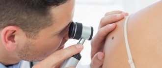
An important diagnostic point when examining skin formations is to perform dermatoscopy.
Modern technologies have allowed doctors and patients to “monitor” the formation using its mapping. This is a procedure for fixing moles, which makes it possible to evaluate the dynamics of changes in structures, sizes and the appearance of new formations.
Results and discussion
According to the literature [1], melanoma is extremely rare in children aged 0–12 years—0.05% of cases; in children aged 12–18 years—in 0.5% of cases. Melanocytic formations, which cause the greatest caution among doctors and concern among parents, most often belong to the group of large and giant congenital nevi and special types of nevi (nevi of special localizations and Spitz/Reed nevus).
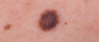
Congenital melanocytic nevi (CMN) are nevi that are present in a child at birth or appear in the first 2 years after birth (late congenital) [2, 3]. Congenital nevi occur in approximately 2–3% of newborns, regardless of ethnicity [4, 5]. Small and medium-sized UMNs are relatively common (Fig. 1, 2) , large or giant UMNs occur in approximately 1 case per 20,000–50,000 newborns [5-8].
Rice. 1. Small congenital melanocytic nevus: clinical (a) and dermoscopic (b) picture.
Rice. 2. Medium congenital melanocytic nevus: clinical (a) and dermoscopic (b) picture.
In 2012, a standardized classification of UMN was proposed, based on the size of the tumor [9]: small - less than 1.5 cm, medium - from 1.5 to 20 cm, large - from 20 to 40 cm, giant - from 40 to 60 cm and more.
Along with the size, it was proposed to evaluate the number of “satellite” nevi (S1: <20, S2: 20–50, S3: >50), color inhomogeneity (C1, C2), surface unevenness (wrinkled surface - R1, R2), the presence of nodes ( N1, N2), presence of hair (N1, N2) [10].
Congenital nevus grows in proportion to the child’s height, and the final diameter of the formation must be calculated by multiplying the existing diameter by the following factors: 1.7 for the head, 3.3 for the legs and 2.8 for other locations [10].
The clinical picture of UMN can be varied. At the beginning of their development, congenital nevi are papules or thin plaques with uniform pigmentation. Over time, nevi become more elevated with light, dark or spotty pigmentation, a wavy, rough, warty surface. Nodules may form on the surface of the UMN (especially large or giant ones), causing concern due to its rapid growth and black or red color. Such changes require a biopsy of the lesion to exclude melanoma [4, 5, 8, 11, 12].
In medium and large congenital nevi, erosions or ulcerations may form due to increased skin vulnerability, which occur in the thickest part of the nevus and self-epithelialize over several days or weeks [13].
UMN are more often mixed, so the dermoscopic picture may contain signs of both epidermal (pigment network, dots, round-shaped globules) and dermal (cobblestone-type globules, structureless areas) origin. VMN are characterized by symmetry, polychrome, and homogeneous structure.
The lifetime risk of developing melanoma in small and medium-sized UMN is less than 1% [14, 15]. In three large retrospective cohort studies of 680 patients with small and/or moderate UMN (mean age at follow-up 10 years, follow-up period 13.5 years), no cases of melanoma were observed [16]. At the Massachusetts Clinical Hospital and the New York University Pigmented Neoplasms Clinic, over 30 years of observation, not a single patient under 20 years of age with melanoma that developed within a congenital nevus with a diameter of less than 5 cm was registered [17].
Melanomas in small and medium-sized UMNs, as a rule, arise after puberty and are located in the epidermis or upper layers of the dermis with the predominant development of the malignant process in the peripheral part of the nevus [16, 18].
In large and giant UMNs, unlike small and medium-sized ones, melanomas more often develop in the dermis or subcutaneous tissue, which precludes their early detection. The risk of developing melanoma in such nevi is 5% or more [19].
The development of melanoma is most likely in patients with UMN in whom the diameter of the adult nevus is expected to be more than 40–60 cm. Additional risk factors for melanoma include location at sites of habitual trauma and the presence of numerous (>20) satellite nevi [19]. Melanomas are less common in patients with UMNs located on the scalp, neck, or extremities.
Large/giant congenital nevi may be associated with neurocutaneous melanosis (NCM). NCM is a congenital condition characterized by large/giant cutaneous UMNs with satellites (S2, S3) and melanocytic tumors of the central nervous system leptomeninges [16].
NCM is divided into asymptomatic and symptomatic [20]. In 4% of patients, NCM occurs with severe neurological symptoms such as seizures, hydrocephalus, and signs of increased intracranial pressure (eg, vomiting, headache), developing by age 2 years. The symptomatic form of NCM has a poor prognosis even in the absence of melanoma development against the background of UMN [21, 22].
Patients with suspected NCM have indications for MRI of the brain and spinal cord [20].
The next group of UMNs, which traditionally causes increased concern among doctors and parents, is represented by acquired nevi in certain anatomical areas. These include nevi of the scalp, genitals and palms/soles.
On the part of specialists, the main reasons for increased attention to nevi of special localizations are the uniqueness of the clinical and dermoscopic picture, monitoring problems and the high frequency of atypical pathomorphological signs [23].
Acquired nevi of the scalp in childhood are predominantly localized in the parietal or occipital regions, their diameter often exceeds 6 mm [5, 24, 25]. Clinically, such nevi are presented as gray-brown nodules or macular-nodular formations with a light brown center and a darker peripheral rim of the “eclipse” type [26] (Fig. 3) .
Rice. 3. Nevus of the scalp: clinical (a) and dermoscopic (b) picture.
Scalp nevi undergo evolutionary changes over time [26–28].
The dermoscopic image of scalp nevi is represented by a predominantly globular model, light or dark brown in color with the presence of punctate vessels or comma-type vessels. Eclipse nevi are characterized by a reticular pattern with perifollicular hypopigmentation [26].
Nevi occurring on the scalp or buttocks during childhood have been reported as an early sign of dysplastic nevus syndrome in individuals at high risk of developing melanoma, especially if there is a family history [29].
In a population-based study, 7% of 1069 Swedish children aged 8–9 years had at least 1 scalp nevus, the presence of which was associated with an almost twofold increase in the total number of nevi compared with children without scalp nevi (p<0.001) [30, 31]. Based on this study, the doctor, if there is a nevus on the scalp, is obliged to conduct a full examination of the entire skin.
As a result, researchers came to a consensus that when various types of acquired or congenital nevi are detected on the scalp, dynamic observation without aggressive diagnostic or therapeutic tactics is advisable [32].
Nevi in the genital area most often appear before the age of 2, can be single or multiple, and look like spots or papules of pink-brown or dark brown color (Fig. 4) . Less common in the genital area are nevi of the “fried egg” type or nevi of the “eclipse” type. These nevi are characterized by evolution with transformation into dermal nevi with a papillomatous surface [33].
Rice. 4. Nevus of the genital area: clinical (a) and dermoscopic (b) picture.
The dermoscopic picture has no peculiarities and corresponds to the clinical type of genital nevus.
In 2014, R. Hunt et al. conducted a retrospective study on the subject of melanocytic nevi in the practice of a pediatric dermatologist. We analyzed data over a 10-year period on 1,159 pediatric patients who underwent a complete examination of the skin, including the genital area. 3.5% had nevi in the genital area, which were not associated with a higher total number of nevi or a family history of melanoma [33].
In children, biopsy or prophylactic removal of nevi of the genital area is not justified in the absence of alarming signs [34].
Acral nevi include nevi localized on the palms, soles and nail apparatus. Clinically, nevi on the thick skin of the palms and soles are represented by spots, less often nodules, of various shades of brown and gray. Dermoscopic features of acral nevi of the palms and soles are “parallel pigmented grooves” and a “lattice pattern” that correspond to the pattern of dermatoglyphs (Fig. 5) . Children are more likely than adults to be diagnosed with the “string of pearls” sign, which corresponds to the excretory ducts of the sweat glands emerging at the top of the comb, or the “fibrillary pattern” found on the supporting areas of the foot [35, 36].
Rice. 5. Acral nevus: clinical (a) and dermoscopic (b) picture.
Acral nevi, which have a subungual location, are longitudinal melanonychia in the form of a light or dark brown stripe, caused by melanocytic proliferation in the matrix of the nail apparatus [37, 38].
In childhood, single dark brown stripes 6 mm wide or more, associated with dystrophy of the nail plate or expansion of the pigmentation zone and dermoscopically characterized by irregular lines in color, width and interval, should be regarded as benign and left under dynamic observation [38-40] . In a similar situation in adults, an incisional biopsy of the subungual matrix is required to rule out melanoma.
The dermoscopic picture of subungual nevi consists of regular parallel lines of different shades of brown. Gradual regression of the acral nevus is accompanied by the appearance of “dots along the lines”; this phenomenon was described in 8 of 15 Japanese children with longitudinal melanonychia [40].
In 2007, a group of researchers [37] proposed a three-step diagnostic algorithm for acquired acral nevi. The basis for changing the existing algorithm was the results of a study confirming that all acral melanomas arise de novo [37, 41], and not against the background of a pre-existing acral nevus. It has been proven that acral nevus does not show signs of melanoma-dangerous; therefore, the authors expressed the idea that follow-up of confidently diagnosed acral nevi is inappropriate.
Spitz/Reed nevus is a special type of benign melanocytic neoplasm that most often develops in childhood. In several large studies, Spitz/Reed nevus was diagnosed in 75% of cases before 20 years of age [42–44].
A Spitz/Reed nevus classically appears as a pink, dark brown, or black nodule, most commonly on the face (especially in young children) or lower extremities. Pigmented forms are usually referred to as Reed's nevus, pink and red - as Spitz's nevus, each of the forms has pathomorphological features. The surface of the nevus can be smooth or papillomatous.
Nevus may be misdiagnosed as wart vulgaris, pyogenic granuloma, dermatofibroma, or juvenile xanthogranuloma [45]. An incorrectly established diagnosis and, as a consequence, inadequate removal of the element can lead to its rapid growth with the formation of atypical clinical and pathohistological signs, which further complicates the determination of tactics for managing the tumor.
Reed's nevus is dermoscopically characterized by a centrally located, structureless area of gray-blue color with peripheral radiance in the form of lines or pseudopodia (metaphorically “exploding star”) [46] (Fig. 6) . The dermoscopic picture of a Spitz nevus is represented by a structureless pink area, punctate, glomerular vessels and chrysalids [47, 48] (Fig. 7) .
Rice. 6. Nevus of Reed: clinical (a) and dermoscopic (b) picture.
Rice. 7. Nevus Spitz: clinical (a) and dermoscopic (b) picture.
It has been demonstrated that Spitz/Reed nevus at different stages of evolution is characterized by different dermoscopic structures [48, 49]. In the growth phase, pseudopodia or globules on the periphery are diagnosed, forming a pattern like an “explosion of a star.” After a few months, the peripheral structures disappear and the formation becomes stable. At this stage, a homogeneous structureless area of brown-black color appears. After a few years, the element begins to lose pigment, undergoing spontaneous involution.
In 2011, Argenziano et al. published data from a study of the natural evolution of Spitz/Reed nevi. According to their study, 80% of nevi underwent complete regression within 25 months. This process did not depend on gender, age and location of elements [6].
In 2010, 175 pediatric dermatologists from the United States of America and Europe provided feedback on their experience in managing Spitz nevi; in total, they analyzed information on 20 thousand cases [45]. Of those surveyed, 96% of doctors believed that typical Spitz nevi were benign, compared with 74% of respondents in a similar study in 2001. 80% of pediatric dermatologists used dermatoscopy to diagnose Spitz nevus, 96% avoided partial biopsy of Spitz nevus. In children with suspected Spitz nevus, clinical doctors indicated the presence of a small stable non-pigmented lesion (49%) and a pigmented formation with a typical “starburst” pattern dermoscopically, 47% of pediatric dermatologists observed involution of Spitz nevus. As a result of observation of 10 thousand children with Spitz nevi, there was never a single case of melanoma and, accordingly, death from a malignant tumor.
Despite the fact that Spitz nevi are benign neoplasms, some morphological features overlap with melanoma.
The dermatoscopic and morphological features of Spitz/Reed nevi lead to controversy regarding appropriate management strategies. With an identical clinical and pathohistological picture, children are much more likely to be diagnosed with Spitz/Reed nevus, while adults are more likely to be diagnosed with melanoma [43, 50].
All researchers who have studied in detail the clinical, dermoscopic and pathomorphological signs of Spitz/Reed nevus testify in favor of the fundamental importance of the patient’s age in determining the tactics for managing the neoplasm [51, 52].
In 1948, Sophia Spitz described “juvenile melanoma”, after which a large number of scientific studies were published revealing the pathohistological picture and variability of Spitzoid neoplasms, proving their benign nature in children. At the same time, there is “Spitzoid melanoma” as a morphological type of melanoma with a special Spitzoid structure. Between these two concepts there is a group of neoplasms with clinical and pathohistological signs of atypia, which are described as “Spitz nevus with atypia”, “atypical Spitz nevus” and “atypical Spitzoid tumor” [53-55].
Atypical Spitzoid neoplasms are a dysplastic type of melanocytic proliferation with borderline histological features indistinguishable from melanoma and uncertain malignant potential [56]. For such tumors, a sentinel lymph node biopsy is performed. A 2014 review found that 39% (119/303) of patients (adults and children) with atypical Spitzoid neoplasms had positive sentinel lymph nodes, but only one of these patients died after five years of follow-up [57]. There is also currently no evidence that further lymph node dissection or adjuvant systemic therapy is of therapeutic benefit in the presence of atypical Spitz nevus [58].
The widespread use of dermatoscopy with the possibility of dynamic control has led to the emergence of new approaches in the management of Spitz/Reed nevus. Many dermatologists have approved the option of long-term monitoring of the clinical and dermoscopic picture of Spitz/Reed nevus at intervals of 3 to 6 months, depending on the age of the children [59-61].
Red, hanging moles - which nevi can scare parents?
Nevus Spitz
Histological examination of UMN shows that this nevus accounts for 1–2% of all UMN. This pink-red or deep black mole has a flat, hemispherical shape. The boundaries are clear, even, and the outlines are regular.
The mole grows, changes quickly, initially appears as a small speck, and mothers often confuse it with dirt on the child’s skin.
Spitz nevus is a benign structure, but clinically and histologically it is the main simulant of melanoma. That is why doctors recommend removing it immediately, or taking it under total control and observation.
Red flags for Spitz nevus:
- size greater than 8 mm;
- rises strongly above the skin level, resembles a knot, a hanging mole;
- independent ulceration, i.e. the appearance of a crust on the surface for no reason;
- pronounced clinical or dermoscopic asymmetry.
Galonevus
This is an acquired melanocytic nevus surrounded by a depigmented rim. It is represented by a red-brown nodule that rises slightly above the skin level. The diameter of the nevus itself is small and is approximately 0.2–1.2 cm, but the whitish rim around it exceeds the size of the nodule itself.
This formation is usually not dangerous and does not require treatment, but dynamic observation and mapping of the element is recommended.
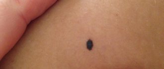
Nevus Reed
A black mole with clear, smooth edges. It is small in size, no more than one centimeter, and is a benign formation.
Nevi of Oto and Ito
They are bluish-gray formations with disturbed pigmentation, located in certain areas. The localization of Oto's nevus is characterized by the periorbital region (in other words, these moles appear on the face, around the eyes), and for Ita - the skin of the neck and shoulders.
Most often, these formations require constant monitoring; laser lightening may be used due to a pronounced cosmetic defect.
Removal methods
Birthmarks need to be removed when:
- localization in a place of increased trauma;
- bleeding, pain;
- changing size, color, shape, structure.
It is necessary to postpone the removal of nevi in children under the following conditions:
- hyperthermia of unknown cause;
- herpes;
- infectious diseases;
- inflammatory process on the skin;
- immunodeficiency.
Nevi are removed after preliminary diagnosis by a dermatologist.
The doctor collects anamnesis, examines the mole with a dermatoscope, and issues a referral for laboratory diagnostics (general and extensive blood tests). If there are no indications, it is better not to remove the formation.
- The medicinal method includes the prescription of cauterizing drugs (cryopharma, stefalin, lapis pencil). Use is allowed only after consultation with a doctor.
- Hardware methods. The choice of removal technique depends on the size and location of the tumor.
| Name | Peculiarities |
| Radio wave | Exposure to the Surgitron apparatus using high frequency waves (up to 4 MHz). Radioknife is used for small moles located on the surface of the skin. The advantage of the procedure is the absence of scars and scars. |
| Cryodestruction | For fresh birthmarks, removal is carried out with a cotton swab moistened with liquid nitrogen. Nevi located in the deep layers of the skin are removed with a cryodestructor. The disadvantage of this method is the danger of damaging healthy cells. |
| Diathermoelectrocoagulation | Removal occurs by high frequency electric current. The advantages of the method are the absence of bleeding and the ability to conduct histology of cut biological material. It is allowed to use electrocoagulation to remove moles on the face, head, and neck. Complete healing of the wound occurs after 7-10 days. |
| Laser | Non-contact and bloodless removal using laser. Impact on the problem area of the skin. |
The procedures are performed under local anesthesia.
- Surgical excision is performed in a hospital setting. The birthmark is removed with a scalpel. Surgery is resorted to in difficult cases when the nevus degenerates into a malignant tumor. The damaged area takes a long time to heal, and there is a possibility of infection of the wound.
- Traditional methods include lightening nevi with chalk mixtures, celandine ointment, and chamomile. Methods help to temporarily disguise a cosmetic skin defect. It is not recommended to use cauterizing substances (acetic essence, table vinegar, iodine) on moles. Uncontrolled use of cauterizing agents poses a health hazard and leads to burns.
You cannot remove the formation yourself (by tying it with hair). It is not recommended to carry out the procedure in a beauty salon. A doctor should remove the nevus. After disposal, the biological material is sent for histology.
After excision of moles, you must adhere to the following rules:
- Avoid contact of the damaged area with water and cosmetics.
- Avoid direct sunlight.
- Do not take wound healing medications without a doctor's prescription.
What to do if a child scratches or tears off a mole?
In most cases, nevi are not a cause for concern. The appearance of moles in a child of any age is absolutely normal. They will gradually increase along with the growth of the child himself, and the location of the moles can be anywhere, including on the head, soles and even the genitals. Moles in children can easily be irregular in shape, have uneven borders, varied color patterns and large size, especially for congenital nevi.
It is also normal that moles will arouse your child's curiosity. If a child scratches or even picks off a mole, do not panic. This is only a reason to contact a dermatologist for dermatoscopy and further observation. So the answer to the question of what to do if your baby picks a mole is very simple: don’t worry and make an appointment with a specialist. ⠀
Parents need to be wary only in the following cases:
- rapid, sudden growth of formation in volume or diameter;
- the appearance of bleeding or a crust on the surface of the mole without previous injury;
- a rare type of mole;
- a large number of moles (>50) or cases of melanoma in the family;
- positive “ugly duckling” symptom (one formation is very different from other moles).
If you or your child just touched a mole, there is no need to worry.