Malignant melanoma has a reputation for complete unpredictability, however, in comparison with other cancer processes, quite a lot is known about the patterns of its progression.
- What does relapse of melanoma look like?
- Causes of melanoma recurrence
- Locations
- Signs and symptoms of melanoma
- Relapse groups
- Frequency of occurrence
- Diagnostics
- Treatment
- Prevention
- Why is relapse dangerous?
- Forecast
What does relapse of melanoma look like?
Recurrent neoplasm differs from primary melanoma not so much in appearance as in the total volume of the lesion. Often secondary formations are very similar to the previously removed maternal one, only there are many more of them, they can be larger or smaller.
In domestic oncology, there are four types of melanoma relapses according to Wagner’s classification:
- local - the tumor is located exactly in the area of the primary operation;
- transit metastases or satellites - next to the scar, but no closer than 2 centimeters, can be localized not only in the skin, but also in the subcutaneous tissue;
- regional metastases - damage by a malignant process to the lymph nodes near the operation area;
- distant metastases - secondary melanoma tumors in other organs and tissues.
The patient can have relapses of any type and in any set, all at once, which is not surprising for such an aggressive tumor.
Causes of melanoma recurrence
It is assumed that the source of development of a recurrent or metastatic tumor is a malignant cell preserved in a blood or lymphatic vessel. The likelihood of the disease returning is affected by the aggressive biology of the melanoma cell and its ability to survive for long periods of time.
It has been noted that the relapse process is typical for young people from 30 to 50 years old, and the timing of progression is associated with gender. For example, damage to regional lymph nodes in women occurs in the first year after removal of melanoma, in men - in the second or third, distant metastases in a man are likely in the first 3 years after treatment, in a woman - after 3 years.
The thickness of the first tumor correlates with the rate of progression; with a thickness of more than 2 mm, 8–9 patients out of ten are likely to develop metastases in the lymph nodes. In clinical studies, a standard approach to the distance from the tumor to the incision line has been developed, depending on the thickness of the tumor node, so at least half a centimeter is retreated at a minimum height, with a nodule thickness of more than 2 mm, 2 centimeters are retreated from it.
As we recently found out, the probability of recurrence has little effect on the volume of the primary operation, but if the volume of excision is insufficient, when malignant cells are found in the edges of the removed tissue during histological examination, a repeat operation is performed no later than 8 weeks or radiation is carried out with preventive drug therapy.
What happens when minimally invasive surgery is stopped?
Everyone is frightened by the results of surgical intervention - the location of the eliminated nevus turns into an open wound. Specialists disinfect it with an antiseptic. They force the patient to be especially careful about hygiene in order to protect the wound from infection until it is completely healed - the formation of a crust of dense consistency.
Everything usually heals within 10-14 days. If, when eliminating a benign neoplasm, the deep layers of the skin were affected, a scar will form (after removal of the mole). This is a normal recovery process. Similar results from such interventions are common. This is due to the individuality of each person’s body.
What causes a scar to appear after mole removal?
Large area of dermis treatment (when the nevus is too large). The healing ability of skin tissue also affects. It is individual for each person. When eliminating nevi of the same size on the body of two different patients, it may turn out that one will have a scar as a result of the recovery period, but the second will not. Other factors that provoke the appearance of a scar:
- the location of a benign tumor above the blood vessels (this leads to the accumulation of an excessive amount of collagen fibers);
- presence of diabetes mellitus;
- infection (healing time increases, risk of scar formation increases);
- constant touching of hands to a fresh open wound;
- not drinking water/vitamins every day (while the recovery period lasts);
- reluctance to use disinfectants and softening ointments.
The likelihood of a scar occurring after mole removal also increases if the patient constantly scratches the scar site, wears uncomfortable clothing that causes injury to the open wound, experiences a sharp surge in hormones, or has unique genetic characteristics that provoke the appearance of a scar.
If you have a scar after removing a mole and you are not satisfied with its presence on your body, then contact a medical center. Most clinics use the laser method of scar removal because it is more effective, painless, and absolutely safe for the body.
Locations
Two thirds of relapses are metastases, one third are nodular formations in the area of the first operation. The location of the maternal tumor affects the fate of the patient:
- every third person operated on for melanoma of the scalp and neck develops tumors in the scar, and in women the problem occurs in the first year after treatment;
- transit metastases have the largest percentage in the group of melanomas of the extremity; this type of progression in women develops five years after excision, and in the male population - in the second and third years after surgery;
- with the initial lesion of the skin of the legs most often, in seven out of ten secondary tumors are found in the lymph nodes, in women in the next year, in men a little later;
- The primary process on the trunk in every fourth person is complicated by distant metastases.
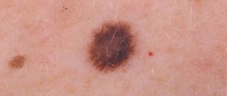
Signs and symptoms of melanoma
Signs of recurrent formation in the skin and soft tissues do not differ from those of a primary tumor; the same diagnostic algorithm is applicable to them: asymmetry and uneven coloring, uneven edges and instability of the condition. The sizes are very variable - from tiny to gigantic, “the size of a baby’s head.”
Symptoms of metastases depend on the location, so if the lungs are affected there may be a cough with shortness of breath, if metastases to the brain - blurred vision, headaches, vomiting, convulsions.
Regional metastasis is manifested by an increase in the package of lymph nodes, followed by tumor growth into soft tissues and blocking of nearby passing vessels, which is complicated by swelling of the limb.
Clinical studies have proven that detecting disease progression before symptoms appear significantly improves life prospects. The course of the disease is always individual, so observation after treatment should be based on the biological characteristics of the malignant tumor. Euroonko does everything for early detection and prevention of health problems.
Relapse groups
In 1985, the domestic oncologist Anisimov proposed, for the convenience of describing the clinical picture, to divide recurrent tumors into six groups:
- The first - round and few formations, often outside the scar and mainly subcutaneously - in the fatty tissue, often fall under the criteria of “transit” metastasis according to Wagner.
- The second is multiple irregular cutaneous and subcutaneous infiltration accompanying vessels and nerves; it is assumed that the external picture of relapse is formed by tumor cells that have taken root in small vessels.
- The third is nodules directly associated with the operation area that have grown from malignant cells remaining in the skin.
- The fourth is polycyclic multiple formations.
- The fifth is a lot of bulging nodules, often on a stalk, like morel mushrooms.
- The sixth is a combination of all five options.
Classification is rarely used in clinical practice, because the assessment of treatment results is based solely on the size of the nodes, and not on their appearance.
Frequency of occurrence
For any type and size of melanoma, the most common progression after surgery is metastases to other organs in almost 60% of patients.
The most dangerous period is the first three years after surgery, but in the first year, 60% are more likely to have metastases in regional lymph nodes. In stages 1 and 2, the incidence of lymph node involvement is low - every fifth person, and very rarely - less than 5% are affected by the postoperative scar.
Due to the development of relapses of the disease, only 60% of patients live longer than five years, but the disease can return after 15 years - in 7%, and after 25 years - in 10%. Every fifth person with an unimportant individual prognosis, however, does not experience a relapse in the next 5 years.
What to do if a mole reappears after removal
If a removed mole re-grows on the body, you should carefully monitor the color and size of the mole. Inspect the operated area daily. If a large scar or scar forms, you should seek help from a doctor. After analyzing the patient, the specialist will select the optimal medications. Medicines will remove the crust on the wounds.
If the recurrence of the nevus is malignant, the following signs are observed:
- pain in the area of the new mole;
- the formation of severe itching of the skin;
- the surface of the body peels off and becomes covered with small cracks;
- blood oozes from the growth;
- education takes on a different color and shape.
If you experience the above symptoms, seek help from your doctor. A medical professional will determine the risks of cancer development and select effective therapy. The main task is to neutralize the effect of metastases, which dynamically spread throughout the human body. Otherwise, complete recovery will become unrealistic.
Diagnostics
The examination is similar to the diagnosis of the primary process. A special feature of diagnosing progression is strict adherence to a schedule of regular examinations, which makes it possible to identify the process at the very beginning and before the appearance of clinical symptoms.
In European countries, it is recommended to visit an oncologist every 2 months during the first year after surgery, quarterly in the second year, and then be monitored without reducing intensity, since most often relapses occur in the first three years.
Early diagnosis of melanoma relapse is an objective reality today. In each clinical case, Euroonko clinics use an individual diagnostic search program, eliminating both the redundancy of the examination and its insufficiency.
Should you be concerned about pigmentation changes?
After surgery to remove a mole, traces of the procedure may be visible on the skin. Light dry spots do not pose a danger to the body. This also applies to the black, dense crust. Over time, the pigmentation of the affected area will be restored.
You should only be wary of dark red and purple spots that cause discomfort. If the operation site is inflamed, moist and hot, this indicates the presence of an inflammatory process. In this case, you should not self-medicate and urgently need to go to the clinic.
Treatment
Treatment of local recurrence in the scar, transit metastases and altered lymph nodes is similar to the tactics for the primary tumor - surgery is optimal, including plastic closure of a large tissue defect.
Adjuvant drug treatment after surgery is the norm; drugs that have not previously been used for prevention are used, although the choice is small: alpha-interferon and ipilimumab.
With metastases to other organs, the question of drug therapy arises, and in the first line they resort to immuno-oncological drugs in combination with targeted drugs in the presence of gene mutations.
The choice of drugs is large, therefore, in the absence of results or progression during second-line treatment, they resort to previously unused drugs. Chemotherapy for relapse of melanoma is used as a last resort, not because of pronounced adverse reactions, the problem is the low result. For superficial formations, radiation therapy is possible.
Causes of relapse after primary removal
The occurrence of relapse is due to a number of reasons and complications. The pathological process is based on the human factor. Incorrect determination of the depth of pigment spots affects the outcome of the operation. It's easy to get an infection into an open wound. The use of antiseptics and medications is a prerequisite for rapid healing of the area.
Some people, after removing a mole, remove the resulting crust. You should wait for the keratinized cells that cover the pink skin. After surgery, select effective wound treatment options. The main task is to prevent inflammation and tumor growth.
Incorrect determination of the depth of pigment tissues
Epiluminescence microscopy magnifies images of pigments 40 times. The procedure allows you to visually study the depth and structure of the formation. The computer records information on the camera, comparing it with the data in the database. In practice, dermatologists do not often resort to cryodestruction for help. Controlling the depth and level of tissue freezing is problematic. If the operation is performed poorly, part of the nevus in the epithelium remains viable. As a result, a relapse will occur and the previously removed mole will grow back. The danger is that damaged structures can transform into atypical cells. It will be difficult to eliminate the pathological process!
Incomplete removal of a mole is fraught with complications and additional disorders. To prevent degeneration, systematically monitor the pigment spot on the skin at home every day.
Incorrect removal method selected
Determining the causes of melanoma development underlies the choice of therapeutic regimen. Options for nevus removal are surgical. Modern technology and equipment allows operations to be carried out through fine tissue dissection. Effective methods and procedures for removing formations:
- surgical intervention;
- use of laser (including during transformations);
- carrying out electrocoagulation;
- radiosurgical approach.
The choice of the appropriate option is carried out personally for each person. Brown structures and cancerous growths are removed with a scalpel. Using this manipulation, nevus tissue is cut out from the deep layers of the dermis. Other moles are removed with laser and liquid nitrogen. The manipulation is carried out carefully, without large losses of blood.
An incorrectly selected treatment regimen is a source of additional disorders. Treating an area of skin with liquid nitrogen can cause severe burns. As a result, wound healing will be prolonged and painful. A person has an increased risk of infection.
Degeneration of nevus
The mechanism of degeneration of a mole is the body’s response to external stimuli. The main activators for a person in this case are prolonged exposure to the sun and daily visits to the solarium. If a person touches the structure with a fingernail, melanoma may develop.
The pathology is dangerous for pregnant women. Hormonal changes affect changes in the structure and cells of the epidermis.
Poor quality surgery is also a source of nevus degeneration. An inaccurate incision or infection entering the wound can cause complications. At the first sign of additional complications, seek help from your doctor.
Self-removal of a mole
At home, it is almost impossible to maintain complete sterility. This factor increases the risk of infectious agents entering a weakened human body. In practice, relapse of the nevus occurs if the growth is not completely removed. Melanoma-prone and malignant structures should not undergo cosmetic procedures.
The formations are eliminated by surgical excision, which is performed in the area of healthy tissue. The disadvantages of scratching or damaging a nevus are:
- a multiple increase in inflammation in size (will grow gradually);
- severe infection;
- repetition and dangerous consequences may occur;
- development of pathologies and complications.
Why is relapse dangerous?
Relapse in melanoma is not only the appearance of a malignant formation in the scar, but also metastases. They most often affect the lymph nodes, lungs, skin, brain and liver. The possibility of removing a recurrent formation opens up prospects for a long life; if the operation is technically impossible, then the present and future will be occupied by drug therapy.
Survival for multiple organ metastases does not exceed six months; the results of drug therapy leave much to be desired, even with the use of innovative immuno-oncological drugs, which rarely promise more than two years of life.
Removing scars after mole removal - laser method
Cosmetologists use this method when pharmacological drugs, dermabrasion and other methods are powerless. Its essence is a minimally invasive manipulation that involves vertical laser penetration into the deep layers of the dermis, vaporizing tissue, increasing blood flow and stimulating collagen synthesis.
The procedure lasts 15 minutes. You don't need to seriously prepare for it. The result of the manipulation will be a noticeable improvement in the skin. All skin irregularities will be eliminated, the epidermal tissue will become more elastic, and scars will be completely smoothed out.
The task of a cosmetologist who aims to remove a scar after removing a mole at the request of a patient is the following: he needs to correctly select the parameters of the laser equipment so that the effect is optimal. To do this, a specialist needs to analyze the structure of the epidermis, as well as its humidity/type.
Forecast
Every year, melanoma is diagnosed in an average of 15 people out of every hundred thousand adults, approximately 3 patients die, and with a fairly stable mortality rate in the last quarter century, men began to die more often. Gender determines a lot in the prognosis of the disease, other things being equal, but young people experience the disease with less difficulty.
The probability of death with a late relapse is many times lower than with an early relapse. Early detection of progression promises better treatment results.
It is extremely difficult to predict the course of melanoma, because even a common process is not considered absolutely fatal; this malignant disease does not often live up to expectations. Don’t guess “to be or not to be”, contact specialists if you have problems, or better yet, before they appear - we will always help.
| Read more about dermatological studies at Euroonko clinics | |
| Consultation with a dermatologist-oncologist | from 5,100 rub. |
| Skin examination using the German FotoFinder device | RUB 13,400 |
| Diagnosis of melanoma | from 5,100 rub. |
Book a consultation 24 hours a day
+7+7+78
Bibliography
1. Semiletova Yu.V., Anisimov V.V., Vagner R.I. /Treatment of patients with primary skin melanoma. Current state of the problem // Siberian Journal of Oncology. 2010. No. 4. 2. Semiletova Yu.V., Anisimov V.V., Lemekhov V.G. et al./Risk factors for relapse after radical treatment of skin melanoma//Sibir.onko.zhur.; 2012. No. 2 (50) 3. Stroyakovsky D. L., Abramov M. E., Demidov L. V. et al. /Practical recommendations for drug treatment of skin melanoma // Malignant tumors: Practical recommendations RUSSCO #3s2, 2018 (volume 4. de Vries E., Bray FI, Coebergh JW, Parkin DM /Changing epidemiology of malignant cutaneous melanoma in Europe 1953–1997: rising trends in incidence and mortality but recent stabilizations in western Europe and decreases in Scandinavia// Int J Cancer 2003; 107. 5. MacKie RM, Bray C., Vestey J., et al./Melanoma incidence and mortality in Scotland 1979– 2003 // Br J Cancer 2007;96.
Ways to remove stains
Pink and white spots do not require treatment. Natural pigmentation of cells occurs gradually. If personal hygiene rules are not followed and the wound is not properly cared for, a permanent white pigment spot may form on the skin.
To avoid possible complications, do not subject the removal site to mechanical damage and do not get it wet . Peeling off the crust and being in a humid environment can contribute to infection of the wound or increase the time of skin regeneration.
Limit your time in direct sunlight . If possible, protect the surgical site with clothing or tape for 3-5 months.
Photo 2. The crust formed at the site of the mole should be preserved for as long as possible. Source: Flickr (Haw Yu)
If the mole was not completely removed or, after accidentally removing the crust, obvious signs of suppuration appeared, the wound bleeds and does not heal for a long time, be sure to contact a specialized specialist .
The doctor will determine the cause of the pathology and prescribe appropriate treatment. In some cases, repeat surgery will be required.