Causes Symptoms Diagnostics Treatment Advantages of treatment at MGK Prices
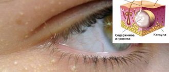
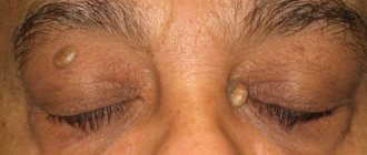
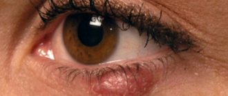
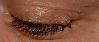
Xanthelasmas of the eyelids look like small single or multiple flat yellowish plaques located at the inner corner of the upper (usually) or lower eyelid. Xanthelasmas are benign formations that are not prone to malignancy; their appearance is associated with general disorders of lipid metabolism.
Prices for treatment for xanthelasma
The cost of surgical treatment of xanthelasma starts from 2000 rubles. (in the presence of a single formation up to 5 mm). The final cost of removal will depend on the number and size of formations, as well as the volume of therapeutic and diagnostic procedures performed.
You can find out the cost of a particular procedure by calling (499) 322-36-36 or online using the appropriate form on the website; you can also read the “Prices” section.
Go to the "Prices" section
Make an appointment
Blepharitis
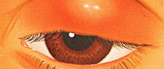
A common disease of the eyelid margins is blepharitis. It can be caused by a lack of vitamins, weakened immunity, diabetes and infections developing in the gastrointestinal tract, nasopharynx or oral cavity. Also unfavorable factors are poor hygiene, dirt, and dust in the air.
Types of blepharitis
There are several types of blepharitis, differing in symptoms.
- Scaly (seborrhea). The eyelids become red and thick, there is itching, photophobia, and narrowing of the palpebral fissure. White or yellowish scales appear on the eyelashes, at the very base. They look a lot like dandruff. Under them, the skin of the eyelids is reddish, sometimes blood vessels are visible, and the eyelids itch. Increased response to dust and wind. My eyes get very tired in the evening. Without treatment, the situation worsens and the disease becomes protracted.
- Ulcerative. Severe form, accompanied by pain. Most often appears at a young age. Scales and bleeding crusts form on the eyelids. The crusts fall off along with the eyelashes, and pus is released from the formed holes. New eyelashes begin to grow in a different direction. In some cases, scars and entropion of the eyelids form. If treatment is not started in time, inflammation will spread from the eyelids to the cornea and conjunctiva.
- Meibomiev. Characterized by inflammation of the meibomian glands. They are located in the cartilage of the eyelids. When pressed, a yellowish liquid appears, and purulent discharge appears in the corners of the eyes. Often the disease is accompanied by conjunctivitis.
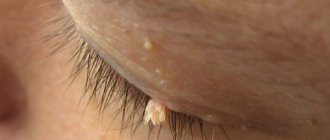
- Demodectic. The cause of this blepharitis is the Demodex mite, which lives in the eyelash follicles. All people have it, but it is active only under certain conditions. Most often this is due to decreased immunity. The disease is characterized by the following symptoms: severe itching, which is especially disturbing in the morning, after sleep. The eyelids thicken and turn red. White flakes, similar to a collar or muff, form at the base of the cilia.
- Allergic. This blepharitis is often accompanied by conjunctivitis. The cause of the disease is the body’s sensitivity to medications, cosmetics or other substances. Often allergies occur due to contact with pollen, wool, dust, fluff and household chemicals. Blepharitis begins suddenly, the eyelids swell, tears flow, there is pain in the eyes, and photophobia is present. In the chronic form, the eyelids are very itchy. Exacerbation often occurs during the flowering period.
Blepharitis is also classified by location:
- anterior marginal, when only the area of cilia growth suffers;
- posterior marginal, when the meibomian glands become inflamed, which provokes inflammation of the conjunctiva and cornea;
- angular, when inflammation of the corners of the eyes is observed, occurs with purulent discharge, thickening of the eyelids and the formation of ulcers.
Symptoms
Xanthelasmas of the eyelids are rounded plaques slightly protruding above the surface of the skin, pale yellow in color, and their consistency is soft. The size of such formations can vary from several millimeters to 1.5 cm; the elements themselves can be either single or multiple. In some patients, xanthelasmas may appear as a solid yellowish stripe extending onto the bridge of the nose.
Xanthelasmas in the lower eyelid area (xanthomas) are rarely isolated; they usually occur in addition to the same formations located on the upper eyelid and are a manifestation of xanthomatosis. Patients with xanthelasmas of the eyelids do not present any complaints; the formations are painless and represent mainly a cosmetic defect. Once occurring, xanthomas and xanthelasmas remain for life, very slowly increasing in size.
Pediculosis
Dandruff on eyelashes is possible due to lice. With the appearance of parasites on the hair, a characteristic itching occurs. Particles of dead epithelium and waste products of lice fall on the eyelashes and are perceived as dandruff. You can also see the nits themselves, because they, like lice, feel great on the scalp, eyebrows and eyelashes, in the beard and mustache.
Pediculosis occurs not only on hair that lacks proper hygiene, but also on absolutely clean hair. The main route of infection is contact (from person to person, from objects, clothing, combs, bed).
It is not difficult to cure the disease; the main thing is to be systematic and follow the doctor’s advice.
Diagnostics
Patients with xanthelasma of the eyelids need to consult not only an ophthalmologist, but also an endocrinologist and a dermatologist. The examination must include a general blood test, a study of lipid metabolism (level of lipoproteins and cholesterol in the blood serum).
The characteristic appearance of the formations usually does not pose any difficulties in making a diagnosis. In addition to xanthelasmas and xanthomas, the patient is often diagnosed with obesity, hypertension, metabolic syndrome or diabetes mellitus. Xanthelasma of the eyelids must be differentiated from other similar skin diseases (syringoma, pseudoxanthoma), incl. malignant tumors.
Complications of the disease
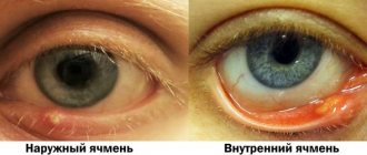
Internal stye (like external stye) can often be accompanied by inflammation of nearby lymph nodes, swelling and hyperthermia, headache and general malaise. There are cases when the inflammatory process of the meibomin glands causes purulent inflammation of the orbit, thrombophlebitis of the orbital veins, thrombosis of the cavernous sinus, inflammation of the meninges and septicemia - dangerous conditions that can lead to death. Usually, this happens when trying to squeeze pus out of an abscess, because... the blood of the eyelids flows to the veins of the orbit, and then to the sinuses of the brain.
Sometimes several styes form at once, which merge into a single abscess. In this case, the likelihood of serious complications becomes much higher.
Treatment of xanthelasma of the eyelids
Since xanthelasmas and xanthelomas in most cases accompany lipid metabolism disorders, liver disease, etc., it is imperative to treat the underlying disease. It is recommended to follow a diet limiting the consumption of animal fats and replacing them with vegetable fats. If hypercholesterolemia is detected, drugs that normalize lipid metabolism (statins, lipoic acid, berlition) are prescribed. The use of drugs that normalize liver function (choleretic agents, Essentiale, multivitamins) is indicated.
There is no drug treatment for xanthelasma of the eyelid; the formation (at the request of the patient) is removed using a laser, electrocoagulation or surgery. The intervention is performed under local infiltration anesthesia. During removal, a specialist uses tweezers and miniature scissors to separate the base of the plaque, then treats the skin in the wound area with an antiseptic. If the size of the lesion to be removed is not large, the edges of the wound are treated with a solution of iron albuminate, which promotes effective healing. In the case of a more extensive wound surface, its edges are treated with electric current (diathermy) or cosmetic sutures are applied.
Popular questions
How is a chalazion different from a stye?
Etiology. Chalazion is a non-infectious disease, while barley (hordeolum) is caused by opportunistic bacteria that are constantly present in the human body and are activated under certain conditions. Most often, the development of barley is provoked by Staphylococcus aureus.
External signs and sensations. Barley always develops on the ciliary edge, chalazion - at some distance from it. Barley is accompanied by severe hyperemia, pain, and local fever. With chalazion, these signs are absent or slightly expressed. The stye suppurates and a yellowish spot appears in its center. Chalazion suppurates only when a secondary infection occurs.
Exodus. Barley almost always breaks through, after which relief comes. Chalazion very rarely breaks out on its own.
Is chalazion dangerous?
Timely diagnosis, medical supervision and treatment appropriate to the form and severity of the disease eliminate the danger of chalazion. However, if medical care is neglected, complications may arise, some of them quite severe.
Complications of chalazion:
- conjunctivitis, blepharitis, abscess formation due to infection;
- mucous cyst of the eyelid;
- visual impairment due to prolonged pressure on the cornea due to large formations;
- fibrosis of the cartilage of the eyelid, loss of eyelashes;
- inflammation of the subcutaneous fatty tissue of the orbit of the eye.
What to do if a child’s chalazion ruptures?
After a breakout of the chalazion capsule in a child, it is necessary to treat the breakout site and surrounding tissues with antibacterial agents to prevent infection and the development of complications. To receive qualified help, you need to see a doctor. If it is not possible to visit a pediatric ophthalmologist in the near future, you should wash the wound surface with an antiseptic solution, such as furatsilin. To do this, soak a cotton pad generously in the solution and rinse the eye moving from the outer to the inner corner. You need to rinse several times, each time using a new cotton pad. After this, you can put antibacterial drops into your eye. Before using them, you should carefully study the instructions to see if the product is suitable for use in children, and also pay attention to the expiration date, including after opening. It is better to use a freshly opened bottle.
What are the reasons for the recurrence of chalazion in a child?
Basically, relapse occurs with concomitant diseases of the body that were the cause of chalazion, other ophthalmological pathologies, as well as with incomplete enucleation of the capsule during surgical removal. Frequent recurrences of chalazion are a reason for histological examination of its contents in order to exclude adenocarcinoma of the meibomian gland.
Sources:
- Recurrent chalazions in children. Serge Doan. Service d'ophtalmologie, hôpital Bichat et Fondation Adolphe-de-Rothschild, Paris, France.https://pubmed.ncbi.nlm.nih.gov/32237654/
- A comparison of intralesional triamcinolone acetonide injection for primary chalazion in children and adults. Jacky WY Lee, Gordon SK Yau, Michelle YY Wong, Can YF Yuen. 1Department of Ophthalmology, Caritas Medical Centre, 111 Wing Hong Street, Kowloon, Hong Kong Special Administrative Region, Hong Kong.https://pubmed.ncbi.nlm.nih.gov/25386597/
- Assessment of the condition of the meibomian glands in children of different ages. Bobryshev V.A. FORCIPE, 2022. p.632-633.
The information in this article is provided for reference purposes and does not replace advice from a qualified professional. Don't self-medicate! At the first signs of illness, you should consult a doctor.
Advantages of xanthelasma treatment and prices at MGK
By contacting the Moscow Eye Clinic, you can be assured of a quick and reliable diagnosis of xanthelasma of the eyelid and its effective treatment. Removal of xanthelasma of the eyelid can only be performed by a specialist with appropriate professional skills. To avoid possible complications, this procedure should be entrusted to an experienced doctor. At the Moscow Eye Clinic you will be able to undergo all the necessary tests, based on the results of which the attending physician will recommend you the most effective treatment methods. The Clinic employs specialists with extensive professional experience who enjoy well-deserved respect both among colleagues and among patients.
Removal of xanthelasma of the eyelid is performed on an outpatient basis under local anesthesia. During the operation, sterile instruments and disposable consumables are used, which eliminates the risk of infectious complications.
Author:
Yakovleva Yulia Valerievna 5/5 (1 rating)
Honey. portal:
Other causes of dandruff on eyelashes
Not only viral or bacterial infections can cause the formation of white or yellowish flakes on the eyelashes. Also at risk of dandruff:
- untimely, inadequate care;
- use of low-quality cosmetics;
- poor nutrition;
- general weakness of the body.
To get rid of a disease, you need to eliminate the root cause. For example, normalize your diet, give up foods that have a detrimental effect on the condition of your skin and hair (spicy, salty, fatty and smoked foods).
If it's a cosmetic issue, you need to change it. You should trust proven products, choose products according to your skin type, and check the expiration date. Sometimes it is beneficial to completely avoid cosmetics.
It is recommended to establish a rational daily routine, spend more time outdoors, avoid stress, get enough sleep, and give up bad habits.
Tips for caring for eyelashes
Proper eyelash care is very important to prevent dandruff.
- Wash your face twice a day. There is no need to use soap; it is better to buy foam, gel or milk.
- Carefully remove mascara, shadows and other dyes from eyelashes and eyelids. Use special products, not just washing.
- Periodically apply burdock or castor oil to your eyelashes. This activates hair growth and helps get rid of scales.
- To soften the skin and provide nutrition to the follicles, you can use lotions with string, St. John's wort, calendula, and burdock.
