Causes Symptoms Diagnostics Treatment Advantages of treatment at MGK Prices
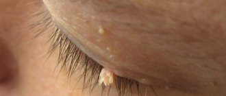
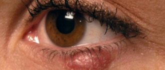
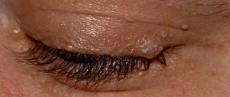
Xanthelasmas of the eyelids look like small single or multiple flat yellowish plaques located at the inner corner of the upper (usually) or lower eyelid. Xanthelasmas are benign formations that are not prone to malignancy; their appearance is associated with general disorders of lipid metabolism.
Prices for treatment for xanthelasma
The cost of surgical treatment of xanthelasma starts from 2000 rubles. (in the presence of a single formation up to 5 mm). The final cost of removal will depend on the number and size of formations, as well as the volume of therapeutic and diagnostic procedures performed.
You can find out the cost of a particular procedure by calling (499) 322-36-36 or online using the appropriate form on the website; you can also read the “Prices” section.
Go to the "Prices" section
Make an appointment
How to remove xanthelasmas
As noted earlier, the most atraumatic and safe method of eliminating tumors is laser removal. At the beginning of the session, the affected skin area is treated with an antiseptic; if necessary, if the patient has a low pain threshold, a local anesthetic can be used. Then the doctor begins targeted heat treatment of the plaque, making a sufficient number of “passes” with the laser to completely remove it. Having evaporated the tumor completely, the doctor re-treats the skin with an antiseptic, and the patient can go home.
View price
Symptoms
Xanthelasmas of the eyelids are rounded plaques slightly protruding above the surface of the skin, pale yellow in color, and their consistency is soft. The size of such formations can vary from several millimeters to 1.5 cm; the elements themselves can be either single or multiple. In some patients, xanthelasmas may appear as a solid yellowish stripe extending onto the bridge of the nose.
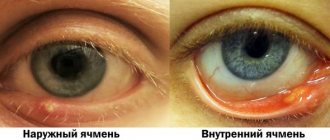
Xanthelasmas in the lower eyelid area (xanthomas) are rarely isolated; they usually occur in addition to the same formations located on the upper eyelid and are a manifestation of xanthomatosis. Patients with xanthelasmas of the eyelids do not present any complaints; the formations are painless and represent mainly a cosmetic defect. Once occurring, xanthomas and xanthelasmas remain for life, very slowly increasing in size.
What symptoms accompany the appearance of yellow spots?
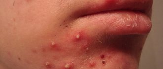
As a rule, the appearance of a yellow spot on the protein is accompanied by other reactions of the body. Symptoms are local and general. If the cause of the discoloration of the sclera is an ophthalmological disease, the following conditions may develop against its background:
- inflammation of the eyelids;
- itching sensation and severe pain in the eye area;
- increased sensitivity to bright light;
- the appearance of discharge from the eyes;
- blurred vision.
If the cause of the yellow spot on the sclera is a systemic disease, the above symptoms include loss of appetite, feeling tired, nausea, chills, fever and increased body temperature.
Diagnostics
Patients with xanthelasma of the eyelids need to consult not only an ophthalmologist, but also an endocrinologist and a dermatologist. The examination must include a general blood test, a study of lipid metabolism (level of lipoproteins and cholesterol in the blood serum).
The characteristic appearance of the formations usually does not pose any difficulties in making a diagnosis. In addition to xanthelasmas and xanthomas, the patient is often diagnosed with obesity, hypertension, metabolic syndrome or diabetes mellitus. Xanthelasma of the eyelids must be differentiated from other similar skin diseases (syringoma, pseudoxanthoma), incl. malignant tumors.
Eye diseases
The second reason why yellow spots may appear on the protein is diseases of the organs of vision. They do not have serious irreversible consequences and do not lead to complete loss of vision. Most often, yellow spots appear due to the following diseases:
- pinguecula. A benign yellow growth in the conjunctival area. Yellow spots appear symmetrically: in the corners of the eyes and on the outside. Elderly people are especially susceptible to the disease, since one of the reasons for its development is age-related processes in the mucous membrane of the eye;
- pterygium. Thickening and proliferation of the mucous membrane in the upper corner of the eye. This pathology appears as a yellow triangular spot. In most cases, it does not cause any discomfort. In some situations, the pterygium may change color to bright pink or scarlet. The disease occurs due to aggressive environmental influences. Most often, the cause of its development is a dry, hot climate, ultraviolet radiation and exposure to strong winds on the eyes;
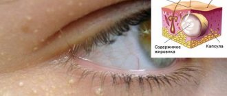
- dermoid cyst of the conjunctiva. Congenital benign neoplasm. It appears in the form of a dense capsule containing a fatty substance inside. Over time, the cyst grows and increases in volume;
- conjunctival cyst. Manifests itself in the form of epithelial growth with fluid inside. The cyst may have a yellow tint and externally look like a small spot on the conjunctiva;
- nevus. Similar in nature to moles on the skin. This is a yellow or brown spot that can be located in any area of the sclera, including its inner or outer corner. When a nevus grows, it poses a serious threat to vision;
- Horner-Trantas spots. They occur due to an allergic reaction of the body. They appear in the form of small “grains” localized on the white of the eye around the cornea. The impetus for development is allergic conjunctivitis or keratitis.
Treatment of xanthelasma of the eyelids
Since xanthelasmas and xanthelomas in most cases accompany lipid metabolism disorders, liver disease, etc., it is imperative to treat the underlying disease. It is recommended to follow a diet limiting the consumption of animal fats and replacing them with vegetable fats. If hypercholesterolemia is detected, drugs that normalize lipid metabolism (statins, lipoic acid, berlition) are prescribed. The use of drugs that normalize liver function (choleretic agents, Essentiale, multivitamins) is indicated.
There is no drug treatment for xanthelasma of the eyelid; the formation (at the request of the patient) is removed using a laser, electrocoagulation or surgery. The intervention is performed under local infiltration anesthesia. During removal, a specialist uses tweezers and miniature scissors to separate the base of the plaque, then treats the skin in the wound area with an antiseptic. If the size of the lesion to be removed is not large, the edges of the wound are treated with a solution of iron albuminate, which promotes effective healing. In the case of a more extensive wound surface, its edges are treated with electric current (diathermy) or cosmetic sutures are applied.
Causes of xanthelasma of the eyelids
The cause of the development is considered to be metabolic disorders, most often lipid metabolism, the presence of problems with the heart, liver, pancreas in people suffering from obesity, diabetes, and atherosclerosis. Growth occurs slowly, mostly observed in older women. Can be inherited. They will not disappear on their own, so it is necessary to eliminate them. There are several options.
The plaque is affected by:
- liquid nitrogen for several seconds until the cells are completely destroyed;
- targeted laser, painless, bloodless, without scarring or unpleasant consequences;
- radio waves contactlessly evaporate unsuitable layers;
- promptly using scissors and tweezers using local anesthesia; the edges of the wound are treated with medications or cauterized with electrodes.
In order to prevent a secondary occurrence, you need to look for the abnormalities in the body responsible for this and cure them.
Advantages of xanthelasma treatment and prices at MGK
By contacting the Moscow Eye Clinic, you can be assured of a quick and reliable diagnosis of xanthelasma of the eyelid and its effective treatment. Removal of xanthelasma of the eyelid can only be performed by a specialist with appropriate professional skills. To avoid possible complications, this procedure should be entrusted to an experienced doctor. At the Moscow Eye Clinic you will be able to undergo all the necessary tests, based on the results of which the attending physician will recommend you the most effective treatment methods. The Clinic employs specialists with extensive professional experience who enjoy well-deserved respect both among colleagues and among patients.
Removal of xanthelasma of the eyelid is performed on an outpatient basis under local anesthesia. During the operation, sterile instruments and disposable consumables are used, which eliminates the risk of infectious complications.
Author:
Yakovleva Yulia Valerievna 5/5 (1 rating)
Honey. portal:
Methods for removing xanthelasma
There are many methods for removing formations, such as chemical cauterization, cryodestruction, electrocoagulation, etc. However, these methods do not guarantee the total elimination of xanthelasma and the prevention of relapse, but only stop the development of this disease. The most effective methods are:
- laser removal is a safe method of layer-by-layer removal of tumor tissue using a laser beam, in which only affected tissue is removed and healthy areas of the skin are not damaged, which significantly reduces the duration of the rehabilitation period; after laser removal there are no scars left, another advantage is the immediate cauterization of the affected capillaries;
- surgical excision - removal under local anesthesia using a scalpel; this method is suitable for eliminating large xanthelasmas and involves the application of cosmetic sutures; The duration of the rehabilitation period is determined by the area of the affected tissue.
Xanthelasma
Laser removal of xanthelasma
Plaques in blood vessels. Lymphologist's View | Part 1
Good afternoon. We hope that the coronavirus situation will soon subside and we will return to normal. Today we will talk about atherosclerosis and cholesterol plaques.
It's no secret that diseases of the cardiovascular system are the main cause of death - in 50% of cases, according to international statistics. These diseases mainly include heart attacks and strokes. They have a common reason for their development. The name of this cause is atherosclerosis.
Table of contents
- What is atherosclerosis?
- How is an atherosclerotic plaque formed?
- Where can atherosclerosis develop?
- Lymphatec approach to the treatment of atherosclerosis
- Video performances
What is atherosclerosis?
Atherosclerosis is a multifactorial disease. Accompanied by pathologies of the arteries of our organs. Lipid and protein substances accumulate on the vessel wall. Then they grow with connective tissue and even become calcified. As a result, the vessel narrows and blood flow is disrupted. Due to poor circulation, organs suffer.
A lot of attention is paid to bad cholesterol. It is believed that this is what causes the plaques to appear. Is this true and should we be afraid of cholesterol? Cholesterol is used as a building material for every cell in our body. And the brain generally consists of cholesterol for the most part. Regardless of whether we eat foods high in cholesterol or not, cholesterol is always present in the lumen of blood vessels.
Atherosclerosis is believed to be an age-related disease. But the trouble is that it is a silent disease. At the initial stages it is asymptomatic. This can go on for many years. As it progresses, it can cause a number of completely different symptoms and ultimately leads to serious consequences: myocardial infarction or stroke. It is clear that when it comes to the blood vessels of the brain or heart, atherosclerosis manifests itself more clearly.
It is very important to understand that atherosclerosis is not characterized by the appearance of one plaque in one place. This is a systemic disease.
How is an atherosclerotic plaque formed?
The first picture shows the structure of the artery in cross section. The inner lining of the vessel (endothelium) consists of endothelial cells that fit tightly together and line the entire inner surface. Next is the middle shell, which consists of either elastic fibers or muscle fibers. And finally, the loose outer shell (adventitia).
Figure 1. Cross-sectional structure of the vessel. Normal state of the interstitium.
What happens during the development of atherosclerosis? The internal junction of endothelial cells is disrupted, and gaps appear in the inner wall of the vessel. We remember that cholesterol is always present in the lumen of the vessel. You already guessed what happens next: cholesterol rushes into the gaps in the endothelium as a building material. Calcium salts can join cholesterol, it begins to become overgrown with connective tissue, and can calcify. And his growth doesn't stop there. Plaques grow, blocking the lumen of the vessel more and more.
Figure 2. Disruption of interstitial structure. The appearance of an atherosclerotic plaque.
For the normal functioning of a large vessel, each level of its wall must be both supplied with blood through small blood vessels and drained through lymphatic vessels. It is these small lymphatic vessels that drain the entire thickness of the artery wall, and they remove everything pathological to larger lymphatic collectors and further to the lymph nodes.
Figure 3. Blood supply to the vessel wall and drainage through the lymphatic system.
Once the lymphatic system stops draining the artery wall, plaque begins to grow. Thus, atherosclerosis is not a cholesterol problem, but a problem of disruption of the lymphatic system, which stops draining the vessel and the plaque grows.
Where can atherosclerosis develop?
Everyone knows the pathologies of the organs of the head and neck, which are supplied with blood in the carotid artery basin. Most often, we observe the occurrence of plaques in this region: in the common, internal and external carotid arteries. And through this blood line, the brain, visual organs, thyroid gland and others are supplied with blood. A catastrophe can develop in the vessels of the heart, in any artery of this pool. Finally, very often we observe the development of atherosclerosis in various places in the blood vessels of the lower extremities.
At first we see mild symptoms. If we are talking about the head and neck, it could be noise in the head, dizziness, headaches. Atherosclerosis of the coronary arteries can manifest as coronary heart disease. Sharp pain occurs in the lower extremities when walking or exercising. There is a need to stop and wait.
Lymphatec approach to the treatment of atherosclerosis
We have a solution to treat this disease even in the early stages, without waiting for the plaque to grow. We can influence the lymphatic system so that it drains the vessel walls and removes atherosclerotic plaques.
Let us give an example of two similar clinical cases in our clinic. Both patients with obliterative, i.e. advanced atherosclerosis of the lower extremities. The surgeon's view: both patients require urgent amputation. One patient was 60 years old, the second was 80.
The second patient agreed to our treatment. He passed away at the age of 92, and he lived with his leg for 12 years thanks to our treatment technology.
Another patient agreed to amputation. Today we continue to accept patients for treatment after a preliminary analysis of the situation.
You can learn about treatment cases from our articles.
- Atherosclerosis of leg vessels. Case of Yuri's treatment
- Atherosclerosis of the vessels of the neck and heart. Analysis of Valery's treatment.
Video performances
And finally, remember that atherosclerosis is a silent, asymptomatic disease. Please, if you are over 40, take care of your health. Carry out an ultrasound examination of the blood vessels, primarily the neck and legs. If necessary, conduct a study of the heart vessels. Today all research methods are available. And if you or your loved ones already have a health problem such as atherosclerosis, do not ignore it.
LEARN ABOUT TREATMENT POSSIBILITIES