Removing a mole is a serious procedure, after which the area of skin requires special care. Usually the wound heals quickly, but if after removal of the mole a tubercle or other neoplasm appears, it is recommended to immediately contact a specialist in order to examine it and exclude or prevent melanoma.
The recovery period after mole removal may be accompanied by changes in the areas.
How they appear
Moles are divided into congenital and acquired.
Congenital nevi
This species represents a defect of embryonic development. Because of it, the transition of melanoblast cells into the skin is disrupted. Nevi appear due to the accumulation of these cells.
Moles appear in the first years of a person’s life.
Small-diameter nevi almost never degenerate. In larger ones, this process is most likely, so if there are such nevi, you need to be constantly monitored by specialists. It is forbidden to irradiate these nevi under ultraviolet light, and, if possible, it is better to remove them in a timely manner.
Acquired nevi
Nevi form in new places on the skin, changing contours, relief, and color. It is necessary to monitor these processes and have follow-up examinations with a doctor.
The appearance of moles can be genetic and can be passed on from parents to children.
Often moles begin to appear due to changes in the endocrine system associated with pregnancy or teenage changes. Also, the number of moles increases due to excessive exposure of the skin to sunlight, so excessive exposure to solariums and sunbathing negatively affects health.
By adolescence, the number of nevi increases, but by old age it decreases.
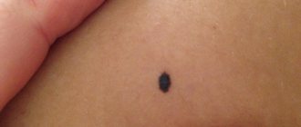
A lump appeared in place after the removal of a mole: what to do?
After removing a mole, you need to treat the area and minimize the negative impact from the outside on it.
After removing a mole with a laser or as a result of surgery, one of the decisive periods begins, on the normal course of which the health of the entire body depends. Removal of a nevus is necessarily accompanied by a long rehabilitation period, during which it is necessary to properly handle, care for and monitor all changes at the site of removal.
If this area of the skin is treated correctly, not overheated in the sun and minimizes the influence of chemical and physical factors on it, it will slowly heal and there will be no trace left of the intervention. However, not everything always goes well and often after such operations unpleasant consequences arise, such as:
- A scar appeared at the site of removal. The situation is typical and does not pose any threat. There is no need to do anything, the scar is a natural phenomenon that will disappear on its own after some time. There's no need to worry.
- Redness after mole removal is an adequate reaction of the body to surgery. But if the redness around the mole does not go away over a long period of time, it is important to consult a specialist.
- A lump appeared at the site of the removed mole. If any formations appear, it is recommended to see a doctor immediately. Often, compaction under a mole signals the initial stage of melanoma (skin cancer) and the development of malignant tumors. They may be benign, but it is better to consult a doctor immediately in order to eliminate all health risks.
On average, the wound healing process takes 2-3 weeks.
During this period, it is important to carefully observe all changes in the skin at the site of removal, wipe the wound with products prescribed by specialists, and if it is noticed that something new has formed, immediately consult a doctor. If healing occurs normally, a small scar will remain after a couple of weeks, which will soon disappear.
Diagnostics
It is necessary to diagnose melanoma in time and determine the stage of its development in order for the treatment of the disease to be successful.
First, an anamnesis is collected, the doctor learns about changes in the nevus.
Do not take a biopsy or expose the mole to chemical and electrical effects unnecessarily.
To diagnose you need to do:
- Histological examination (a superficial smear is taken if there is bleeding or cracks, provided that the tumor is removed immediately after the diagnostic results);
- Epiluminescent microscopy (examination of a mole using a special apparatus to determine its structure);
- CT (image recording, data processing, comparison with standards).
Differences between a compacted mole and atheroma, cyst
An atheroma or cyst is a ball-shaped neoplasm that is localized under the skin. The lump consists of sebum, the structure is dense and tough. The skin over a lipoma or lump under a nevus can be easily folded, but this cannot be done over a cyst. The dermis changes color above the atheroma. The size of the cyst varies from 5 mm to 5 cm. It does not cause pain if you do not scratch or touch it. The seals do not reach large sizes.
When infected, the atheroma breaks through and the contents penetrate into the dermis, leading to an abscess. Doctors have found 2 reasons leading to the formation of atheromas:
- lack of hygiene;
- metabolic disorders.
Doctors advise removing cysts to reduce the risk of developing atheromatosis.
How nevi are removed
A nevus is removed when it becomes a cosmetic problem or is cancerous. The removal method is selected based on cosmetic indications or oncological indications.
Cosmetic
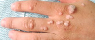
If cosmetic problems arise, the nevi are removed:
- Surgical intervention;
- Method based on liquid nitrogen;
- Using electric currents;
- Using laser surgery;
- Radiosurgical method.
Surgical intervention
It is well suited for removing large nevi. A disadvantage may be the presence of postoperative marks.
Adenoma
A liquid nitrogen
Also called cryodestruction. With it, the tissue is destroyed by cold. The nevus turns into a dry scab, under which new tissue then grows. The removal site is automatically protected from infections. This is how moles that are flush with the skin are removed. If the pathological tissues are not completely destroyed, then the procedure must be performed again.
Electricity
The method is also called electrocoagulation; it affects the tissue with high-frequency currents around the site of removal. Next, the material is sent for histology. The wound heals under the crust, forming a small scar.
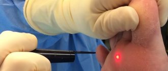
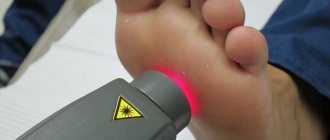
Laser removal
Moles are removed with laser on parts of the body that are most often exposed, on the face. The laser acts precisely on the area, leaving a small removal diameter and preserving the tissue around the nevus. The crust protects the wound from infection.
Radiosurgery
The method is non-contact. In this case, tissues are excised with a special radio knife under radio wave exposure. It is often used in cosmetology. It is possible to remove both benign and malignant tissues. Simultaneously with removal, it has a disinfecting and hemostatic effect. There are no scars left after this operation.
Oncological indications
If there is a suspicion or it is definitely established that the nevus has degenerated into melanoma, then within the healthy tissues they are completely excised and examined for histology.
Causes of a red spot
Various marks on the surface of the skin remain after various methods of mole removal. There are several reasons for the appearance of hypertrophic scars:
- Depth and size of the removed mole. The likelihood of a bump appearing on the skin after excision of a birthmark increases in the presence of large moles and nevi.
- method . After laser coagulation, a red spot appears in rare cases, since during the procedure only the upper layer of the epidermis is affected. But after surgical removal with a scalpel, pink and red spots may remain that do not go away for 6-8 months.
- wound care rules The rehabilitation period after surgical removal of a mole is of great importance. Failure to follow the rules may cause red spots to appear. Most often they occur when the wound is exposed to sunlight.
Scar stretching and inflammatory processes can also have an impact and significantly increase the risk of a hypertrophic spot.
A red spot (hypertrophic scar) after removal of a mole in some cases requires treatment. Reasons: removal of a large mole, scalpel removal, improper care after removal.
Treatment is prescribed until the scar becomes rough. Prescribed administration of corticosteroids, local agents to reduce the formation of scar tissue, cryotherapy, peeling, dermabrasion; occasionally surgical excision of a rough scar.
At the same time, fibroblasts are activated, cells begin to form more actively and collagen synthesis increases. The appearance of hypertrophic scars occurs against the background of increased collagen breakdown and the development of fibrous tissue.
Before removing a nevus, regardless of the method chosen, it is recommended to perform a biopsy of the mole followed by histological examination for cellular atypia.
If the analysis did not reveal signs of malignant degeneration, then the red spot after removal of the nevus should not cause alarm.
Cosmetologist, dermatologist, trichologist
Kalinina Olga Alexandrovna
4 years of experience
Possible complications
The nevus began to harden and a round thickening formed under it - this indicates the development of an inflammatory process in the dermis, improper functioning and distribution of subcutaneous fat, disturbances in metabolic processes, and hormonal imbalances.
Growths under the skin in the area of the birthmark indicate the appearance of a lipoma, cyst, abscess, foreign body inside, enlarged lymphatic and rheumatoid nodes.
Thickening of the dermis under nevi indicates the development of superficial malignant tumors - sarcoma. Such neoplasms are voluminous. In the initial stages they do not cause discomfort to the patient; they gradually decompose and become inflamed. The growth of the skin under the tumor is the first signal of the development of a cancerous tumor - melanoma.
The lump under the mole should be examined by a doctor. Malignant cells penetrate deep into the body, spreading through the lymph nodes into organs and systems.
When should you see a doctor and is it dangerous?
Rounded tubercles under nevi on the shoulders, head, cheekbones, arms and legs signal that changes are occurring in the body. The following symptoms should be the reason for contacting a dermatologist:
- The thickening became hard, the mole swelled.
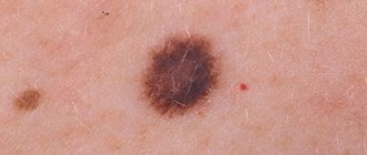
- The nevus has changed its halo, color, structure, and shape. The boundaries have become fuzzy.
- The neoplasm and thickening of the dermis underneath changes in size and increases.
- The lump itches, hurts, becomes inflamed, and fluid or blood leaks from it.
- Cracks appear on the surface and hairs fall out on the surface of the stain.
A number of these reasons are the reason for going to the hospital.
The appearance of symptoms indicates the development of melanoma. The doctor will decide what to do next with the mole and what method of removal to choose.
How to get rid of hardening
If a mole is hard and itchy, doctors advise removing it. Operations are performed in a hospital. Surgeons use the following methods:
- Surgical. The doctor uses a scalpel to excise the protrusion on the dermis and send the tissue for histological examination. The operation is used to remove large tumors with a deep root system and is performed under anesthesia.
- Laser. The doctor acts on the thickening with a laser. The method is painless, healing occurs in a short time.
- Cryodestruction. Liquid nitrogen is applied to the neoplasm. It becomes covered with a crust, which gradually disappears. The disadvantage of the procedure is manifested in possible damage to healthy areas of the skin.
- Electrocoagulation. A high-frequency current is applied to the mole and lump; the procedure is performed with local anesthesia. The tumor is sent for histology.
- Radio waves. The tubercle is removed using a radio knife.
If there is a lump under a mole and it hurts, you cannot try to eliminate it yourself using traditional medicine.
Thickening of the dermis under the birthmark can also be observed in a child, in which case the formation should be seen by a doctor immediately.