Definition of the term “keloid scar”
The elimination of nevi and other formations on the skin with the modern level of development of medical technologies makes this procedure almost painless and quick. In most cases, the patient does not even have scars on the skin.
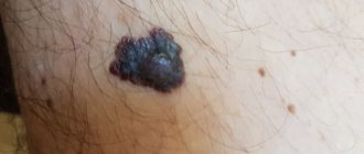
In cases where there is a deviation in the healing process of wounds after removal of moles, complications appear in the form of a scar (keloid scar).
The skin in this area acquires a compacted structure in the form of convex tubercles and differs in color from normal (bluish or dark pink).
Laser mole removal
In most cases, moles do not cause inconvenience, and therefore do not need to be removed.
The process of hypertrophic formation is long-term, so the skin heals up to one year, and the scar appears as a compaction within six months. The time it takes for a raised scar to appear is influenced by the size of the sanitized skin area.
After removal of tumors during the first weeks, the skin protects itself by forming a thin layer of epithelial tissue. Under the influence of unfavorable factors, disturbances in the growth of this layer occur.
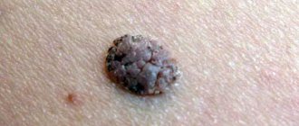
As a result, epithelial tissue grows and thickens with a color change to a pale shade. Then the tissue swells, and the resulting colloidal tissue grows. The wound hurts and itches, gradually these symptoms go away, and the neoplasm takes on convex, hard shapes.
Lymph node excision
After the diagnosis of melanoma is confirmed, it is necessary to check the lymph nodes close to the tumor (they are called regional) - perhaps the tumor cells have managed to spread into them. Computed tomography, MRI, PET scan, and fine-needle biopsy of lymph nodes are performed. If they are also affected, they are excised. If, according to the examination, the regional lymph nodes are not affected, a sentinel biopsy, or sentinel lymph node biopsy, can be performed. A radiopharmaceutical or fluorescent dye is injected into the area of the melanoma.
It penetrates the lymphatic vessels and spreads through them to the lymph nodes. The lymph node that receives the dye first is called the sentinel node. It is removed and a biopsy is performed. If malignant cells are found in it, then they most likely have spread to other regional lymph nodes. Sentinel biopsy is especially useful in cases where the melanoma is thicker than 1 mm. It is currently unknown whether a patient can be cured by removing lymph nodes affected by melanoma. At the very least, it helps prolong life and reduce pain and other symptoms.
Characteristic symptoms of formation
In most cases, the formation of keloid scars at the site of a removed mole occurs no earlier than after 1.5-2.0 weeks. The epithelial tissue continues to grow and thicken.
Patients in most cases cannot independently understand the types of scars. Thus, a hypertrophic scar in an inflamed state itches and hurts, and the patient’s condition worsens.
Unlike a hypertrophic scar, a keloid scar remains in an inflamed state for a short time, the painful symptoms go away, and the scar begins to grow.
How to care for a removed mole
Thanks to modern medicine, today there are many safe ways to get rid of moles.
In the early stages, with timely consultation with specialists, there is a high probability that the keloid scar will be removed and the skin will acquire a healthy appearance. The older the scar, the more difficult it is to fight it.
An experienced surgeon can identify the symptoms of a keloid scar. You should not self-medicate; if there is the slightest deviation in the patient’s health after the destruction of a mole, it is better to turn to doctors for qualified help.
Symptoms of scar formation are accompanied by the following conditions:
- the presence of pain , which can manifest itself even outside the former mole;
- increased sensitivity ;
- the scar becomes red and the surrounding skin becomes inflamed;
- smooth scar tissue forms ;
- feeling of constant itching and burning;
- a steady and constant increase in the size of raised scars;
- lack of growth at the site of removal;
- the formation protrudes above the surface of the skin.
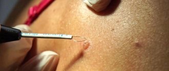
What is dermatoscopy
Dermatoscopy is one of the modern methods for diagnosing neoplasms formed on the surface of the skin.
Such symptoms may occur over several years in different patients. This depends on the general health of the person. The scar continues to be hard and elastic.
Keloids most often form on the following areas of the body:
- cervical region;
- ears (lobes);
- rib cage;
- joint area.
Ignorance leads to the formation of rough scars and the argument that when the wound heals, it must itch, does not stand up to criticism.
Keloids, even in their neglected and old form, do not pose a threat to the patient’s health. A specialist must decide on the course of treatment (removal) of the scar.
Price for melanoma removal in Moscow
The cost depends on many factors: the volume of the operation, studies that the patient may require, the pricing policy of the clinic, experience, qualifications and status of the surgeon, additional treatment methods before and after surgery. We cooperate with the best oncology centers in Moscow, which perform operations to remove melanomas on the back, face and other parts of the body. We will recommend a good clinic where you can get treatment for stage 4 melanoma:
Causes
Doctors have not yet developed a consensus on the reasons for the formation of keloids.
One of the reasons for the formation of keloid tumors is a violation of the integrity of the skin during surgery.
Epithelial tissue grows rapidly due to the excess collagen content in the body. Excess collagen leads to the appearance of a red keloid scar at the site of nevus removal.
Removing moles with an electric knife
Situations are often observed when ordinary moles, under the influence of certain circumstances, transform into malignant neoplasms.
Experts consider the following factors to be important in the formation of keloids:
- Heredity is one of the main ways of development of many pathological changes in the body. The presence of keloid scars in parents increases the likelihood of the patient developing them after mole removal.
- Hormonal imbalances. The development of connective tissue under the influence of hormonal imbalance can lead to the formation of keloids.
- Careless and improper handling of the wound after mole removal. Under no circumstances can the protective crust be removed or picked out. Such actions are fraught with infection, pathological changes in epithelial tissues, and delayed healing of the wound.
- Damage fresh , healed wound, skin overstrain. This is facilitated by uncomfortable clothes (small, tight, rubbing, etc.), and excessive tanning.
- of collagen in the body leads to the growth of epidermal tissue in excessive volumes.
- Pregnancy period .
- Endocrine system , which works with disturbances.
The appearance of keloid scars is chronic in nature and requires periodic therapeutic courses of treatment.
Consequences of melanoma removal
Possible consequences of surgical interventions in patients with melanoma:
- Common complications for any surgery: pain, bleeding, risk of infection.
- Relapse is possible. Moreover, melanoma can recur not only in the place where it was before surgery, but also in other organs in the form of metastases.
- After removal of large melanomas, noticeable scars remain. Extensive defects require plastic surgery - transplantation of a skin flap from another part of the body.
- After removal of lymph nodes, some patients experience lymphedema - swelling due to impaired lymph drainage.
Basic treatment methods
The basis for success in removing keloid scars is timely treatment.
Self-medication is dangerous with complications!
Attention
Despite the fact that our articles are based on trusted sources and have been tested by practicing doctors, the same symptoms can be signs of different diseases, and the disease may not proceed according to the textbook.
Pros of seeing a doctor:
- Only a specialist will prescribe suitable medications.
- Recovery will be easier and faster.
- The doctor will monitor the course of the disease and help avoid complications.
find a doctor
Do not try to treat yourself - consult a specialist.
Specialists in their arsenal have a variety of methods and means of treating keloid scars.
Method of using laser
Using a laser beam, the newly formed keloid scar after mole removal is polished. The procedure consists of treating the surface of the keloid scar layer by layer with a laser. Treatment occurs in a gentle manner until an area of skin with a normal healthy shade is reached.
Why can our articles be trusted?
We make health information clear, accessible and relevant.
- All articles are checked by practicing doctors.
- We take scientific literature and the latest research as a basis.
- We publish detailed articles that answer all questions.
The treatment area is controlled; healthy tissues are not exposed to laser radiation. After applying the laser beam, either a small depression or an almost indistinguishable spot appears, which at first you want to scratch.
This method has a significant drawback - there is a high probability of remission of the keloid at the site of destruction of the mole.
Using patches and dressings
A technique that uses tight bandages and plasters after mole removal minimizes the likelihood of keloid scars enlarging.
Tight bandaging does not allow the keloid to grow freely.
Hormonal therapy
This treatment method is used only after prescription by the attending physician. The specialist determines acceptable medications and their dosage. Corticosteroids in the form of a suspension are injected approximately once a month.
Treatment using this method involves the introduction of medications directly into the colloid tissue. This helps prevent further growth and increase in the density of the keloid scar. The technique is used both basic and additional in combination with other treatment methods.
According to statistical observations, the use of hormonal drugs reduces the recurrence of scarring by 70 to 90%.
Available creams and pharmaceutical products
Pharmacy chains offer ready-made ointments and creams for the treatment of keloid scars after mole removal. In terms of their effectiveness, they are inferior to the methods listed above.
The keloid scar will not resolve; at best, the skin will lighten and soften a little. These drugs do not cause allergic reactions, which allows them to be used by pregnant women, lactating women, and children.
Cold removal
Cryodestruction allows you to use cold to remove a keloid scar formed after the destruction of a mole. In this case, the scar is not completely eliminated; its surface becomes not convex, but flat and almost indistinguishable. The procedure is painful.
Microwave therapy
The scar is destroyed by alternating exposure to heat and cold. The temperature difference allows you to achieve a positive result for a long period of time.
Surgical intervention
If using the above treatment methods the proper result is not achieved, then they resort to surgical excision of the keloid scar. Using a scalpel, the scar is removed, followed by the application of a tight bandage or silicone patch. In addition to surgical treatment, hormonal therapy is prescribed.
What to do after papilloma removal: doctors' recommendations
Physical impact inevitably leads to a change in the condition of the skin at the site of papilloma localization. Swelling, redness, and pain may occur in this area - these phenomena are considered normal and disappear after 3-4 days. It is impossible to predict the exact duration of the recovery period, since it depends on the location, size and number of tumors, characteristics of the body, quality of care, destruction method used, and doctor’s qualifications.
How long does it take for a wound to heal after papilloma removal on average:
surgical intervention using a scalpel – up to 30 days, with the inevitable formation of a scar at the site of exposure;- cryodestruction – 15-18 days. The first hours after surgery are accompanied by hyperemia, swelling, and the formation of an epidermal bladder - these symptoms disappear within a day;
- electrocoagulation – 12-14 days. Completion of the operation during the day is accompanied by slight swelling and redness;
- laser removal – 7-10 days;
- radio wave destruction – 4-7 days. As with the use of a laser, redness and swelling in the treated area remain for no more than a day, after which a scab begins to form.
Complete healing after removal of papillomas, including complete skin regeneration and restoration of pigmentation, may require several months.
Why moles appear on the face
There are several groups of moles based on the characteristics of their internal structure, appearance or size. Moles are distinguished:
- Large, the size of which is more than 10 cm;
- Medium from one and a half to 10 cm;
- Small - from 0.5 mm to one and a half cm.
Small moles, as a rule, are absolutely harmless to the body, but large and medium ones can be quite dangerous, since there is always a risk that a mole or birthmark may develop into a malignant tumor. According to statistics, about 30% of cases end this way, especially if there are other unfavorable factors: taking certain (certain) medications, poor lifestyle or prolonged exposure to the open sun under direct sunlight. Therefore, removing a large mole on the face is a necessity.
In most cases, moles appear almost immediately after the baby is born. Usually, by the age of 8, moles are already fully formed, and at a later age they can only change slightly in size. But sometimes it happens that a mole appears in adulthood. This occurs due to the concentration of pigment cells in certain layers of the skin. Such moles are divided into the following groups:
- Intradermal formed inside the dermal layer of the skin;
- Epidermal are located in the middle layer of the skin;
- Mixed - moles located on the border of the dermis and epidermis.
Mixed moles are usually flat, while intradermal and epidermal moles look like small peas.
Why do newly acquired nevi appear?
- Due to any hormonal imbalances in the body;
- During certain periods of life, the human body is most predisposed to the formation of moles (puberty, pregnancy, lactation, transition to adulthood - after 50 years, nevi often appear on the face and neck, etc.);
- The effect of the sun on the skin (including the effect of solarium lamps);
- Prolonged treatment with drugs or hormonal drugs;
- Frequent massage.