Quincke's edema (or angioedema) is an allergic disease in which sudden swelling of the subcutaneous tissues and mucous membranes occurs.
Most often, Quincke's edema affects the face, mouth, respiratory tract and gastrointestinal tract. But it can also affect joints and internal organs. Damage to the larynx (due to the risk of difficulty breathing) and the brain (can lead to serious neurological disorders) is considered especially dangerous. Pathology of this type can develop in almost any person, since there are many predisposing factors: from food allergies to acute infectious diseases.
In half of the cases it accompanies urticaria, and in 11% of cases it develops in isolation. Sometimes the cause of sudden swelling cannot be found, and then it is called idiopathic. It is noteworthy that Quincke's edema is a kind of family disease, and is often transmitted, for example, from parents to children. “Inherited” a person receives a deficiency of certain enzymes that should fight substances responsible for edema.
Edema can develop, pass or recur1.
Symptoms of Quincke's edema
Symptoms depend on location and are especially acute at the onset of the disease, developing at high speed - from a few minutes. External manifestations of the pathology are severe swelling of the nasolabial triangle, tongue and tonsils.
It is believed that the form of the disease becomes most dangerous at the moment when the trachea, larynx and pharynx are exposed to an allergic reaction. In this case, the following may be observed:
- hoarseness of voice;
- difficulty breathing up to suffocation;
- "barking" cough.
If medical attention is not promptly provided and further swelling is not prevented, the outcome can be fatal2.
Less commonly, with Quincke's edema, loose stools, vomiting and painful attacks appear, localized in the abdominal area. Symptoms of an “acute abdomen” indicate the spread of the disease to the gastrointestinal tract.
If the brain is “affected” during an allergic reaction, the symptoms will resemble nervous system dysfunction:
- aphasia;
- hemiplegia;
- nausea, vomiting and severe dizziness;
- frequent seizures.
If the reactions go further, then the urinary system will come under the influence of the disease. Acute cystitis develops, accompanied by prolonged urinary retention. Swelling of the genital organs is possible.
Pathology results
With timely first aid there are no consequences for the body. Ignoring the developing symptoms of Quincke's edema causes anaphylactic shock, even death.
For patients prone to spontaneous allergic reactions, doctors recommend adhering to certain rules of prevention:
- exclusion from the daily menu of foods that can cause an attack;
- preventing contact with external allergens;
- use of antihistamines during the flowering season of plants.
Timely treatment of chronic diseases will help reduce the risk of angioedema.
Quincke's edema in children
Quite often, young children suffer from laryngeal edema. The following factors influence this:
- feeding an infant with artificial food;
- frequent use of medications;
- allergic reaction of a household type (to shampoo, bed linen conditioner or powder);
- abuse of multivitamins and potent antibiotics.
For children under 3 years of age, non-allergic Quincke's edema is typical. This is due to heredity and often manifests itself in combination with bronchial asthma or urticaria. The narrowing of the pharynx occurs quickly.
- the first stage manifests itself almost imperceptibly, without shortness of breath;
- the second causes pale skin, the nasolabial triangle turns blue, and the heart rate increases;
- at the third stage, respiratory failure occurs. Severe sweating appears, the child has difficulty breathing;
- the fourth stage is the most difficult. It is accompanied by a slow heartbeat, shallow breathing and loss of consciousness.
At the first manifestations of swelling of the larynx, it is necessary to consult a specialist, since the lack of qualified assistance in this case can be fatal.
Symptoms
Angioedema of the eye, as a rule, can be distinguished from other pathologies by its very rapid progression and the fact that the swollen skin does not change color. The swollen tissues are dense. It can affect both the upper and lower eyelids, or both at the same time. As a rule, there is no itching, but in some cases, when allergies are accompanied by hives, the skin may itch. There is no pain when pressing, which distinguishes swelling from inflammatory swelling. This is what Quincke's edema looks like on the eyelids in the photo.
The allergic reaction may affect one eye or both. It can spread to the cheek, mouth, neck. The palpebral fissure narrows, intraocular pressure increases. With massive Quincke's edema, serious consequences can develop:
- conjunctival chemosis;
- the appearance of infiltrates on the cornea;
- secondary glaucoma;
- displacement of the eyeball.
The main danger of allergic edema of the eyelid in a child or adult is that the process can become generalized and also affect the larynx, digestive tract, and genitourinary tract. If the membranes of the internal organs are affected, the patient complains of severe pain, diarrhea, and vomiting. Uneven distribution of fluid in the facial area can lead to permeation of the membranes of the brain. This leads to loss of consciousness, severe headache, and convulsions.
This is what allergic swelling of the eyelid that has spread to the entire face looks like in the photo:
Diagnosis of Quincke's edema
Diagnosis includes examination of the patient and collection of anamnesis (predisposing factors).
During the examination, the specialist pays attention to:
- location, its size, density and color, temperature of the edema and surrounding tissues, the presence of itching or burning, time of development;
- the condition of the respiratory tract, voice, swallowing process to eliminate danger to life;
- blood pressure and pulse rate, body temperature, etc.
At the second stage, predisposing factors are assessed. These include:
- cases of edema in the family;
- the presence of blows, injections, injuries in the patient in the last few hours;
- taking estrogen-containing drugs during pregnancy;
- nutritional and medicinal factors;
- frequent hospitalizations with signs of an “acute abdomen” (may indicate internal edema).
You should also find out whether the patient suffered from oncology or autoimmune diseases. Only after analyzing all the complaints and information will it be possible to determine the nature of the edema - whether it is acquired or hereditary.
First Aid Basics
The appearance of primary symptoms of angioedema requires calling an ambulance, regardless of the severity of clinical signs. The list of first aid measures includes:
- avoiding contact with allergens;
- applying a heating pad with ice to the area of swelling;
- taking activated carbon or Polysorb to cleanse the body of ingested allergens;
- use of lozenges with antihistamine properties: Suprastin, Zodak, Zyrtec, Clarotadine, Fenistil;
- the use of vasoconstrictor drugs for swelling of the nasal mucosa - Naphthyzin, Rinonorma.
The arriving specialists will inject the victim with hormonal medications based on prednisolone. After relief of the main symptoms of Quincke's edema, the ambulance team will take the patient to the nearest hospital with an allergy department.
Emergency care for Quincke's edema
Given the speed at which swelling develops, you should immediately call an ambulance. Before the ambulance arrives, you need to act as follows:
- calm the patient and sit him in the most comfortable position;
- It is necessary to remove everything unnecessary from the patient. It is necessary to ensure a flow of fresh air;
- limit direct contact with the irritant. If the swelling is caused by an insect bite, you need to get rid of the sting;
- administer antihistamines. In this case, injections will be the most effective. But if it is not possible to get an injection, you need to take the drug. This is the only way to reduce the intensity of the reaction. Among the latest generation of antihistamines, Cetrin® stands out. Its action is aimed at blocking histamine, a substance responsible for the occurrence of an allergic reaction. Therefore, the drug works regardless of the cause that caused Quincke's edema and can help relieve an attack3.
Causes
The pathogenesis of allergic Quincke's edema, regardless of the cause, occurs after a person comes into contact with an allergen, after which the active production of immunoglobulins begins. Histamine is released into the blood, which dilates the capillaries and increases their permeability, which is why the first symptoms of angioedema appear: swelling of the skin and mucous membranes.
Causes of angioedema:
- Food;
- animal wool and feathers;
- bites of various types of insects;
- chemical compounds and substances;
- house or street dust;
- medical supplies;
- chronic stress;
- hereditary causes of angioedema.
Treatment of Quincke's edema
Direct treatment of the disease takes place in the inpatient allergy department. In order to begin treatment, it is necessary to determine the cause of the allergic reaction. The patient is prescribed a mandatory diet that excludes foods containing honey and chocolate, berries and citrus fruits and other strong allergens; Prescribe drugs with an antihistamine effect in combination with hormonal drugs - dexamethasone or, for example, prednisolone ointment. To remove the allergen, treatment is accompanied by the prescription of polysorbs.
The above complex of medications is supplemented with agents that normalize the functioning of the body’s nervous system, for example, calcium or ascorbic acid.
Self-treatment of Quincke's edema, without consultation and supervision of a qualified specialist, is impossible.
Bibliography
- Goryachkina G. A., Differential diagnosis of various forms of urticaria and Quincke's edema / G. A. Goryachkina, E.Yu. Borzova // Pulmonology and allergology. - No. 3. – 2004. – 13 p.
- Attending physician [electronic resource]. – Access mode: https://www.lvrach.ru. – Luss L.V., Modern ideas about the pathogenesis and principles of treatment of urticaria and angioedema. (Last access date 08/25/17)
- Instructions for use of the medicinal product for medical use Cetrin®. – Registration number: P N013283/01.
Prevention of angioedema of the eyelids
To reduce the risk of recurrent angioedema, the patient must strictly follow the recommendations received from the attending physician. In most cases, they come down to a number of simple rules that can be implemented at home:
- In the first days after emergency medical care, do not use hot water (only warm water - up to 37 degrees);
- Use exclusively soft, skin-friendly soap;
- Apply cold or cool compresses as symptoms subside;
- Strictly adhere to a hypoallergenic diet (especially in case of food-related angioedema of the eyelids);
- Take only those medications prescribed by your doctor;
- Do not resort to self-treatment of any other pathologies and consult a specialist before each use of medications;
- Avoid stressful situations;
- Take care to prevent fungal, parasitic and viral infections (including influenza);
- Be wary of any impact on previously affected areas of the skin around the eyes (refuse to use cosmetics);
- In case of hereditary angioedema of the eyelids, it is imperative to carry out preventive treatment before carrying out planned operations (including dental ones);
- Limit the intensity of physical activity.
All these recommendations, if strictly followed, will help avoid possible relapses.
Complications
If the condition is not stopped at the first signs of edema, this can lead to the development of severe complications:
- breathing problems - due to sudden or progressive swelling of the respiratory organs;
- dizziness and fainting;
- collapse is a condition close to fainting, which is accompanied by a sharp drop in blood pressure and a rapid deterioration in the functioning of important body systems.
Complications of angioedema can be avoided with timely medical care. After relief of long-term effects of edema, no swelling is observed.
1.General information
If you see a person’s face rapidly swelling, especially in the area of the eyelids and lips, the first thing that should come to mind is Quincke’s edema.
Acutely developing angioedema (neurovascular) edema is named after the German physician Heinrich Quincke, who gave the first detailed clinical description in 1882 (casuistic references to similar conditions have been found before). It is interesting that the term “Quincke’s edema” is used mainly in German- and Russian-speaking medicine, while in international English terminology they usually talk about “angioedema”.
Angioedema is one of the specific types of hypersensitivity reaction and can occur in any of us, regardless of age and gender. However, the predominance of young women (20-30 years old) among those with diagnosed episodes of Quincke's edema is sometimes reported. On the other hand, there is a form of angioedema (or rather, three subtypes) with a hereditary predisposition, first described by W. Osler six years after the publication of G. Quincke; In the presence of a family history, men are more often affected.
According to statistics, in the United States alone, angioedema causes emergency room calls about one hundred thousand times each year, and it consistently leads in the total number of allergic reactions and conditions that lead to hospitalization.
Sign up for a consultation
A must read! Help with treatment and hospitalization!
Relevance
According to statistics from a number of European countries (Germany, England, France, etc.), from 10 to 30% of the population suffers from allergic diseases [1]. One of the manifestations of a human allergic reaction is Quincke's edema (QE) - an acute disease characterized by the appearance of clearly limited swelling of the skin, subcutaneous tissue, and the mucous membrane of various organs and systems of the body. OK was first described by Heinrich Quincke in 1882, but only almost 100 years later (in 1963) the pathogenesis of this disease was determined. For a long time, OK was presented as an exclusively allergic reaction. The progressive development of allergology and immunology has gradually expanded knowledge about OK. This disease is divided into hereditary and acquired forms. It was found that OK can be a manifestation of allergy and pseudo-allergy. It has also been found that this disease can develop as a result of taking angiotensin-converting enzyme inhibitors (ACE inhibitors) (captopril, enalapril). It should be noted that in 45% of patients it is not possible to identify the causative factor for the development of OK [2]. Thus, over time, a number of human pathological conditions with identical clinical manifestations, but with different etiologies and pathogenesis, began to be classified as a separate nosological form. This introduced significant uncertainty into the clinical work of specialists.
Today, according to ICD-10, OK is classified as angioedema (AO) with identification code T78.3. There are: I. Allergic A.O. (giant urticaria); II. Pseudoallergic edema (nonspecific histamine liberation); III. Complement-dependent edema: 1. Hereditary A.O. (HAE): a) HAE with a quantitative deficiency of C1 inhibitor (HAE type I); b) HAE with a functional deficiency of the C1 inhibitor (HAE type II); c) HAE, in which the level of C1 inhibitor and its function in patients are normal (HAE type III); 2. Acquired A.O. (PAO): a) immunocomplex (PAO type I); b) autoimmune (PAO type II); c) when administering drugs that activate the complement system; IV. Idiopathic A.O..
I
.
Allergic AO
. The most common occurrence in everyday clinical practice is A.O. Up to 80% of the causes of AO are the development of an allergic reaction of type I - reagin (IgE-dependent type). Various factors can lead to the development of allergic AO: medications, food products, some dietary supplements, stinging insect venom, latex (gloves, rubber urinary catheters, endotracheal tubes, intravenous catheters), epidermal allergens - saliva, cat and dog dander, cosmetics . Allergic AO is characterized by a clear connection between exposure to the allergen and the development of the reaction, an acute onset - usually the reaction develops 15-30 minutes after contact with the allergen, rapid development of edema, and a combination with urticaria. Swelling has a characteristic appearance - it is usually dense, asymmetrical, painless swelling, may be pale pink in color and not different from unchanged skin. It is localized mainly in places with well-developed subcutaneous fat (on the face these are most often the lips, eyelids, in the oral cavity - the soft palate, tongue, tonsils). Involvement of the mucous membrane of the respiratory system (swelling of the larynx, trachea, bronchi) is especially dangerous due to the threat of asphyxia. It is possible that AO may develop as the onset of a generalized anaphylactic reaction - anaphylactic shock, which is manifested by generalized itching, urticaria, lacrimation, sneezing, bronchospasm, swelling of the tongue, larynx, pharynx, hoarseness, hypersecretion of mucus in the bronchi, nausea, vomiting, cramping abdominal pain, diarrhea, tachycardia, arterial hypotension, cardiac arrhythmia, development of acute cardiovascular failure, seizures, respiratory arrest, coma. In this case, death occurs from swelling of the larynx and cardiac arrhythmia.
II. Pseudoallergic edema
. The clinical manifestations of pseudoallergic edema are in many ways similar to those of allergic edema, however, the release of allergy mediators occurs in a non-immune way. With this mechanism of edema development, an important role is played by foods and medications that change the metabolism of mediators, the formation of excess leukotrienes, bradykinin, dysbacteriosis, namely muscle relaxants (thiopental, tubocurarine), opiates, antibiotics, radiocontrast agents, local anesthetics, plasma substitutes, plasma, albumin, protamine directly affects mast cells, increasing the level of histamine in the blood; fish, cheese, smoked meats, wine, beer, chocolate, nuts, berries (strawberries, raspberries), citrus fruits - contain a large amount of histamine in their composition; non-steroidal anti-inflammatory drugs (NSAIDs), nutritional supplements (tartrazine), salicylates, sulfites, nitrates - lead to the formation of excess leukotrienes D4, C4, E4, which have significant vasodilatory and chemotactic activity; taking ACE inhibitors is a fairly common cause of the development of AO (from 4 to 8% of all AO) due to the accumulation of bradykinin, which increases vascular permeability. It was noted that 0.1-0.7% of all patients taking ACE inhibitors had an episode of AO [3, 4].
III. Hereditary angioedema (HAE)
is an extremely rare disease, its prevalence may vary in different regions and is approximately 1:50,000 people. Such a low prevalence leads to extremely low awareness among doctors about the disease and, as a consequence, to its poor and late detection, as well as to incorrect management tactics and selection of therapy. HAE is a disease whose main clinical manifestations are recurrent swelling of the mucous membranes and deep layers of the dermis of various locations [5, 6].
The development of edema is caused by an increased release of the mediator bradykinin (a low-molecular nanopeptide that causes an increase in the permeability of the vascular wall and extravasation of plasma) due to a deficiency or impairment of the functional activity of the C1-inhibitor (C1-INH). Most often, the development of the disease is based on a mutation in the SERPING1
[7].
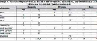
In order to diagnose HAE with C1-ING deficiency, indicators of the level of C1-ING and its functional activity, as well as the level of the C4 component of complement in the blood are used. HAE type I (85% of cases) is diagnosed when the C1-ING level is less than 30% of normal. HAE type II (15% of cases) is diagnosed when a decrease in the functional activity of C1-ING is detected by more than 50% of the norm with a normal or increased level of C1-ING. To make a diagnosis, changes must be recorded in at least 2 studies, which must be repeated at intervals of 1-3 months. The level of complement C4 in patients with HAE I and II is usually reduced (<50% of normal), but the low sensitivity and specificity of this test limit its diagnostic value, so this indicator can only be used as a screening marker for selecting patients due to with its availability in routine practice and low price [5] (Table 1).
Table 1. Diagnostic indicators for hereditary angioedema types I and II
Thus, the mechanism for the development of edema in HAE is due to the excessive activity of complement and Hageman factor with the formation of bradykinin and C2-kinin, which increase vascular permeability. This explains the lack of effect in patients from taking antihistamines and glucocorticosteroids (GCS) (Table 2).
Table 2. Differential diagnosis of hereditary and allergic angioedema
Type III HAE was initially described in women and then in men.
The mechanism of its development is unclear. The C1-ING level and its function in patients are normal. It is possible that the disease is associated with increased production of bradykinin and a slowdown in its destruction due to a decrease in ACE (kininase) activity under the influence of estrogens.
Clinical manifestations of HAE.
The main clinical manifestations of HAE are peripheral edema, abdominal attacks (caused by swelling of the intestinal wall) and upper respiratory tract (URT) edema. Almost all patients with HAE suffer from recurrent peripheral edema, the most common clinical manifestation of the disease. A characteristic feature of edema is the absence of urticaria and changes in the skin (redness, temperature) over the edema. There may be tingling, burning, and soreness at the site of swelling. The most common localization is the upper and lower extremities. Unlike histamine edema, edema caused by bradykinin increases more slowly, but also regresses more slowly. On average, swelling persists for about 72 hours [8].
The next most common symptom (traced in more than 50% of patients) is abdominal attacks, which can occur either together with peripheral edema or in isolation, which significantly complicates diagnosis. The clinical picture can vary from mild discomfort to symptoms of an “acute abdomen”, which become the reason for unnecessary surgical interventions. Abdominal ultrasound and CT can detect swelling of the intestinal area and free fluid in the abdominal or pelvic cavity [9].
The greatest threat to the patient's life is edema of the upper respiratory tract: swelling of the larynx, tongue and palate. Clinically, this is manifested by breathing and swallowing disorders, dysphonia, and stridor. The possibility of swallowing movements should be assessed and the upper respiratory tract accessible to examination should be examined. The time from the onset of symptoms of respiratory failure to complete asphyxia is unpredictable and can range from 20 minutes to 14 hours. Edema of the upper respiratory tract may be the first clinical manifestation of the disease [1, 8].
Trigger factors for the development of HAE may include emotional stress, physical stress, mechanical trauma (even the most minor, such as pressure from a belt or shoe), infectious diseases, the use of certain drugs (certain contraceptives, ACE inhibitors or angiotensin II receptor antagonists), insect bites , a number of food products, weather changes [1].
Factors associated with an increased risk of developing urgent edema include many medical procedures: dental procedures, invasive methods of examination and treatment, surgical interventions (especially those associated with the need for intubation anesthesia). Swelling associated with these procedures usually occurs within 2 days after the procedure. It is very important to remember that manipulations performed in the past without complications do not guarantee the safety of each subsequent medical intervention [10-12].
The presence of uncompensated chronic diseases (for example, caries, tonsillitis, cholecystitis) is also a trigger factor for the development of edema, therefore, in the absence of adequate correction of such diseases, a vicious circle is formed that progressively worsens the course of the underlying disease [13]. In this regard, the presence of a clear protocol for patient management before medical intervention is of great importance to minimize the risk of complications and, consequently, to reduce disability and mortality. In addition, the presence of such a plan significantly increases the chance of patients to receive timely, quality medical care. The enormous importance of timely diagnosis and adequate treatment for such conditions is clearly demonstrated in the study by K. Bork et al.: out of 70 examined fatal cases from asphyxia during airway obstruction, 63 patients were not diagnosed, and only 7 patients had the disease verified [ 8].
Recommendations for invasive medical interventions in patients with HAE
According to available data, based on the principles of evidence-based medicine, when carrying out invasive medical interventions, patients with HAE are recommended to follow the following general rules: use a drug for short-term prophylaxis (C1-esterase inhibitor - benert) to prepare for planned and urgent interventions 1-6 hours before procedures. In its absence, fresh frozen plasma and danazol can be used as second-line drugs [1, 5]. Basic therapy should not be interrupted (if the patient is receiving it) [5].
It is recommended to avoid, if possible, intubation anesthesia and give preference to other types of anesthesia [14], to prefer conduction anesthesia to general anesthesia [14], to ensure the possibility of resuscitation measures, in particular to restore airway patency [14].
You should make sure that the patient is not receiving drugs prohibited for HAE: ACE inhibitors, angiotensin II receptor blockers, estrogen-containing drugs.
It is important to remember that since edema in HAE is caused by the action of bradykinin, this group of edema is insensitive to the use of systemic corticosteroids, antihistamines and adrenaline; therefore, it is not advisable to use these drugs for short-term prevention and relief of edema [1, 5].
According to the latest edition of international recommendations for the management of patients with HAE, the only drug for short-term prevention based on the principles of evidence-based medicine (both for planned and urgent medical intervention) is plasma C1-ING concentrate [1]. This is a C1-esterase inhibitor concentrate obtained from donor blood, which affects all stages of pathogenesis in the treatment of HAE. The only C1-esterase inhibitor drug registered in Russia is benert. Berinert confirmed its effectiveness and safety in clinical trials both abroad and in the Russian Federation. Throughout the world, benert is a C1-ING drug with more than 30 years of experience in use (more than 500 thousand courses of treatment). Data on its safe use in all age categories of patients, including children and pregnant women, have been published. Therefore, if there is a choice, this drug should always be preferred, especially if extensive medical intervention is planned or it is not possible to avoid intubation anesthesia [1, 5].
C1-ING concentrate should be used for prophylactic premedication as close as possible to the beginning of the procedure. According to the instructions adopted in the Russian Federation, the prophylactic dose of the drug before an invasive procedure or surgery is 1000 IU for an adult and 15-30 IU/kg for a child. The drug is administered intravenously [5]. Carrying out such premedication allows you to minimize the risk of possible life-threatening edema, however, it must be remembered that even the use of a concentrate of donor C1-ING esterase from a person does not provide an absolute guarantee: there are reports of clinical cases and series of cases where edema developed even after relatively minor operations against the background premedication [1, 14]. Therefore, even when premedicating during surgery, the patient must have with him means to relieve acute edema [1, 5].
Unfortunately, in routine practice in Russia, the use of benert may be limited due to the low supply of patients and medical institutions with this drug. In such a situation, it is necessary to prepare the patient for the planned intervention with the help of second-line drugs: danazol, fresh frozen plasma, antifibrinolytics.
During planned medical interventions, danazol can be used as a short-term prophylactic medication. This drug belongs to the pharmacological group of attenuated androgens. To date, a lot of experience has been accumulated in the use of attenuated androgens in HAE. Danazol is prescribed according to the following regimen: 200 mg/day 7 days before and 4-5 days after surgery (if the patient was on basic therapy with danazol, then the dose is increased 2 times from the original) with further cancellation or transition to the previous dose of basic therapy [5 , 12]. It should be remembered that attenuated androgens have many side effects and there are categories of patients for whom the use of these drugs is contraindicated, despite their effectiveness. However, short courses of danazol, as a rule, do not have clinically significant consequences for the patient’s health [12].
In the absence of danazol and benert, the only available agent that can reduce the risk of edema during medical intervention is native or fresh frozen plasma, which also donors the C1-esterase inhibitor. It is prescribed in a dose of 250.0 ml 1-6 hours before the procedure. In addition, during planned surgery, it is possible to use danazol and plasma together. Since plasma is not standardized for the content of C1-ING and may contain components such as blood coagulation factor XII, prekallikrein, high molecular weight kininogen, its transfusion may not always lead to the expected effect. Cases have been described of not only the lack of effect from plasma administration, but also increased edema. In addition, the safety of plasma in comparison with that of C1-ING drugs is significantly lower, which is associated with a higher risk of transmission of vector-borne infections, as well as allosensitization [1].
The use of antifibrinolytics as short-term prophylactic drugs is not currently recommended, at least as monotherapy [1]. A number of experts believe that it is possible to use aminocaproic acid as an additional method of preventing the development of edema: in addition to danazol, it is recommended to administer aminocaproic acid by intravenous drip of 200.0 ml [5]. Also, before performing dental procedures, rinse the mouth with a 5% solution of aminocaproic acid.
Much more often than with medical interventions, patients encounter everyday trigger factors: these can be stressful situations (exams, moving, personal experiences), menstruation, infectious diseases, insect bites, physical activity. The upcoming effects of some trigger factors are often known in advance, so it is possible to prepare for them. If it is impossible to prevent C1-ING, it is recommended to prescribe danazol (or increase its dose) according to the regimen recommended for preparation for medical procedures. In some patients (with previously proven effectiveness), it is possible to prescribe or increase the dose of tranexamic acid. Prescribing or doubling the dose of the drug for the period of infectious diseases (starting from the prodromal stage) is also considered effective.
Any preventive premedication does not exclude the development of “breakthrough” attacks, so there should not only be a supply of drugs (berinert or icatibant) to relieve edema, but also in case of insufficient response to pharmacotherapy, the possibility of intubation. The patient should be left under the supervision of an anesthesiologist-resuscitator when the first signs of compression of the upper respiratory tract appear [1, 14].
Today, the main problem in the management of patients with HAE in our country is the low awareness of doctors about this disease and, as a consequence, its low detection rate. Patients have been treated for years with incorrect diagnoses, prescribed inappropriate therapy (or not prescribed at all), and denied relevant medical care. In our practice, we often encounter patients in whom the development of edema after surgical procedures is regarded as drug intolerance to local anesthetics; because of this, they undergo many painful procedures without anesthesia (which can also lead to edema) or are denied assistance. Fortunately, today doctors have enough drugs in their arsenal to minimize the risk of complications due to medical manipulation.
Acquired C1-ING deficiency is a rarer pathology than HAE. PAO can occur due to pronounced utilization and consumption of normal C1-ING (PAO type I) or the synthesis of autoantibodies against C1-ING, which impairs its function (PAO type II). PAO occurs in tumors (lymphoproliferative diseases, etc.), autoimmune and infectious diseases [15].
IV. Idiopathic A.O.
The diagnosis of idiopathic AO is made if the cause of AO is not found. This form is characterized by the absence of a family history of the disease. In recent large studies, idiopathic AO accounted for up to 40% of all cases of isolated AO. Idiopathic A.O. characterized by dense pasty whitish swelling, without itching, pain, or hyperemia. The development of idiopathic AO is associated with trauma, sometimes surgical, tissue compression (for example, during a handshake), minor bruise, hypothermia, emotional stress, and the menstrual cycle. Young and middle-aged women are most often affected. The increase in idiopathic AO occurs within 48-72 hours, followed by spontaneous reverse development within 3-4 days. In the absence of exacerbation, patients are practically healthy.
The greatest danger is posed by idiopathic AO of the larynx, which can cause death in patients from asphyxia. Death is most likely between the ages of 30 and 40 years. Possible idiopathic AO of the mucous membrane of the gastrointestinal tract, which is manifested by severe abdominal pain, vomiting bile, and watery diarrhea. This localization of idiopathic AO can simulate the picture of an “acute abdomen”, while there is no rigidity of the abdominal wall, fever and leukocytosis. In such cases, unjustified surgical intervention can cause progression of idiopathic AO up to death [7].
In conclusion, I would like to note that in modern planned ENT surgery, such a complication as angioedema is quite rare. However, given the fatal nature of its consequences, more detailed detailing of the patient’s medical history, timely prediction and verification of the type of angioedema are necessary. This can serve as the key to adequate perioperative prevention and etiopathogenetic therapy, allowing one to avoid such a formidable complication as laryngeal edema, which is the cause of death from asphyxia.
The authors declare no conflict of interest.
The authors declare no conflicts of interest.
Information about authors
Kryukov A.I. — Doctor of Medical Sciences, Prof., Director of the State Budgetary Institution “NIKIO named after. L.I. Sverzhevsky" DZM, head. Department of Otorhinolaryngology "Russian National Research Medical University named after. N.I. Pirogov" Ministry of Health of the Russian Federation, Chief Freelance Otorhinolaryngologist of Moscow, Honored. scientist of the Russian Federation; https://orcid.org/0000-0002-0149-0676
Kunelskaya N.L. - Doctor of Medical Sciences, Prof. Department of Otorhinolaryngology l/f FSBEI HE "RNIMU named after. N.I. Pirogov" Ministry of Health of the Russian Federation, deputy. Director for Scientific Work of the State Budgetary Institution "NIKIO named after. L.I. Sverzhevsky" DZM; e-mail; https://orcid.org/0000-0002-1001-2609
Tsarapkin G.Yu. - Doctor of Medical Sciences, Ved. Researcher, Head of the Department of Pathology of the Upper Respiratory Tract and Aesthetic Rhinofacial Surgery, State Budgetary Institution of Healthcare "NIKIO named after. L.I. Sverzhevsky" DZM; e-mail; https://orcid.org/0000-0003-2349-7438
Tovmasyan A.S. - Candidate of Medical Sciences, Senior Researcher, State Budgetary Institution "NIKIO named after. L.I. Sverzhevsky" DZM, e-mail; https://orcid.org/0000-0002-1214-4939
Lapchenko A.A. - Candidate of Medical Sciences, Head of the Department of Otorhinolaryngology No. 32, City Clinical Hospital No. 1 named after. N.I. Pirogov" DZM; e-mail; https://orcid.org/0000-0001-6729-8815
Kishinevsky A.E. - Junior Researcher, State Budgetary Institution "NIKIO named after. L.I. Sverzhevsky" DZM; e-mail; https://orcid.org/0000-0002-6700-3308
Gorovaya E.V. — otorhinolaryngologist of the KDO, junior researcher in the department of pathology of the upper respiratory tract and aesthetic rhinofacial surgery of the State Budgetary Healthcare Institution “NIKIO named after. L.I. Sverzhevsky" DZM; e-mail; https://orcid.org/0000-0003-2072-5415
Aleksanyan T.A. — Candidate of Medical Sciences, otorhinolaryngologist, plastic surgeon; e-mail; https://orcid.org/0000-0002-9164-6282
How to quote:
Kryukov A.I., Kunelskaya N.L., Tsarapkin G.Yu., Tovmasyan A.S., Lapchenko A.A., Kishinevsky A.E., Gorovaya E.V., Aleksanyan T.A. Angioedema. Classification, diagnosis, prevention, treatment tactics. Bulletin of Otorhinolaryngology
. 2019;84(3):68-73. https://doi.org/10.17116/otorino201984031