Expert tested
Kuznetsova Ekaterina Olegovna Nutritionist, gastroenterologist, cosmetologist
Publication date: March 11, 2022
Review date: November 09, 2022
Radio wave removal of moles, warts and papillomas using the Surgitron device Surgitron or radioknife is a radio wave surgery device manufactured in America.
Surgitron or radio knife is used in dermatology, cosmetology, gynecology and urology. More often, the technique is used to remove warts, moles and papillomas. The device is also used for cervical erosions and for bloodless biopsy. Radioknife helps get rid of scarring after abortion and childbirth. After its use there is no pain, swelling, or infection.
The Surgitron treatment method is called radio wave surgery, radio wave coagulation or radio wave excision.
Advantages of Surgitron
Surgitron is a high-frequency device. The main advantage of such devices is the minimal traumatic effect on tissue.
The radio wave incision is performed without the physical manual pressure or crushing of tissue cells inherent in electrosurgical, pulsed, medium- and low-frequency devices, instruments made of the finest wire (a special tungsten alloy that does not heat up under the influence of radio waves), called surgical electrodes that emit high-frequency radio waves.
The specific interaction of radio waves with cells allows for careful dissection while preserving the underlying tissue. Radio waves, absorbed by intracellular fluid, affect only the superficial layers of tissue - there is no risk of damage to laterally and deeper located organs and tissues.
This is why radio wave treatment is often used on the face, cervix and genitals, especially in nulliparous women. That is, where the skin or mucous membrane is very delicate and thin, and where scar formation is undesirable.
Contraindications to radio wave removal
Like any surgical intervention, radio wave removal of keratomas also has its contraindications, in which the use of this method is highly undesirable:
- You should refrain from using a radioknife if the patient has been diagnosed with diabetes.
- Radio wave removal of tumors for pregnant and lactating women is excluded.
- The procedure should be approached with extreme caution if the patient is diagnosed with cardiovascular failure.
- Removal of the keratoma using the radio wave method should be postponed to a later date if the patient has recently suffered from ARVI, influenza or other similar diseases.
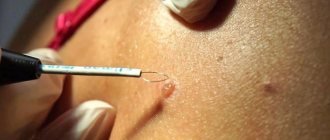
How to remove moles, nevi, papillomas and other skin formations
First, the skin is treated with an antiseptic. Then local anesthesia is performed with an anesthetic (lidocaine, novocaine). The anesthetic is injected into the skin with a syringe or by applying a special anesthetic cream.
When the anesthetic has begun to take effect, the doctor takes the loop waveguide, turns on the Surgitron device and removes the formation using a radio wave.
Everything happens very quickly and painlessly. Moreover, if the neoplasm then needs to be sent for histological examination, then the mole or papilloma is removed at the root.
Kuznetsova Ekaterina Olegovna
Nutritionist, gastroenterologist, cosmetologist
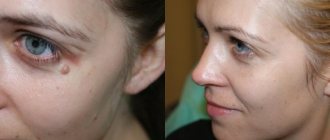
You can also remove a mole or papilloma using Surgitron using smooth fractional movements, removing the formation tissue layer by layer. After such removal, virtually no traces will remain on the skin.
After removal, the wound is smoothed with a ball waveguide. In addition, tissues are coagulated to stop bleeding.
The final stage is treating the tumor removal site with an aqueous solution of chlorhexidine or fucorcin (or another antiseptic).
Advantages and disadvantages of radio wave removal of keratomas
The method of radio wave surgery has recently become increasingly popular. This is quite natural, since this procedure for getting rid of tumors on the skin boasts a huge number of advantages compared to other methods.
First of all, the radioknife allows the surgeon to regulate the radiation power, thereby controlling the depth of penetration of the waves. As a result, skin formations are removed very carefully, without damaging surrounding tissues. As a result, unaesthetic scars do not appear at the site of removed tumors.
A tangible advantage of the radio wave method of removing keratomas is the absence of pain. Since the radiation is directed, it does not irritate nerve endings and does not provoke muscle contraction. Therefore, during the procedure the patient practically does not feel any discomfort.
Since the radioknife not only cuts, but also coagulates tissue, the removal process is bloodless. This eliminates the development of inflammatory processes and infection. In addition, the likelihood of developing postoperative edema and other complications is minimized.
Another undeniable advantage of this method is the ability to remove tumors located in hard-to-reach places. Also, the removed tissue can be sent for histological analysis to exclude the possibility that malignant degeneration has begun.
In fact, the radio wave method of removing keratomas has only one drawback: it is used only in cases where it is necessary to remove a formation of small diameter. If the area of the keratoma is large enough, other methods are usually used.
How is radio wave exposure performed with Surgitron in gynecology?
As with dermatological treatments, anesthesia is first performed. The doctor then removes the mass on the cervix with a loop electrode, either in one pass or, more often, in several passes.
After this, the wound is treated with a ball electrode, radio wave coagulation of the bleeding vessels is carried out and the wound on the cervix is smoothed.
At the end of the operation, the doctor treats the wound with an aqueous solution of chlorhexidine or another aqueous antiseptic.
The entire operation takes only 7-10 minutes.
Types of keratomas
Dermatologists distinguish several types of keratomas. For example, after forty years, so-called senile formations may appear - spots of a white or grayish tint, the diameter of which may increase over time. They occur most often on the neck, face and on the back of the hands. Sometimes these spots can become crusty and inflamed.
Content:
- Types of keratomas
- In what cases should a keratoma be removed?
- The essence of the radio wave method
- Advantages and disadvantages of radio wave removal of keratomas
- Contraindications to radio wave removal
- How is radio wave removal performed?
- How to behave after radio wave removal
- Possible complications
Solar keratomas appear most often in men with fair skin. They occur in those areas of the body that are most often exposed to sunlight. These neoplasms can be considered as harbingers of skin cancer, and therefore require mandatory medical consultation.
One of the most dangerous is seborrheic keratoma - a yellowish-brown spot that over time begins to peel and bleed, causing inflammation. These formations also require mandatory medical monitoring, as do horny keratomas, which are most prone to transform into malignant tumors. Follicular keratomas are the least common. Women are more susceptible to them than men. These neoplasms usually appear on the upper lip and scalp and are pinkish-gray nodules.
Rehabilitation after radio wave exposure in gynecology
In the first day after exposure, there may be a slight nagging pain in the lower abdomen. You can take a Nurofen tablet to relieve pain.
Wound healing occurs under the fibrin film within 7-10 days. From 4-5 days, partial rejection of the fibrin film begins. From this time on, the woman may have slight bleeding from the vagina. They should not be abundant and should not have an unpleasant odor. The discharge can last up to 14-20 days, gradually decreasing.
After surgery, it is recommended to insert suppositories with antiseptics into the vagina (for example, Depantol).
After the operation, you cannot have sex, take a hot bath or go to the sauna, play sports, or do heavy physical work for a month.
Types of tumors removed by radio wave method
Removal of neoplasms using the radio wave method includes the removal of moles, vascular mesh, keratosis, papilloma, fibroma. It is necessary to get rid of tumors, especially if there are certain indications. Each growth on the skin is fraught with a certain danger:
Warts
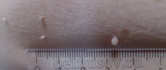
Warts are neoplasms that arise at the junction of the two upper layers of skin - the epidermis and dermis. The formation rises above the surface of the skin and has a dense stratum corneum. Children are frightened by the fact that warts appear because they touch frogs and toads, and they allegedly infect them with an unknown virus. Warts are actually a type of human papillomavirus.
Sometimes warts appear in adolescence (juvenile warts) during a period of rapid hormonal changes and weakened immunity. Neoplasms in children are eliminated on their own as soon as adolescence passes and immunity is restored. Plantar warts are dangerous. In appearance, they resemble calluses, but constant friction threatens injury and further degeneration into a more aggressive form.
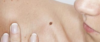
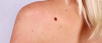
Moles
Moles are benign neoplasms found in almost every person. Moles or naevus maternus are made up of melanin-producing cells. They are dangerous due to their degeneration into basal cell cancer cells and melanoma, a malignant skin tumor. Moles larger than 1 cm in size, with swelling and pigmentation of varying intensity should be removed.
There are various ways to get rid of nevi and moles on the skin, but one of the safest methods is to remove tumors using the radio wave method.
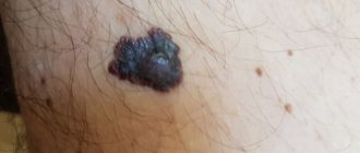
Keratoma
Keratoma is a benign proliferation of epidermal cells (keratinocytes). Due to the exposure of the skin to ultraviolet light, the keratinized cells stop exfoliating, and they accumulate and become keratinized. Keratomas often become injured and bleed. They can degenerate into cancer, so it is recommended to remove them.
Fibroma
Cutaneous fibroma looks like a dense sac localized under a fold of skin. If its height does not exceed 2 cm, it is better not to touch the fibroma.
Papillomas
Papillomas are skin formations that are the result of infection with the human papillomavirus. About 90% of the world's population is infected with HPV, but not all neoplasms have a risk factor. It all depends on the strain of the virus, and sometimes skin manifestations go away on their own. But they have an unaesthetic appearance, so they are often removed from open areas of the skin.
Condylomas
Condylomas are viral tumors on the mucous surfaces of the body. Transmitted only through sexual contact. Some strains are especially dangerous because in 99.9% of cases they cause cervical cancer in women.
Condylomas on the surface of the uterus provoke miscarriage, and in men they cause bladder cancer. Removal of such tumors is carried out using the radio wave method, because it is the most effective and safe.
What to do after the procedure
The radioknife acts on the tumor, evaporating the tissue. A crust forms at the site of exposure, which quickly heals without leaving a scar. After the procedure, the wound should be treated with a solution of vodka and iodine, and after the crust falls off, with Panthenol wound spray.
If you have at least once applied for removal of tumors, avoid being in direct sunlight. Moles, keratomas and other formations indicate that the skin has a low protective reaction to ultraviolet radiation; melatonin is produced unevenly, accumulating in damaged areas. Even when going outside normally, you should use creams with a high SPF content.
Be sure to strengthen your immune system, because HPV indicates a weakening of the body’s immune functions. After the procedure, you should visit a dermatologist every year.
Keratoma removal methods
There are different ways to treat skin growths classified as keratoma:
- Removal of keratoma by using liquid nitrogen.
A widespread and affordable method that allows you to remove several keratomas in one session, however, it has a number of disadvantages: rough scars remain, a long recovery period, and complications are possible.
- Removal of keratoma by electrocoagulation.
By applying layer-by-layer current to the tissue around the keratoma and the vascular walls, it is possible to prevent bleeding and infection. The crusts disappear 7–10 days after the procedure. After removal of large defects, subtle, light-colored scars remain. Small keratomas disappear without a trace.
- Radio wave method.
Recognized as milder in impact, but no less traumatic.
- Laser removal of keratoma. One of the most effective modern techniques. Its essence lies in the painless “evaporation” of the tumor by exposure to high temperature. Thanks to the cooling system equipped with the laser equipment, the patient does not experience discomfort. It does not matter where the keratoma is located. Removal of keratoma using a laser is possible on any part of the body, including the hairline. The wound heals in just 1–2 weeks. There are practically no traces left. There are no age restrictions on the use of the laser method.
Keratoma removal Price
Keratoma, the removal of which by laser is relatively inexpensive, can be removed at different prices: the price range for laser keratoma removal is determined by the number of skin defects to be removed, their depth, complexity and other characteristics.
Our hair removal and cosmetology center “SkinLazerMed” specializes in a wide range of procedures that involve laser technology. We guarantee high-quality and painless keratoma removal on the same day of treatment.
How is the Laser Keratoma Removal procedure performed?
Removing a keratoma in this way is based on a laser beam affecting the affected area of the skin.
- Before starting the procedure, the dermatologist collects an anamnesis, finds out the characteristics of the patient’s health condition, possible contraindications to these manipulations, and tolerability of medications. These data allow you to determine the optimal power of the laser beam and the duration of its exposure, which allows you to perform the removal in one visit.
- A powerful cooling system is built into the equipment intended for exposure, this explains the comfort of laser keratoma removal. However, if the patient wishes, a subcutaneous injection of an anesthetic can be given to ensure that no discomfort occurs during the session.
- When it comes to a disease such as keratoma, laser removal is represented by stepwise “evaporation” of the affected tissue until the beam reaches a healthy layer of skin. During this procedure, bleeding occurs extremely rarely, but a small wound appears on the affected area. It is treated with antiseptic medications.
- The removed formation - keratoma, laser removal of which should be carried out only by a highly qualified specialist, should be monitored by a doctor for several more weeks.
The SkinLazerMed clinic will perform laser keratoma removal at an affordable price. The procedure is quick, effective and completely painless.