All people have moles on their bodies.
Among them there may be a birthmark that looks like a wart, called a warty nevus.
The danger of such a neoplasm is that it is capable of degeneration into a malignant form.
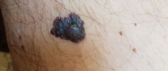
A warty (verrucous) nevus can be recognized by the presence of a bumpy surface.
The mole looks like a head of cabbage or has the appearance of a garland.
Nevus is dangerous, since such neoplasms often degenerate into a cancerous tumor.
A mole may appear some time after the baby is born, and sometimes it forms much later.
- Initially, a convex formation appears on the surface of the child’s skin. As it grows, the formation darkens and becomes brown or yellowish.
- As the child grows, the mole also becomes more noticeable, but there is no growth in width. The size of the nevus increases due to the proliferation of epithelial tissue that it affects.
- Over time, the surface of the neoplasm becomes keratinized and the mole becomes convex. As a result of injury to the upper layer, the neoplasm becomes larger.
In most cases, moles are not dangerous, but sometimes they become so as a result of degeneration into a malignant form.
Why does a warty nevus appear?
Scientists have come to the conclusion that the formation of moles begins at the embryonic stage of development.
Factors that provoke their formation in humans are:
- imbalance of female sex hormones in a woman during pregnancy
- the presence of infectious pathologies during pregnancy
- the influence of unfavorable environmental factors on a woman’s body during pregnancy
- hereditary factors
As a result of these reasons, the development process of melanoblasts, from which melanocytes are formed, is disrupted.
Melanocytes, concentrating in certain areas of the skin, turn into nevocytes.
Their difference from melanocytes is that they do not have processes that contribute to the spread of pigment.
Cryodestructive method for raised moles
This technique is based on exposing mole tissue to extremely low temperatures. The procedure is quite simple; it involves touching the mole with a medical instrument or a regular cotton swab dipped in liquefied nitrogen. When this touch occurs, the cells freeze. This leads to their rupture as a result of the expansion of water freezing in them. After these manipulations, their life activity becomes impossible, and they die. After the procedure, a patch of healthy skin remains at the site of the mole without noticeable traces of intervention.
The cryodestruction session is short - just a few minutes, after which the patient is sent home. When removing large nevi, the procedure can be painful and require the use of an anesthetic.
Can a nevus turn into cancer?
A birthmark that forms in the upper layer of the skin poses a risk of degeneration.
Malignancy of a warty nevus can occur with repeated damage.
Please note the following changes:
- mole color change
- hair loss on a mole
- change in size towards larger tumors
- appearance of asymmetry
- presence of discharge from the nevus
- uneven edges
If at least one of the signs appears, you should immediately visit a doctor.
Laser therapy to remove moles
Today, this procedure is most in demand due to the large number of advantages: the absence of marks on the skin, bleeding, and the risk of infection in the postoperative period. One cannot fail to note the rapid recovery, which is not available for most procedures for removing the skin defects in question.
During the procedure, the laser beam acts exclusively on the tissue of the mole, which guarantees maximum accuracy. Healthy skin tissue is not affected, even if the CO2 laser light reaches it. Under the influence of the laser, the cells in the mole warm up to a temperature of 70-80 °C, which leads to their peculiar evaporation on the upper layers of the nevus. Evaporation is carried out layer by layer until the laser beam reaches the lower layers of the mole and eliminates it completely.
The only disadvantage of this method is the fact that the removal of large convex nevi sometimes requires several sessions, which means that the patient will have to visit the operating room more than once, and he will see the result only months after the start of therapy.
Types of warty nevus
There are localized forms of nevus manifestation or systemic (verrucous).
Localized - most moles are found in this form.
Signs of its manifestation:
- The diameter of the neoplasm does not exceed 1 cm.
- elements appear separately or are located so close that they can merge
- localization zone is clearly limited
- the keratinized surface of the nevus often has cracks
- moles are dark brown or maroon in color. In very rare cases, nevi are light brown or light pink in color.
- the area affected by moles is often devoid of hair
Systemic form of the disease - the incidence of pathology is approximately 20% of cases.
A distinctive feature of a nevus is the presence of a “garland” of neoplasms.
Such elements are located at close distances relative to each other and “stretch” along the surface of the skin along the entire body.
This form of nevus has a number of features:
- the elements spread along large vessels, and they can also be in different places of the body
- The surface layer of moles may differ in shade. Elements may have different color intensities
- the length of one garland can reach up to 20 cm.
- When feeling the moles, it is noted that they have a dense structure
- the surface of each mole is lumpy and tortuous. As a person ages, these symptoms become more pronounced.
Each type of warty mole can be frequently injured.
Characteristic manifestations of verrucous nevus
The neoplasms rise above the skin by 2 cm, but sometimes they can be higher.
Moles look like a cluster of a large number of papilloma-like growths, which are located very close to each other and can merge into a single whole.
The surface of the new growths is slightly rough and quite dense.
The color of verrucous nevus varies from flesh-colored to dark brown.
Growths can appear on any part of the skin surface.
Both single neoplasms and large numbers can be observed, including they can be fused and have an individual shape, size and color.
Verrucous nevus is characterized by the presence of elements on one side of the body (right or left).
When located on the extremities, the localization of neoplasms is noted in the area of large vessels and nerve bundles.
Keratotic nevus grows very slowly, possibly the growth of new elements.
An increase in the size of moles is more often observed in the longitudinal direction than in the transverse direction.
The presence of moles does not cause discomfort, they do not cause pain or itching, and do not interfere with any movements.
Their presence on the body has a pronounced cosmetic disadvantage, especially if they are located on open areas of the body or on the face.
Basic recommendations after removal of nevi
Regardless of the reason that prompted the patient to remove a raised mole, some restrictions should then be followed. These include avoiding contact with water for a week, avoiding sunbathing for several weeks, and using sunscreen in the future. The latter is relevant for all people who have many large convex nevi on the body. If for small tumors tanning for 10-15 minutes is acceptable, then for large moles even it can provoke the formation of cancer. If you stay on the beaches under an umbrella and don’t forget about ultraviolet protection, you don’t have to worry about skin cancer.
Attention!
This article is posted for informational purposes only and under no circumstances constitutes scientific material or medical advice and should not serve as a substitute for an in-person consultation with a professional physician.
For diagnostics, diagnosis and treatment, contact qualified doctors! Number of reads: 3291 Date of publication: 09.11.2018
Dermatologists - search service and appointment with dermatologists in Moscow
Diagnosis of keratotic nevus
Only a specialist can identify the presence of a warty nevus.
Based on external data and a conversation with the patient, the doctor can make a preliminary diagnosis.
For a more accurate diagnosis, laboratory tests will be required.
When examining a nevus, the doctor pays attention to the presence of the following features:
- coloring of moles
- size of tumors
- Locations of birthmarks
- shape of moles
- absence or presence of hair on the surface of the neoplasm
To confirm the diagnosis, laboratory tests are performed:
- oncocytology. A scraping is taken from the surface of the nevus. The disadvantage of the technique may be a traumatic factor, which can provoke the development of complications such as infection or degeneration into melanoma. Therefore, this study is prescribed for those patients whose moles have been traumatized;
- Lumenescence microscopy technique. Its essence is to apply a special composition to the neoplasm, after which the birthmark is examined under a microscope. This research method is considered to be the safest, since the nevus is not injured during the procedure;
- conducting a computed tomogram , which allows you to examine the internal structure of moles using x-rays;
- blood tests . The blood is examined for tumor markers - specific proteins that are produced when the body is prone to cancer;
- histology . A piece of tissue is taken for examination. The resulting biomaterial is subjected to histological examination for malignancy of the nevus. This technique is informative enough to confirm the development of a malignant process.
Radio wave removal of convex moles
This technique is based on the use of high-frequency waves, which, by causing the cells of moles to warm up, actually cut them off from the skin. As with laser removal, during this procedure the blood vessels are sealed, which ensures the absence of bleeding both during the removal of the mole and during the process of tissue regeneration. At the same time, the wound is disinfected and gently cauterized, which significantly accelerates the healing of the postoperative wound. As for the disadvantages, the main one is considered to be the high probability of burns to the skin surrounding the mole if it is accidentally affected. It is not uncommon for patients to experience scarring from thermal injury. It is worth saying that this rarely happens in high-level clinics, where experienced doctors work, therefore, when it becomes necessary to extract convex tumors using this method, it is better to choose a trusted medical center.
Treatment of warty nevus
The basis of treatment for keratotic nevus is its removal using one of the most suitable methods.
The choice of method depends on the location of the warty nevus, its shape, and size.
The specialist decides which destruction method will be most effective.
To get rid of the manifestations of skin defects, the following techniques can be used:
- use of liquid nitrogen (cryotherapy)
- application of electric current (electrocoagulation)
- removal by radio waves (radiotherapy)
- destruction with a laser beam (laser therapy)
How to remove a warty nevus
Removal of a verrucous nevus is carried out using one of the following methods.
Surgical excision of moles is a classic method of ridding a patient of skin tumors.
After anesthesia and antiseptic treatment of the skin and surface of the nevus, it is cut out with a scalpel.
Then the wound is treated with a disinfectant and a sterile gauze bandage is applied.
This technique has negative aspects: infection of the wound surface and the formation of a postoperative scar are possible.
Removal of a nevus using radio wave radiation - this technique has many advantages:
- the ability to control the depth of exposure to radio waves, which avoids damage to healthy tissue around the tumor
- absence of bleeding due to their coagulation
- the procedure is painless
- the procedure is carried out very quickly
- radio waves can be used on any area of the skin
- There are no negative consequences after surgery
Laser therapy is one of the most common.
The laser beam can be used for keratotic moles on the scalp, as well as in the facial area.
Benefits of using laser:
- it is impossible for infection to penetrate at the site of exposure
- Coagulation of the wound eliminates the development of bleeding
- after removing the growth, a crust remains
- the risk of scarring is minimal
Removing nevus with liquid nitrogen: the use of low temperatures allows you to remove small tumors.
The procedure is painless.
Pain appears some time after exposure to nitrogen.
A disadvantage of the technique is also the need for repeated sessions, since the technique does not allow accurately calculating the time of exposure of the pathological tissue to nitrogen.
Electrocoagulation method - high frequency currents are used for treatment.
Before removal, the doctor administers local anesthesia.
As a result of exposure to electric current, a crust forms at the site of removal, which protects the wound from infection.
How is mole removal carried out in the clinic?
In modern medical centers, specialists have various methods for eliminating raised moles:
- Laser therapy;
- Cryodestruction;
- Electrocoagulation;
- Radio wave method;
- Surgical method.
In any case, before using any technique, a primary examination is carried out. At this stage, the condition of the mole and surrounding skin is assessed, anamnesis is collected, and other pathologies are identified. If cell degeneration is suspected, the doctor will prescribe a series of tests.
If the decision is made to have surgery on a raised mole, the patient is referred for blood tests for HIV, hepatitis and clotting ability.
Preliminary observation in the ward, as well as postoperative observation, is not required. However, after the procedure, the patient must come to the clinic for a final examination. If stitches were applied, as, for example, when removing moles with a scalpel, you need to come for dressings and treat with an antiseptic at home.
Features of care for verrucous nevus
If elements of a wart nevus are found on a patient’s body, doctors suggest removing it to remove the cosmetic defect.
If the patient for some reason does not want or cannot do this, then he must follow a number of rules that will help avoid the development of complications.
The basic rules are as follows:
- Eliminate factors that may cause overheating of moles. Prohibited: visiting saunas, baths, spa treatments.
- In the warm season, avoid being under the rays of the sun during the hours of its greatest activity: after 10 a.m. and before 4 p.m. Avoid visiting the solarium. It has been proven that protective agents cannot prevent the development of melanoma;
- Before taking hormonal medications, you should first consult with a specialist.
- Monitor the condition of the nevus elements and consult a doctor at the first symptoms of malignancy of moles.
You should pay attention to the following changes:
- accelerated nevus growth
- the appearance of unpleasant sensations that were not there before: pain, itching, burning, etc.
- change in the color of the neoplasm, it may become multi-colored
- appearance of peeling
- formation of cracks on the nevus
- hair loss
- asymmetrical growth, torn edges
- acquisition of granularity by a mole
- formation of outgrowths
- the appearance of discharge of various types
If at least one of the above symptoms appears, you should immediately consult a doctor.
If treatment turns out to be timely, then birthmarks will not pose a danger to a person in terms of malignancy.
And modern treatment methods will eliminate the presence of discomfort, both physical and cosmetic.
It is important to understand that self-medication can cause irreparable harm to health.
Removal of tumors should only take place in a medical facility and be carried out by a specialist.
Histology after removal of a convex nevus
In many situations, the removed mole is sent for histological analysis. Its results help confirm or refute suspicions that an oncological disease has arisen in the body of a nevus. This possibility should not be neglected, since the presence of cancer cells is fraught with very serious consequences for a person, including death. The patient can receive histology results during his last visit to the clinic, when the doctor makes a final examination of the operation site.
This applies not only to surgical, but also to any other removal of raised moles - all of the listed methods require the presence of a certain amount of biomaterial to be examined. Some doctors prefer to play it safe and send human tissue for analysis, even if the suspicion of a cancerous tumor is very slight. This indicates a responsible attitude towards your work; you should definitely obtain the results of the analysis.
Why is a warty nevus dangerous?
This type of mole among other nevi is the safest.
A certain group of factors can contribute to its degeneration.
The possibility of infection due to injury to a mole can provoke the development of an inflammatory process.
If a keratotic nevus is localized on the head or in another area of increased risk of injury, they should be removed as quickly as possible after their formation.
In some cases, manifestations of a systemic warty neoplasm can serve as a sign of hydrocephalus and other lesions of the nervous system, as well as melanoma.
In this case, therapy should be carried out simultaneously by several specialists: oncologist, neurologist and dermatologist.
Forms and complications of nevi
There are melanocytic (containing pigment cells - melanocytes) and non-melanocytic nevi (formed by cells other than melanocytes), which include epidermal nevus, nevus of the sebaceous glands, Becker's nevus and some other benign formations, vascular nevi of the skin.
Melanocytic nevi
There are acquired and congenital melanocytic nevi.
Acquired melanocytic nevi (AMN) are benign skin tumors. As a rule, they do not have a tendency to undergo malignant transformation. Special forms of acquired melanocytic nevi include Spitz nevus, Reed's nevus, spotted nevus (spilus), halonevus (Setton), and blue nevus (Jadassohn-Tiche) lying in the deep layers of the skin.
Atypical (dysplastic) nevi (AN) are classified into a separate group. Unlike common acquired melanocytic nevi, atypical melanocytic nevi may have some clinical characteristics of melanoma, such as asymmetry, ill-defined borders, multiple colors, or a size greater than 6 mm. Their main difference from a malignant tumor is stability, absence of changes over time and similar characteristics in one person. Atypical melanocytic nevi have a relatively increased risk of malignant transformation and require careful monitoring.
Congenital melanocytic nevi occur as a developmental defect (hemartoma) and are usually present at birth. Large congenital melanocytic nevi are also associated with an increased (but still low) risk of malignant transformation. Very rarely, they can be associated with a pathological accumulation of melanocytes in the central nervous system (neurocutaneous melanosis). Congenital melanocytic nevi with the expected size in an adult (over the course of life they will increase in proportion to the child’s growth) also require increased attention and medical supervision.
Nevi of Ota and Ito also have a melanocytic structure, but they are not based on excessive cell division, but on their accumulation in the deep layers of the skin.
Preventive measures for warty nevus
Verrucous nevus can provoke the development of oncological pathology.
But with proper prevention it is possible to avoid complications.
Prevention of complications is as follows:
- eliminating the possibility of injury to neoplasms
- Seeing a doctor for excision of growths
- you need to forget forever about saunas and solariums. Even the use of sunscreen cosmetics cannot protect against the development of melanoma
- in hot weather, avoid going outside to prevent overheating of tumors
- stay indoors after 10 a.m. and before 4 p.m.
- If at least one of the signs of malignancy appears, immediately go to the doctor
- Regular visits to a specialist to monitor moles